pdf - Lipedema Simplified
advertisement
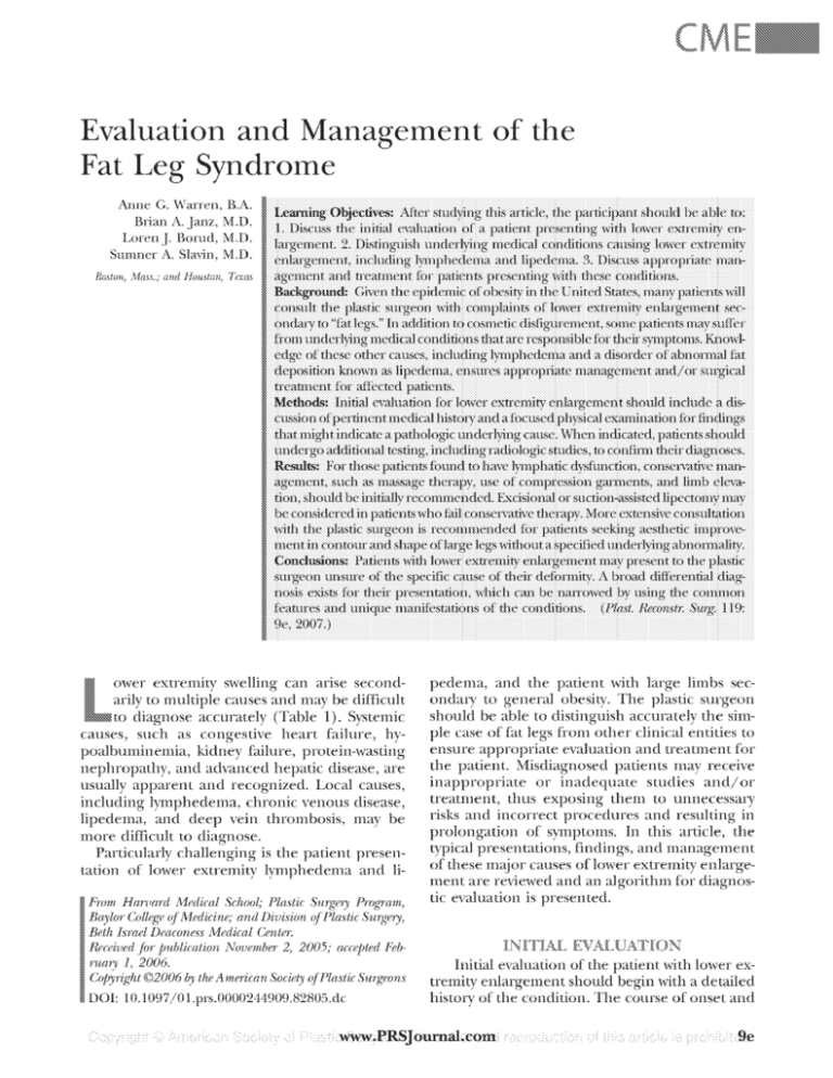
Evaluation and lVIanagement of tl1e Fat I_Jeg Syndro1ne Anne G. ~Warren, B.A. Brian A. Janz, M.D. Loren J Bomd, M.D. Sumner A. Slavin, M.D. Boston) ]\![ass.; and J!ouston; Texas ~ ower extremity swelling can arise secondarily to multiple causes and may be difficult ~to diagnose accurately (Table 1). Systemic causes, such as congestive heart failure, hypoalbuminemia, kidney failure, protein-wasting nephropathy, and advanced hepatic disease, are usually apparent and recognized. Local causes, including lymphedema, chronic venous disease, lipedema, and deep vein thrombosis, may be more difficult to diagnose. Particularly challenging is the patient presentation of lower extremity lymphedema and 1i- ~ ~From Haruard 1\/Iedical School; Plastic Surgery Program, pedema, and the patient \Vith large limbs secondary to general obesity. The plastic surgeon should be able to distinguish accurately the simple case of fat legs from other clinical entities to ensure appropriate evaluation and treatment for the patient. Misdiagnosed patients may receive inappropriate or in adequate studies and/ or treatment, thus exposing them to unnecessary risks and incorrect procedures and resulting in prolongation of symptoms. In this article, the typical presentations, findings, and management of these me:~ or causes oflower extremity enlargement are reviewed and an algorithm for diagnostic evaluation is presented. Collecre O'fr l'vJedicine; and Division O'frPlastic SurrYm, :s:~ Ba"lor J b' . . () ..' ~ Beth Israel Deaconess Niedical Center. ~ ruarv- I, 2006. ~ Received for jmblication November 2, 2005; acce[1ted Feb:& ~ ~ Copyright @2006 by the American Society ofPlastic Surp;eons ~DOl: l0.1097/0l.prs.0000244909.82805.dc - Initial evaluation of the patient ·with lower extremity enlargement should begin with a detailed history of the condition. The course of onset and ·~ . .\.Vww.PRSJournal.com Plastic and Reconstructive Surgery • January 2007 Table 1. Causes of Lower Extremity Swelling Systemic causes ·Increased capillary permeability (sepsis) Congestive heart failure Renal failure Hypoalbuminemia Prcitein-losing nephropathy Drug-induced edema Regional causes Chronic venous disease Deep vein thrombosis Lip edema Lymphedema Postoperative edema Cellulitis Baker's disease Cyclical and idiopathic edema Increased capillary permeability (burns) Myxedema the presence of associated symptoms can be quite revealing of the underlying abnormality, particularly when the patient describes an unremarkable development of symptoms consistent '"'rith generalized obesity and weight gain. Given the high incidence of obesity (defined as a body mass index > 30) in the United States, the number of patients complaining of lower extremity enlargement that can be attributed to obesity-related changes is not uncommon. Patients may notice a gradual progression in the size of their legs and may be unaware of similar changes in other regions of their body as their weight increases. Patients with pathologic causes, however, wi11 t)1JicaHy describe more dramatic presentations, such as leg swelling accompanied by pain, skin texture changes, or episodes of cellulitis. Any other significant medical history should also be elicited, with particular attention paid to history of malignancy, nodal dissection or other surgical procedures, and history of trauma. Physical examination should include thorough assessment of any asymmetry between leg volumes, which can be determined by circumferential measurements of the limbs at several points along the limb, including the maximal diameters at the level of the calf: the thigh just superior to the knee, and the thigh-buttock junction. Any edema of the legs or feet should be noted, as should the texture and consistency of the edema when present. Dermal abnonnalit-y, such as peau d' orange skin changes, should also be documented. The need for subsequent diagnostic evaluation is determined by relevant clinical findings. The patient's overall body habitus and the inability on physical examination to localize fatty tissue excess to a specific region should suggest gener- lOe alized obesity as the cause of their condition. However, particularly in patients who are overweight but not yet obese and in cases where lower extremity enlargement is symptomatic beyond psychological distress, other pathologic conditions may be responsible and further workup is incticated. Lymphedema and lipedema, tlvo related but distinct disorders leading to lower extremity swelling, have unique clinical features and findings on diagnostic testing and should be excluded as possible causes. Lymphedema refers to localized swelling, t:}1Jically of an extremity, secondary to lymphatic dysfunction. Causes of the condition are typically classified as primary or secondary. Primary lymphedema is further subdivided according to age at onset. Milroy's disease, or congenital hereditary lymphedema, describes lymphedema that is present at birth and is associated with certain genetic endothelial cell receptor mutations. 1•2 Less common are f2unilial lymphedema praecox (Meige's disease), which presents during puberty, and lyrnphedema tarda, which is the spontaneous onset of lymphedema in early adulthood. Secondary lymphedema is much more common and is typically secondary to malignancy or its treatment in the Western world, although the infectious disease filariasis is overwhelmingly responsible tor cases of ly1nphedema worldwide. 3 The classic characteristics oflymphedema and its distinction from lipedema are described in Table 2. Patients frequently report a history of prior cancer and subsequent surgical lymphatic disruption or radiation therapy. Swelling is typically unilateral, nonpainful, and may be associated '"ith recurrent episodes of cellulitis. Although earlystage lymphedema may stili be soft and pitting, by the time patients are accurately diagnosed and present to the plastic surgeon, the disease has frequently progressed to a state of fibrosis and Table 2. Clinical Features of Lymphedema and Lipedema Characteristic Sex Family history Cellulitis Bilateral involvement Foot involvement Malleolar bt nad Nature of swe'lling Pitting edema Tenderness Lymphedema Both Rare Occasional Occasional Common Absen1 Finn Variable Rare Lipedema Female Common Rare Common Never Present Soft Rare Common Volume 119, Number 1 • Fat Leg Syndrome nonplttmg edema (Fig. l). Lower extremity lymphedema can often be diagnosed by the positive Stemmer sign, 1 in which the dorsal skin of the foot cannot be pinched because of the significant fluid accumulation in the subcutaneous tissues. Although clinical presentation usually suggests lymphedema, the diagnosis is t)1Jically confirmed by lymphoscintigraphy (Fig. 2). This technique has largely supplanted the more invasive procedure of lymphangiography as a means of delineating ly1nphatic vessel function. 5 Computed tomography or magnetic resonance imaging may also be helpful in the initial evaluation if the diagnosis of lymphatic dysfunction is not readily apparent; findings include dermal thickening and fluid accumulation in the subcutaneous fat 6 (Table 3). Magnetic resonance imaging is typically recommended because of its high sensitivity in differentiating clinical causes. 7 On gross examination of excised lymphedema tissue, subcutaneous fibrous stranding is seen. Microscopically, chronic lymph stasis causes increased fibroblast Fig. 1. Bilateral !ower extremity lymphedema. The patient is a 40-year-old man who spontaneously developed bilateral lower extremity lymphedema 2 years before presentation. He had no previous history of surgical procedures to the groin or other predisposing risk factors aside from obesity. Conservative therapy with compression garment use and bandaging was recommended. Fig. 2. Abnormallymphoscintigram in severe bilateral !ower extremity lymphedema from the patient described above. After tracer injection into the first dorsa! web spaces of the feet, no evidence of lymphatic drainage from the injection sites into the lymph nodes of the !ower extremities is seen at either 3 or 5 hours of delay. and keratinocyte production, in addition to collagen deposition proliferation. Increased adipocyte deposition is also seen in these patients, which has been attributed to a physiologic response to a chronic inflammatory state. Many cases of lymphedema are best managed conservatively, particularly if they are mild or moderate. Compression garments, massage therapy, manually1nphatic drainage, and exercise have all proven effective in case series. 8 ·9 Serial monitoring through circumferential measurements of the affected limb or the use of newer technologies such as bioimpedance 10 or perimetry can be useful in documenting clinical response to these therapies. Given the prolonged, and in many situations indefinite, course of treatment using conservative therapies, numerous surgical techniques have been developed as a means of "curing" lymphedema. Surgical debulking through skin and subcutaneous excision has been the most popular surgical approach and can provide good long-tem1 benefits. 11 The use of suction-assisted lipectomy with subsequent continuous compression gam1ent use has also been described, with excellent outcomes. 12- 14 Lipedema refers to a syndrome of localized adipose deposition in the buttocks and lower extremities that is seen almost exclusively in women. It was first described by Allen and Hines in 1940, E· who later characterized the condition as one of "fat legs and edema." In their series on 119 patients diagnosed with lipedema, 16 they reported that the adiposity was seen bilaterally, progressed gradually, and was accentuated by activity and warm weather. Many of their patients complained of significant aching and pain in their lower extremities, particularly below the knee. Although the m<Yority of their patients were overweight, a He Plastic and Reconstructive Surgery • January 2007 Table 3. Diagnostic Findings in Lymphedema and Lipedema Test Lymphangiogram Lymphoscintigram CT scan MRI Lymphedema Lipede:ma Abnormal Abnormal with delayed lymphatic flow, subdermal collateralization, dermal backflow Calf skin thickening, thickening of the subcutaneous tissues, increased fat density, thickening of the perimuscular aponeurosis, honeycomb appearance caused by fibrous and edematous stranding of fat Skin thickening, subcutaneous tissue thickening, honeycomb appearance Normal Normal, with mild lymphatic flow delay seen in some patients Diffuse and homogenous lipomatous hy1;ertrophy of subcutaneous tissue Normal skin thickness, increased fatty tissue CT, con1puted to!nographic; MRt rnagnetic resonance i1naging. significant number were of normal weight and were found to have disproportionately enlarged lower extremities. In addition, those that were ovenveight reported no change in lower extremity size with dieting or weight loss. The typical onset of presentation of signs and symptoms associated with lipedema is during or soon after puberty. A family history of "similarappearing fat legs" is frequently elicited from the patientY'· 17 Patients describe progressive enlargement of their lower extremities with associated sensations of heaviness and discomfort. They also express significant frustration and embarrassment at the social stigma of the condition and have frequently attempted numerous diuretic, compressive, and dietary therapies to no avaiL On physical examination, patients are found to have bilateral enlargement of the legs, thighs, and buttocks (Fig. 3). The skin is soft and the edema is typica11y nonpitting. The subcutaneous tissue excess begins abruptly above the malleoli, causing a region of sharp demarcation between normal and abnormal tissue at the ankle, leading to a ring-like deformity in contour (Fig. 4). This sparing of the feet is unique to lipedema among the various causes for lower extremity edema 18 and allows the physician to distinguish the condition from lymphedema, as the Stemmer sign will be absent in cases of lipedema (Fig. 5). This dramatic characteristic also allows for distinction benveen lipedema and obesity-associated large legs, which can be difficult to differentiate when evaluating the rest of the lower extremities (Fig. 6). The diagnosis of lipedema can usually be made clinically, although patients frequently undergo more extensive radiologic evaluation as part of a workup for lymphedema or vascular disease. The disting11ishing radiologic findings seen in patients \Vith lipedema6·7•19 are described in Table 3. Pathologically, the subcutaneous tissue seen in these patients is soft and lacks the fibrotic elements so fre- 12e Fig. 3. Lower extremity enlargement consistent with lipedema. The patient is a 66-year-o!d woman who reported abnormal appearance to her legs and thighs bilateraiiysincetheageof 5 years. She complained of chronic lower extremity pain, fatigue, and easy bruising. She had not worn clothing that exposed her legs throughout her life because of embarrassment at her condition. A prior lymphoscintigram showed normal lymphatic function. quently seen in patients with lymphedema. Histology reveals no specific abnonnalities, 20 •21 aside from edematous adipose cells with moderate hyperplasia. 22 One case series also described a high prevalence of microlymphatic aneurysms in affected limbs. 23 Lipedema is a difficult condition to treat, particularly becasue patients have frequently been misdiagnosed and consequently received prior in- Volume 119, Number 1 • Fat Leg Syndrome Fig. 4. Contour deformity of the ank!e in l!pedema. The excess subcutaneous fat seen in the !ower !eg ceases abruptly at the level of the malleoli, sparing the feet and leading to a ring-like appearance of the ankle. Fig.6. Large lower extremities secondary to obesity. The patient is a 46-year-old woman who described 5 years of gradual enlargement of her lower extremities. She had a history of two gastric bypass procedures and had subsequently lost 75 pounds, although she was stiil quite overweight (body mass index of 29.3). A lymphoscintigram was performed to exclude spontaneous onset of lymphedema, which was negative. Afterfu rtherweig ht loss and plateau, the patient chose to undergo bilateral medial thigh lifts. erative outcomes, 17 •2'1 although reports using suction lipectomy have been more promising. 17 ·25 Fig. 5. Negative Stemmer sign in li pedema. The dorsa I skin of the foot is easily elevated because of the absence of edema or fibro-sis. appropriate therapy. Although dietary and lifestyle changes may provide some improvement in the subset of obese patients, these efforts are usually ineft"txtive. The use of compression garments has shown some success, 20 although this may be most effective in patients with long-standing lipedema who have developed subsequent secondary lymphedema caused by progressive mechanical insufficiency of the lymphatic system. Surgical therapy has traditiona11y involved surgical de bulking procedures, which have shown varied postop- Based on clinical presentation, patients should undergo further evaluation when indicated to determine appropriate management and therapy. Patients who are suspected of having lymphedema should have lyrnphoscintigraphy testing to confirm lymphatic dysfunction or obstruction. Those with presumed venous abnormality should be referred for vascular surgery. vVhen lipedema or fat legs secondary to obesity are suspected or diagnosed, further discussion with the plastic surgeon can be held to discuss potential surgical management. ·when lyrnphoscintigraphy demonstrates abnormal lymphatic flow, thus confirming a diagnosis of lymphedema, initial conservative therapy involving compression bandaging and gannent use should be prescribed. Patients found to have lipedema may be counseled on the use of these compressive tech- 13e Plastic and Reconstructive Surgery • January 2007 Lower Extremity Enlargement ~, Clinical Exam • • , Lvmghedema Lipedema Non-pitting edema involving digits; cellulitis, peau d"orange, and/or(+) Obesitv-associated large legs Symmetric, soft, nonpitting bilateral edema: no swelling in the feet Symmetric, soft, bilateral enlargement; obese or overweight patient • Ste.mn1er's siffi t CT! MRl: Thickened skin, "honeycombing" of tluid in fibrous tissue CT I MRI: Bilateral subcutaneous l~'lt hypertrophy Lymphoscinti graphy: Abnormal lymphatics Lymphoscintigraphy: Nonnal lymphatics • (+) * H • • Conservative therapy Reassess (+) (-) • • Consider liposuction • CT/tviRI: Normal UIS: No evidence of thrombosis •• Consider cosmetic surgery, inclL!Lling liposuction or excisional surgery (i.e. thigh lift) Reassess Fig. 7. Algorithm for evaluation and management of lower extremity enlargement. niques as well, although affected patients should be advised that these therapies '"'ill likely not be particularly effective and that surgical treatment through excisional and/ or suction-assisted lipectomy may be required for more significant improvement. Those patients vvitb fat legs associated vvith obesity should be counseled on modifying their lifestyles and be given the option of future cosmetic procedures if they are able to achieve sufficient weight loss to become better surgical candidates, which is typically considered after patients have lowered their body mass index below 30. Patients '"'ith lower extremity enlargement may present to the plastic surgeon having been given one of many diagnoses that may or may not accurately represent their condition. Using key 14e differences in the clinical presentations of the causes allows for the most effective and efficient management; an algorithm to guide this process in managing lymphedema, lipedema, and obesityrelated large lower extremities is presented in Figure 7. Knowledge of the common clinical features of these conditions and their unique radiologic manifestations ensures appropriate management for affected patients and shapes surgical decisionmaking and planning. Sumner A.. Sla:vin, M.D.~ 1101 Beacon Street~ Brookline, Mass. 024,16 ~ sslavin@ bidmc .harvard. ed u ~ None of the authors has a financial interest in any of the products, devices, or drugs mentioned in this article. Volume 119, Number 1 • Fat Leg Syndrome 1. Irrthum, A., Karkkainen, M.J., Devriendt, K., Alitalo, K., and Vikkula, M. Congenital hereditary lymphedema caused by a rnut~_tion that inactivates \lEGFR3 tyrosine kinase. il.rn. J Hum. Genet. 67: 295, 2000. 2. Karkhinen, M. J., Ferrell, R. E., Lawrence, E. C., et al. Missense mutations interfere with VEGFR-3 signalling in primary lymphoedema. Nat. Genet. 25: 153, 2000. 3. Szuba, A., Shin, 'vV. S., Strauss, H. Vi., and Rockson, S. The third circulation: Radionuclide lymphoscintigraphy in the evaluation of lymphedema. J Nucl. Aled. '11: 13, 2003. 4. Stemmer, R. A clinical symptom for the early and differential diagnosis of lymphedema. Fasa 5: 261, 1976. 5. \Vitte. C. L., 1/v'itte, 1v1. H,~ lJnger, E. C., et aL A.dvances in imaging of lymph flow disorders. Radiographies 20: 1697, 2000 6. Dimakakos, P. B., Stefanopoulos, T., i\ntoniades, P., Antonicm, A., Gouliamos, A., and Rizos, D. MRI and ultrasonographic findings in the investigation of lymphedema and lipedema. Int. Swg. 82: 411, 1997. 7. Duewell, S., Hagspiel, K. D., Zuber,]., von Schulthess, G. K., Bollinger, A., and Fuchs, 'N. A. Swollen lower extremity: Role of MR in1aging. Radiology 184: 227, 1992. 8. Hinrichs, C. S., Gibbs,J. F., Driscoll, D., et al. The effectiveness of complete decongestive physiotherapy for the treatment of lymphedema following groin dissection for melanoma. J Su~r;. Oneal. 85: 187, 2004. 9. Cheville, A. L, McGarvey, C. L, Petrek, J. A., Russo, S. A., Taylor, M. E., and Thiadens, S. R. Lymphedema management. Sernin. Radiat. Oneal. 13: 290, 2003. 10. Cornish, B. H., Bunce, I. H., Ward, L. C., Jones, L C., and Thomas, B . .J. Bioelectrical impedance for monitoring the efficacy of lymphoeden1a treatn1ent progran1mes. Breast Cancer Res. Treat. 38: 169, 1996. 11. Miller, T. A. Surgical approach to lymphedema of the arm after mastectomy. Am. J Surg 148: 152, 1984. 12. Brorson, H. Liposuction in arm lymphedema treatment. Scand J Surg. 92: 287, 2003. 13. Brorson, H. Liposuction gives complete reduction of chronic large arm lymphedema after breast cancer. Acta Oneal. 39: 407, 2000. 14. Greene, A. K., Slavin, S. A., and Borud, L J. Treatment of lower extremity lymphedema with suction assisted lipectomy. ?last. Reconstr. Surg 118: 118e, 2006. 15. iiJlen, E. V., and Hines, E. A., .Jr. Lipedema of the legs: A syndrome charanerized by fat legs and orthostatic edema. Proc. Staff A1eet. Mayo Clin. 15: 184, 1940. 16. Wold, L E., Hines, E. A., Jr., and Allen, E. V. Lipederna of the legs: A syndrome characterized by fat legs and edema. Ann. Intern. 1\1ed. 34: 1243, 1951. 17. Rudkin, G. H., and l'viiller, T. A. Lipedema: A clinical entity distinnfrom lymphedema. Plast. Reconstr. Sur_g. 94:841, 1994. 18. Harwood, C. A., Bull, R H., Evans, J., and iv!ortirner, P. S. Lymphatic and venous function in lipoedema. Br. J Derrnatol 134: 1, 1996. 19. Monnin-Delhom, E. D., Gallix, B. P., Achard, C., Bmel,J. M., and.Janbon, C. High resolution unenhanced computed tomography in patients with swollen legs. Lyrnphology 35: 121. 2002. 20. Beninson, .J., and Edelglass, J. W. Lipedema: The non-lymphatic masquerader. Angiology 35: 506, 1984. 2l. Stallworth, .J. M., Hennigar, G. R., .Jonsson, H. T., Jr., and Rodriguez, 0. The chronically swollen painful extremity: A detailed study for possible etiological factors. JA..lvL4.. 228: 1656, 1974. 2~) Bilancini, S., Lucchi, M., Tucci, S., and Eleuteri, P. Func-tional lymphatic alterations in patients suffering from lipedema. Angiology 46: 333, 1995. 23. i\mann-Vesti, B. R, Franzeck, LT. K., and Bollinger, A. Microlymphatic aneurysms in patients with lipedema. Lyrnphoiogy 34: 170, 20(l1. 21. Rank, B. K., and Wong, G. S. Lipoedema. Aust. N Z J S1ng. 35: 166, 1966. 25. Chen, S. G., Hsu, S.D., Chen, T. M., and Wang, H.J. Painful fat sy11elrome in a male patient. B-r. J Plast Swg. .57: 282, 2004. Hie

