Ch 29) Molecules and Solids
advertisement
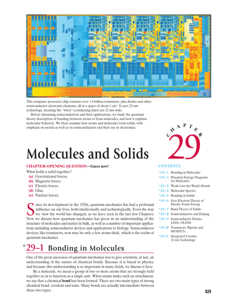
This computer processor chip contains over 1.4 billion transistors, plus diodes and other semiconductor electronic elements, all in a space of about 1 cm2. It uses 22-nm technology, meaning the “wires” (conducting lines) are 22 nm wide. Before discussing semiconductors and their applications, we study the quantum theory description of bonding between atoms to form molecules, and how it explains molecular behavior. We then examine how atoms and molecules form solids, with emphasis on metals as well as on semiconductors and their use in electronics. H A P T E C 29 CHAPTER-OPENING QUESTION—Guess now! What holds a solid together? (a) Gravitational forces. (b) Magnetic forces. (c) Electric forces. (d) Glue. (e) Nuclear forces. S ince its development in the 1920s, quantum mechanics has had a profound influence on our lives, both intellectually and technologically. Even the way we view the world has changed, as we have seen in the last few Chapters. Now we discuss how quantum mechanics has given us an understanding of the structure of molecules and matter in bulk, as well as a number of important applications including semiconductor devices and applications to biology. Semiconductor devices, like transistors, now may be only a few atoms thick, which is the realm of quantum mechanics. * 29–1 R Molecules and Solids CONTENTS *29–1 Bonding in Molecules *29–2 Potential-Energy Diagrams for Molecules *29–3 Weak (van der Waals) Bonds *29–4 Molecular Spectra *29–5 Bonding in Solids *29–6 Free-Electron Theory of Metals; Fermi Energy *29–7 Band Theory of Solids *29–8 Semiconductors and Doping *29–9 Semiconductor Diodes, LEDs, OLEDs *29–10 Transistors: Bipolar and MOSFETs *29–11 Integrated Circuits, 22-nm Technology Bonding in Molecules One of the great successes of quantum mechanics was to give scientists, at last, an understanding of the nature of chemical bonds. Because it is based in physics, and because this understanding is so important in many fields, we discuss it here. By a molecule, we mean a group of two or more atoms that are strongly held together so as to function as a single unit. When atoms make such an attachment, we say that a chemical bond has been formed. There are two main types of strong chemical bond: covalent and ionic. Many bonds are actually intermediate between these two types. 829 * Covalent Bonds Nucleus (⫹1e) Nucleus (⫹1e) FIGURE 29–1 Electron probability distribution (electron cloud) for two H atoms when the spins are the same: S = 12 + 12 = 1. FIGURE 29–2 Electron probability distribution (cloud) around two H atoms when the spins are opposite (S = 0): in this case, a bond is formed because the positive nuclei are attracted to the concentration of the electron cloud’s negative charge between them. This is a hydrogen molecule, H 2 . Nucleus (⫹1e) Nucleus (⫹1e) To understand how covalent bonds are formed, we take the simplest case, the bond that holds two hydrogen atoms together to form the hydrogen molecule, H 2 . The mechanism is basically the same for other covalent bonds. As two H atoms approach each other, the electron clouds begin to overlap, and the electrons from each atom can “orbit” both nuclei. (This is sometimes called sharing electrons.) If both electrons are in the ground state (n = 1) of their respective atoms, there are two possibilities: their spins can be parallel (both up or both down), in which case the total spin is S = 12 + 12 = 1; or their spins can be opposite (ms = ± 12 for one, and ms = – 12 for the other), so that the total spin S = 0. We shall now see that a bond is formed only for the S = 0 state, when the spins are opposite. First we consider the S = 1 state, for which the spins are the same. The two electrons cannot both be in the lowest energy state and be attached to the same atom, for then they would have identical quantum numbers in violation of the exclusion principle. The exclusion principle tells us that, because no two electrons can occupy the same quantum state, if two electrons have the same quantum numbers, they must be different in some other way—namely, by being in different places in space (for example, attached to different atoms). Thus, for S = 1, when the two atoms approach each other, the electrons will stay away from each other as shown by the probability distribution of Fig. 29–1. The electrons spend very little time between the two nuclei, so the positively charged nuclei repel each other and no bond is formed. For the S = 0 state, on the other hand, the spins are opposite and the two electrons are consequently in different quantum states (ms is different, ± 12 for one, – 12 for the other). Hence the two electrons can come close together, and the probability distribution looks like Fig. 29–2: the electrons can spend much of their time between the two nuclei. The two positively charged nuclei are attracted to the negatively charged electron cloud between them, and this is the attraction that holds the two hydrogen atoms together to form a hydrogen molecule. This is a covalent bond. The probability distributions of Figs. 29–1 and 29–2 can perhaps be better understood on the basis of waves. What the exclusion principle requires is that when the spins are the same, there is destructive interference of the electron wave functions in the region between the two atoms. But when the spins are opposite, constructive interference occurs in the region between the two atoms, resulting in a large amount of negative charge there. Thus a covalent bond can be said to be the result of constructive interference of the electron wave functions in the space between the two atoms, and of the electrostatic attraction of the two positive nuclei for the negative charge concentration between them. Why a bond is formed can also be understood from the energy point of view. When the two H atoms approach close to one another, if the spins of their electrons are opposite, the electrons can occupy the same space, as discussed above. This means that each electron can now move about in the space of two atoms instead of in the volume of only one. Because each electron now occupies more space, it is less well localized. From the uncertainty principle with ¢x larger, we see that ¢p and the minimum momentum can be less. With less momentum, each electron has less energy when the two atoms combine than when they are separate. That is, the molecule has less energy than the two separate atoms, and so is more stable. An energy input is required to break the H 2 molecule into two separate H atoms, so the H 2 molecule is a stable entity. This is what we mean by a bond. The energy required to break a bond is called the bond energy, the binding energy, or the dissociation energy. For the hydrogen molecule, H 2 , the bond energy is 4.5 eV. 830 CHAPTER 29 Molecules and Solids * Ionic Bonds An ionic bond is, in a sense, a special case of the covalent bond. Instead of the electrons being shared equally, they are shared unequally. For example, in sodium chloride (NaCl), the outer electron of the sodium spends nearly all its time around the chlorine (Fig. 29–3). The chlorine atom acquires a net negative charge as a result of the extra electron, whereas the sodium atom is left with a net positive charge. The electrostatic attraction between these two charged atoms holds them together. The resulting bond is called an ionic bond because it is created by the attraction between the two ions (Na± and Cl–). But to understand the ionic bond, we must understand why the extra electron from the sodium spends so much of its time around the chlorine. After all, the chlorine atom is neutral; why should it attract another electron? The answer lies in the probability distributions of the electrons in the two neutral atoms. Sodium contains 11 electrons, 10 of which are in spherically symmetric closed shells (Fig. 29–4). The last electron spends most of its time beyond these closed shells. Because the closed shells have a total charge of –10e and the nucleus has charge ±11e, the outermost electron in sodium “feels” a net attraction due to ±1e. It is not held very strongly. On the other hand, 12 of chlorine’s 17 electrons form closed shells, or subshells (corresponding to 1s22s22p63s2). These 12 electrons form a spherically symmetric shield around the nucleus. The other five electrons are in 3p states whose probability distributions are not spherically symmetric and have a form similar to those for the 2p states in hydrogen shown in Figs. 28–9b and c. Four of these 3p electrons can have “doughnutshaped” distributions symmetric about the z axis, as shown in Fig. 29–5. The fifth can have a “barbell-shaped” distribution (as for ml = 0 in Fig. 28–9b), which in Fig. 29–5 is shown only in dashed outline because it is half empty. That is, the exclusion principle allows one more electron to be in this state (it will have spin opposite to that of the electron already there). If an extra electron—say from a Na atom—happens to be in the vicinity, it can be in this state, perhaps at point x in Fig. 29–5. It could experience an attraction due to as much as ±5e because the ±17e of the nucleus is partly shielded at this point by the 12 inner electrons. Thus, the outer electron of a sodium atom will be more strongly attracted by the ±5e of the chlorine atom than by the ±1e of its own atom. This, combined with the strong attraction between the two ions when the extra electron stays with the Cl–, produces the charge distribution of Fig. 29–3, and hence the ionic bond. + – Na Cl FIGURE 29–3 Probability distribution for the outermost electron of Na in NaCl. FIGURE 29–4 In a neutral sodium atom, the 10 inner electrons shield the nucleus, so the single outer electron is attracted by a net charge of ±1e. −10e Last (3s) electron −e +11e x ⫺12 e ⫹17e ⫺4e FIGURE 29–5 Neutral chlorine atom. The ±17e of the nucleus is shielded by the 12 electrons in the inner shells and subshells. Four of the five 3p electrons are shown in doughnut-shaped clouds (seen in cross section at left and right), and the fifth is in the dashed-line cloud concentrated about the z axis (vertical). An extra electron at x will be attracted by a net charge that can be as much as ±5e. ⫺1e * Partial Ionic Character of Covalent Bonds A pure covalent bond in which the electrons are shared equally occurs mainly in symmetrical molecules such as H 2 , O2 , and Cl 2 . When the atoms involved are different from each other, usually the shared electrons are more likely to be in the vicinity of one atom than the other. The extreme case is an ionic bond. In intermediate cases the covalent bond is said to have a partial ionic character. *SECTION 29–1 831 H (⫹) ⫹1e ⫹8e O (⫺) ⫹1e H (⫹) FIGURE 29–6 The water molecule H 2O is polar. FIGURE 29–7 Potential energy pe as a function of separation r for two point charges of (a) like sign and (b) opposite sign. pe The molecules themselves are polar—that is, one part (or parts) of the molecule has a net positive charge and other parts a net negative charge. An example is the water molecule, H 2O (Fig. 29–6). Covalent bonds have shared electrons, which in H2O are more likely to be found around the oxygen atom than around the two hydrogens. The reason is similar to that discussed above in connection with ionic bonds. Oxygen has eight electrons A1s22s22p4 B, of which four form a spherically symmetric core and the other four could have, for example, a doughnut-shaped distribution. The barbell-shaped distribution on the z axis (like that shown dashed in Fig. 29–5) could be empty, so electrons from hydrogen atoms can be attracted by a net charge of ±4e. They are also attracted by the H nuclei, so they partly orbit the H atoms as well as the O atom. The net effect is that there is a net positive charge on each H atom (less than ±1e), because the electrons spend only part of their time there. And, there is a net negative charge on the O atom. * 29–2 Potential-Energy Diagrams for Molecules It is useful to analyze the interaction between two objects—say, between two atoms or molecules—with the use of a potential-energy diagram, which is a plot of the potential energy versus the separation distance. For the simple case of two point charges, q1 and q2 , the potential energy PE is given by (we combine Eqs. 17–2a and 17–5) pe = k Repulsive force (two like charges) 0 (a) r pe r 0 Attractive force (unlike charges) q1 q2 , r where r is the distance between the charges, and the constant k A= 1兾4p⑀ 0 B is equal to 9.0 * 109 N⭈m2兾C 2. If the two charges have the same sign, the potential energy is positive for all values of r, and a graph of PE versus r in this case is shown in Fig. 29–7a. The force is repulsive (the charges have the same sign) and the curve rises as r decreases; this makes sense because if one particle moves freely toward the other (r getting smaller), the repulsion slows it down so its KE gets smaller, meaning PE gets larger. If, on the other hand, the two charges are of the opposite sign, the potential energy is negative because the product q1 q2 is negative. The force is attractive in this case, and the graph of PE (r –1兾r) versus r looks like Fig. 29–7b. The potential energy becomes more negative as r decreases. Now let us look at the potential-energy diagram for the formation of a covalent bond, such as for the hydrogen molecule, H 2 . The potential energy PE of one H atom in the presence of the other is plotted in Fig. 29–8. Starting at large r, the pe decreases as the atoms approach, because the electrons concentrate between the two nuclei (Fig. 29–2), so attraction occurs. However, at very short distances, the electrons would be “squeezed out”—there is no room for them between the two nuclei. Without the electrons between them, each nucleus would feel a repulsive force due to the other, so the curve rises as r decreases further. (b) pe FIGURE 29–8 Potential-energy diagram for the H 2 molecule; r is the separation of the two H atoms. The binding energy (the energy difference between pe = 0 and the lowest energy state near the bottom of the well) is 4.5 eV, and r0 = 0.074 nm. 0 r0 Binding energy This part corresponds to repulsive force 832 CHAPTER 29 Molecules and Solids r This part corresponds to attractive force Lowest energy state There is an optimum separation of the atoms, r0 in Fig. 29–8, at which the energy is lowest. This is the point of greatest stability for the hydrogen molecule, and r0 is the average separation of atoms in the H 2 molecule. The depth of this “well” is the binding energy,† as shown. This is how much energy must be put into the system to separate the two atoms to infinity, where the pe = 0. For the H 2 molecule, the binding energy is about 4.5 eV and r0 = 0.074 nm. For many bonds, the potential-energy curve has the shape shown in Fig. 29–9. There is still an optimum distance r0 at which the molecule is stable. But when the atoms approach from a large distance, the force is initially repulsive rather than attractive. The atoms thus do not form a bond spontaneously. Some additional energy must be injected into the system to get it over the “hump” (or barrier) in the potential-energy diagram. This required energy is called the activation energy. pe Activation energy r0 0 r Binding energy Repulsion Attraction FIGURE 29–9 Potential-energy diagram for a bond requiring an activation energy. Repulsion The curve of Fig. 29–9 is much more common than that of Fig. 29–8. The activation energy often reflects a need to break other bonds, before the one under discussion can be made. For example, to make water from O2 and H 2 , the H 2 and O2 molecules must first be broken into H and O atoms by an input of energy; this is what the activation energy represents. Then the H and O atoms can combine to form H 2O with the release of a great deal more energy than was put in initially. The initial activation energy can be provided by applying an electric spark to a mixture of H 2 and O2 , breaking a few of these molecules into H and O atoms. When these atoms combine to form H 2O, a lot of energy is released (the ground state is near the bottom of the well) which provides the activation energy needed for further reactions: additional H 2 and O2 molecules are broken up and recombined to form H 2O. The potential-energy diagrams for ionic bonds, such as NaCl, may be more like Fig. 29–8: the Na± and Cl– ions attract each other at distances a bit larger than some r0 , but at shorter distances the overlapping of inner electron shells gives rise to repulsion. The two atoms thus are most stable at some intermediate separation, r0 . For partially ionic bonds, there is usually an activation energy, Fig. 29–9. Sometimes the potential energy of a bond looks like that of Fig. 29–10. In this case, the energy of the bonded molecule, at a separation r0 , is greater than when there is no bond (r = q). That is, an energy input is required to make the bond (hence the binding energy is negative), and there is energy release when the bond is broken. Such a bond is stable only because there is the barrier of the activation energy. This type of bond is important in living cells, for it is in such bonds that energy can be stored efficiently in certain molecules, particularly ATP (adenosine triphosphate). The bond that connects the last phosphate group (designated ¬ in Fig. 29–10) to the rest of the molecule (ADP, meaning adenosine diphosphate, since it contains only two phosphates) has potential energy of the shape shown in Fig. 29–10. Energy is stored in this bond. When the bond is broken (ATP S ADP + ¬ ), energy is released and this energy can be used to make other chemical reactions “go.” † The binding energy corresponds not quite to the bottom of the potential-energy curve, but to the lowest quantum energy state, slightly above the bottom, as shown in Fig. 29–8. *SECTION 29–2 PHYSICS APPLIED ATP and energy in the cell FIGURE 29–10 Potential-energy diagram for the formation of ATP from ADP and phosphate (¬). pe r0 r ATP ADP + P Potential-Energy Diagrams for Molecules 833 In living cells, many chemical reactions have activation energies that are often on the order of several eV. Such energy barriers are not easy to overcome in the cell. This is where enzymes come in. They act as catalysts, which means they act to lower the activation energy so that reactions can occur that otherwise would not. Enzymes act via the electrostatic force to distort the bonding electron clouds, so that the initial bonds are easily broken. * 29–3 + C – O 0.12 nm 0.29 nm + – H N 0.10 nm FIGURE 29–11 The C ±—O – and H ±—N – dipoles attract each other. (These dipoles may be part of, for example, the nucleotide bases cytosine and guanine in DNA molecules. See Fig. 29–12.) The ± and – charges typically have magnitudes of a fraction of e. PHYSICS APPLIED DNA Weak (van der Waals) Bonds Once a bond between two atoms or ions is made, energy must normally be supplied to break the bond and separate the atoms. As mentioned in Section 29–1, this energy is called the bond energy or binding energy. The binding energy for covalent and ionic bonds is typically 2 to 5 eV. These bonds, which hold atoms together to form molecules, are often called strong bonds to distinguish them from so-called “weak bonds.” The term weak bond, as we use it here, refers to an attachment between molecules due to simple electrostatic attraction—such as between polar molecules (and not within a polar molecule, which is a strong bond). The strength of the attachment is much less than for the strong bonds. Binding energies are typically in the range 0.04 to 0.3 eV—hence their name “weak bonds.” Weak bonds are generally the result of attraction between dipoles. (A pair of equal point charges q of opposite sign, separated by a distance l, is called an electric dipole, as we saw in Chapter 17.) For example, Fig. 29–11 shows two molecules, which have permanent dipole moments, attracting one another. Besides such dipole;dipole bonds, there can also be dipole;induced dipole bonds, in which a polar molecule with a permanent dipole moment can induce a dipole moment in an otherwise electrically balanced (nonpolar) molecule, just as a single charge can induce a separation of charge in a nearby object (see Fig. 16–7). There can even be an attraction between two nonpolar molecules, because their electrons are moving about: at any instant there may be a transient separation of charge, creating a brief dipole moment and weak attraction. All these weak bonds are referred to as van der Waals bonds, and the forces involved van der Waals forces. The potential energy has the general shape shown in Fig. 29–8, with the attractive van der Waals potential energy varying as 1兾r6. The force decreases greatly with increased distance. When one of the atoms in a dipole–dipole bond is hydrogen, as in Fig. 29–11, it is called a hydrogen bond. A hydrogen bond is generally the strongest of the weak bonds, because the hydrogen atom is the smallest atom and can be approached more closely. Hydrogen bonds also have a partial “covalent” character: that is, electrons between the two dipoles may be shared to a small extent, making a stronger, more lasting bond. Weak bonds are very important for understanding the activities of cells, such as the double helix shape of DNA (Fig. 29–12), and DNA replication H H T G C A T (a) 834 CHAPTER 29 Molecules and Solids – H + ha in C 52 in A A N ha T C G T G C C A C G T T A H 0.290 nm Guanine (G) O – N C + H C 0 .3 C 0 0 nm C C N H N – C N N C + – + 0.290 nm C N O H N – + – H 1.08 nm 54 Cytosine (C) To c A C G T A G c To FIGURE 29–12 (a) Model of part of a DNA double helix. The red dots represent hydrogen bonds between the two strands. (b) “Close-up” view: cytosine (C) and guanine (G) molecules on separate strands of a DNA double helix are held together by the hydrogen bonds (red dots) involving an H ± on one molecule attracted to an N – or C ±—O – of a molecule on the adjacent chain. See also Section 16–10 and Figs. 16–39 and 16–40. (b) (see Section 16–10). The average kinetic energy of molecules in a living cell at normal temperatures (T L 300 K) is around 32 kT L 0.04 eV (kinetic theory, Chapter 13), about the magnitude of weak bonds. This means that a weak bond can readily be broken just by a molecular collision. Hence weak bonds are not very permanent—they are, instead, brief attachments. This helps them play particular roles in the cell. On the other hand, strong bonds—those that hold molecules together—are almost never broken simply by molecular collision because their binding energies are much higher (L 2 to 5 eV). Thus they are relatively permanent. They can be broken by chemical action (the making of even stronger bonds), and this usually happens in the cell with the aid of an enzyme, which is a protein molecule. EXAMPLE 29;1 Nucleotide energy. Calculate the potential energy between a C ± – O – dipole of the nucleotide base cytosine and the nearby H ± – N – dipole of guanine, assuming that the two dipoles are lined up as shown in Fig. 29–11. Dipole moment (= ql) measurements (see Table 17–2 and Fig. 29–11) give qH = –qN = 3.0 * 10 –30 C⭈m = 3.0 * 10 –20 C = 0.19e, 0.10 * 10–9 m qC = –qO = 8.0 * 10–30 C⭈m = 6.7 * 10 –20 C = 0.42e. 0.12 * 10 –9 m and APPROACH We want to find the potential energy of the two charges in one dipole due to the two charges in the other, because this will be equal to the work needed to pull the two dipoles infinitely far apart. The potential energy of a charge q1 in the presence of a charge q2 is pe = kAq1 q2兾r12 B where k = 9.0 * 109 N⭈m2兾C 2 and r12 is the distance between the two charges. (See Eqs. 17–2 and 17–5.) SOLUTION The potential energy consists of four terms: pe = peCH + peCN + peOH + peON where peCH means the potential energy of C in the presence of H, and similarly for the other terms. We do not have terms corresponding to C and O, or N and H, because the two dipoles are assumed to be stable entities. Then, using the distances shown in Fig. 29–11, we get: pe = k B qC qH qC qN qO qH qO qN + + + R rCH rCN rOH rON = (9.0 * 109 N⭈m2兾C 2)(6.7 * 10 –20 C)(3.0 * 10–20 C) ¢ = A9.0 * 109 N⭈m2兾C 2 BA6.7BA3.0B A10–20 CB 2 A10 –9 mB ¢ 1 1 1 1 + ≤ rCH rCN rOH rON 1 1 1 1 + ≤ 0.31 0.41 0.19 0.29 = –1.86 * 10 –20 J = –0.12 eV. The potential energy is negative, meaning 0.12 eV of work (or energy input) is required to separate the dipoles. That is, the binding energy of this “weak” or hydrogen bond is 0.12 eV. This is only an estimate, of course, since other charges in the vicinity would have an influence too. *SECTION 29–3 Weak (van der Waals) Bonds 835 DNA New protein chain of 4 amino acids (a 5th is being added) 1 A C C T G C A A T G T G Growing end of m-RNA A 2 3 C t-RNA 4 5 Ribosome G U U A C A C Anticodons G U A A C G C A U U G C Codon 4 m-RNA Codon 5 * Protein Synthesis FIGURE 29–13 Protein synthesis. The yellow rectangles represent amino acids. See text for details. PHYSICS APPLIED Protein synthesis Weak bonds, especially hydrogen bonds, are crucial to the process of protein synthesis. Proteins serve as structural parts of the cell and as enzymes to catalyze chemical reactions needed for the growth and survival of the organism. A protein molecule consists of one or more chains of small molecules known as amino acids. There are 20 different amino acids, and a single protein chain may contain hundreds of them in a specific order. The standard model for how amino acids are connected together in the correct order to form a protein molecule is shown schematically in Fig. 29–13. We begin at the DNA double helix: each gene on a chromosome contains the information for producing one protein. The ordering of the four bases, A, C, G, and T, provides the “code,” the genetic code, for the order of amino acids in the protein. First, the DNA double helix unwinds and a new molecule called messenger-RNA (m-RNA) is synthesized using one strand of the DNA as a “template.” m-RNA is a chain molecule containing four different bases, like those of DNA (Section 16–10) except that thymine (T) is replaced by the similar uracil molecule (U). Near the top left in Fig. 29–13, a C has just been added to the growing m-RNA chain in much the same way that DNA replicates (Fig. 16–40); and an A, attracted and held close to the T on the DNA chain by the electrostatic force, will soon be attached to the C by an enzyme. The order of the bases, and thus the genetic information, is preserved in the m-RNA because the shapes of the molecules only allow the “proper” one to get close enough so the electrostatic force can act to form weak bonds. Next, the m-RNA is buffeted about in the cell (recall kinetic theory, Chapter 13) until it gets close to a tiny organelle known as a ribosome, to which it can attach by electrostatic attraction (on the right in Fig. 29–13), because their shapes allow the charged parts to get close enough to form weak bonds. (Recall that force decreases greatly with separation distance.) Also held by the electrostatic force to the ribosome are one or two transfer-RNA (t-RNA) molecules. These t-RNA molecules “translate” the genetic code of nucleotide bases into amino acids in the following way. There is a different t-RNA molecule for each amino acid and each combination of three bases. On one end of a t-RNA molecule is an amino acid. On the other end of the t-RNA molecule is the appropriate “anticodon,” a set of three nucleotide bases that “code” for that amino acid. If all three bases of an anticodon match the three bases of the “codon” on the m-RNA (in the sense of G to C and A to U), the anticodon is attracted electrostatically to the m-RNA codon and that t-RNA molecule is held there briefly. The 836 CHAPTER 29 Molecules and Solids ribosome has two particular attachment sites which hold two t-RNA molecules while enzymes bond the two amino acids together to lengthen the amino acid chain (yellow in Fig. 29–13). As each amino acid is connected by an enzyme (four are already connected in Fig. 29–13, top right, and a fifth is about to be connected), the old t-RNA molecule is removed—perhaps by a random collision with some molecule in the cellular fluid. A new one soon becomes attracted as the ribosome moves along the m-RNA. This process of protein synthesis is often presented as if it occurred in clockwork fashion—as if each molecule knew its role and went to its assigned place. But this is not the case. The forces of attraction between the electric charges of the molecules are rather weak and become significant only when the molecules can come close together, and when several weak bonds can be made. Indeed, if the shapes are not just right, the electrostatic attraction is nearly zero, which is why there are few mistakes. The fact that weak bonds are weak is very important. If they were strong, collisions with other molecules would not allow a t-RNA molecule to be released from the ribosome, or the m-RNA to be released from the DNA. If they were not temporary encounters, metabolism would grind to a halt. As each amino acid is added to the next, the protein molecule grows in length until it is complete. Even as it is being made, this chain is being buffeted about in the cell—we might think of a wiggling worm. But a protein molecule has electrically charged polar groups along its length. And as it takes on various shapes, the electric forces of attraction between different parts of the molecule will eventually lead to a particular shape of the protein which is quite stable. Each type of protein has its own special shape, depending on the location of charged atoms. In the last analysis, the final shape depends on the order of the amino acids. * 29–4 Molecular Spectra When atoms combine to form molecules, the probability distributions of the outer electrons overlap and this interaction alters the energy levels. Nonetheless, molecules can undergo transitions between electron energy levels just as atoms do. For example, the H 2 molecule can absorb a photon of just the right frequency to excite one of its ground-state electrons to an excited state. The excited electron can then return to the ground state, emitting a photon. The energy of photons emitted by molecules can be of the same order of magnitude as for atoms, typically 1 to 10 eV, or less. Additional energy levels become possible for molecules (but not for atoms) because the molecule as a whole can rotate, and the atoms of the molecule can vibrate relative to each other. The energy levels for both rotational and vibrational levels are quantized, and are generally spaced much more closely (10 –3 to 10–1 eV) than the electronic levels. Each atomic energy level thus becomes a set of closely spaced levels corresponding to the vibrational and rotational motions, Fig. 29–14. Transitions from one level to another appear as many very closely spaced lines. In fact, the lines are not always distinguishable, and these spectra are called band spectra. Each type of molecule has its own characteristic spectrum, which can be used for identification and for determination of structure. We now look in more detail at rotational and vibrational states in molecules. 3p FIGURE 29–14 (a) The individual energy levels of an isolated atom become (b) bands of closely spaced levels in molecules, as well as in solids and liquids. 2s Isolated atom Atom in a molecule (a) (b) *SECTION 29–4 Molecular Spectra 837 * Rotational Energy Levels in Molecules We consider only diatomic molecules, although the analysis can be extended to polyatomic molecules. When a diatomic molecule rotates about its center of mass as shown in Fig. 29–15, its kinetic energy of rotation (see Section 8–7) is (Iv)2 1 2 , Erot = Iv = 2 2I where Iv is the angular momentum (Section 8–8). Quantum mechanics predicts quantization of angular momentum just as in atoms (see Eq. 28–3): m1 r1 cm r2 r m2 Rotation axis where l is an integer called the rotational angular momentum quantum number. Thus the rotational energy is quantized: FIGURE 29–15 Diatomic molecule rotating about a vertical axis. FIGURE 29–16 Rotational energy levels and allowed transitions (emission and absorption) for a diatomic molecule. Upward-pointing arrows represent absorption of a photon, and downward arrows represent emission of a photon. l=5 ΔE 4h2 I l=3 l=2 l=1 l=0 2 6 hI U2 U2 l(l + 1) (l - 1)(l) 2I 2I U2 is for upper d (29;2) = l. c lenergy state I We see that the transition energy increases directly with l. Figure 29–16 shows some of the allowed rotational energy levels and transitions. Measured absorption lines fall in the microwave or far-infrared regions of the spectrum (energies L 10–3 eV), and their frequencies are generally 2, 3, 4, p times higher than the lowest one, as predicted by Eq. 29–2. ΔE 3h2 I EXERCISE A Determine the three lowest rotational energy states (in eV) for a nitrogen molecule which has a moment of inertia I = 1.39 * 10–46 kg⭈m2. ΔE 2h2 I EXAMPLE 29;2 Rotational transition. A rotational transition l = 1 to l = 0 for the molecule CO has a measured absorption wavelength l1 = 2.60 mm (microwave region). Use this to calculate (a) the moment of inertia of the CO molecule, and (b) the CO bond length, r. APPROACH The absorption wavelength is used to find the energy of the absorbed photon, and we can then calculate the moment of inertia, I, from Eq. 29–2. The moment of inertia is related to the CO separation (bond length r). SOLUTION (a) The photon energy, E = hf = hc兾l, equals the rotational energy level difference, ¢Erot . From Eq. 29–2, we can write h2 3 I 1 h2 I 0 = l(l + 1) ¢Erot = El - El - 1 = 2 2 10 hI (Iv)2 U2 . l = 0, 1, 2, p . (29;1) 2I 2I Transitions between rotational energy levels are subject to the selection rule (as in Section 28–6): ¢l = &1. The energy of a photon emitted or absorbed for a transition between rotational states with angular momentum quantum number l and l - 1 will be Erot = 15 hI ΔE 5h2 I l=4 l = 0, 1, 2, p , Iv = 2l(l + 1) U , ΔE h2 I U2 hc . l = ¢Erot = hf = I l1 With l = 1 (the upper state) in this case, we solve for I: A6.63 * 10–34 J⭈sBA2.60 * 10 –3 mB hl1 U 2l l1 = = hc 4p2c 4p2 A3.00 * 108 m兾sB –46 2 = 1.46 * 10 kg⭈m . I = (b) The molecule rotates about its center of mass (cm) as shown in Fig. 29–15. Let m1 be the mass of the C atom, m1 = 12 u, and let m2 be the mass of the O, m2 = 16 u. The distance of the cm from the C atom, which is r1 in Fig. 29–15, is given by the cm formula, Eq. 7–9: r1 = 838 CHAPTER 29 0 + m2 r 16 = r = 0.57r. m1 + m2 12 + 16 The O atom is a distance r2 = r - r1 = 0.43r from the cm. The moment of inertia of the CO molecule about its cm is then (see Example 8–9) I = m1 r21 + m2 r22 = C(12 u)(0.57r)2 + (16 u)(0.43r)2 D C1.66 * 10 –27 kg兾uD = A1.14 * 10–26 kgB r2. We solve for r and use the result of part (a) for I: r = 1.46 * 10–46 kg⭈m2 B 1.14 * 10 –26 kg = 1.13 * 10–10 m = 0.113 nm L 0.11 nm. EXERCISE B What are the wavelengths of the next three rotational transitions for CO? * Vibrational Energy Levels in Molecules The potential energy of the two atoms in a typical diatomic molecule has the shape shown in Fig. 29–8 or 29–9, and Fig. 29–17 again shows the pe for the H 2 molecule (solid curve). This pe curve, at least in the vicinity of the equilibrium separation r0 , closely resembles the potential energy of a harmonic oscillator, pe = 12 kx2, which is shown superposed in dashed lines. Thus, for small displacements from r0 , each atom experiences a restoring force approximately proportional to the displacement, and the molecule vibrates as a simple harmonic oscillator (SHO)—see Chapter 11. According to quantum mechanics, the possible quantized energy levels are Evib = An + 12 Bhf, n = 0, 1, 2, p, (29;3) where f is the classical frequency (see Chapter 11—f depends on the mass of the atoms and on the bond strength or “stiffness”) and n is an integer called the vibrational quantum number. The lowest energy state (n = 0) is not zero (as for rotation), but has E = 12 hf. This is called the zero-point energy. Higher states have energy 32 hf, 52 hf, and so on, as shown in Fig. 29–18. Transitions between vibrational energy levels are subject to the selection rule ¢n = &1, so allowed transitions occur only between adjacent states†, and all give off (or absorb) photons of energy ¢Evib = hf. (29;4) This is very close to experimental values for small n. But for higher energies, the pe curve (Fig. 29–17) begins to deviate from a perfect SHO curve, which affects the wavelengths and frequencies of the transitions. Typical transition energies are on the order of 10 –1 eV, roughly 10 to 100 times larger than for rotational transitions, with wavelengths in the infrared region of the spectrum A L 10–5 mB. EXAMPLE 29;3 Vibrational energy levels in hydrogen. Hydrogen molecule vibrations emit infrared radiation of wavelength around 2300 nm. (a) What is the separation in energy between adjacent vibrational levels? (b) What is the lowest vibrational energy state? APPROACH The energy separation between adjacent vibrational levels is (Eq. 29–4) ¢Evib = hf = hc兾l. The lowest energy (Eq. 29–3) has n = 0. SOLUTION A6.63 * 10 –34 J⭈sBA3.00 * 108 m兾sB hc (a) ¢Evib = hf = = = 0.54 eV, l A2300 * 10–9 mBA1.60 * 10 –19 J兾eVB where the denominator includes the conversion factor from joules to eV. (b) The lowest vibrational energy has n = 0 in Eq. 29–3: Evib = An + 12 Bhf = 1 2 hf pe 1 SHO ( 2 kx2) 0.1 nm 0 r r0 0.074 nm H2 molecule 4.5 eV FIGURE 29–17 Potential energy for the H 2 molecule and for a simple harmonic oscillator (pe = 12 kx2, with ∑x∑ = @r - r0 @ ). FIGURE 29–18 Allowed vibrational energies for a diatomic molecule, where f is the fundamental frequency of vibration (see Chapter 11). The energy levels are equally spaced. Transitions are allowed only between adjacent levels (¢ = &1). Vibrational quantum number v 5 4 E Vibrational energy 11 hf 2 9 hf 2 3 7 hf 2 2 5 hf 2 E Energy 1 3 hf 2 0 1 hf 2 = 0.27 eV. EXERCISE C What is the energy of the first vibrational state above the ground state in the hydrogen molecule? Forbidden transitions with ¢ = 2 are emitted with much lower probability, but their observation can be important in some cases, such as in astronomy. † *SECTION 29–4 839 * 29–5 (a) (b) (c) FIGURE 29–19 Arrangement of atoms in (a) a simple cubic crystal, (b) face-centered cubic crystal (note the atom at the center of each face), and (c) body-centered cubic crystal. Each of these “cells” is repeated in three dimensions to the edges of the macroscopic crystal. FIGURE 29–20 Diagram of an NaCl crystal, showing the “packing” of atoms. Bonding in Solids Quantum mechanics has been a great tool for understanding the structure of solids. This active field of research today is called solid-state physics, or condensed-matter physics so as to include liquids as well. The rest of this Chapter is devoted to this subject, and we begin with a brief look at the structure of solids and the bonds that hold them together. Although some solid materials are amorphous in structure (such as glass), in that the atoms and molecules show no long-range order, we are interested here in the large class of crystalline substances whose atoms, ions, or molecules are generally accepted to form an orderly array known as a lattice. Figure 29–19 shows three of the possible arrangements of atoms in a crystal: simple cubic, face-centered cubic, and body-centered cubic. The NaCl crystal lattice is shown in Fig. 29–20. The molecules of a solid are held together in a number of ways. The most common are by covalent bonding (such as between the carbon atoms of the diamond crystal) and by ionic bonding (as in a NaCl crystal). Often the bonds are partially covalent and partially ionic. Our discussion of these bonds earlier in this Chapter for molecules applies equally well to solids. Let us look for a moment at the NaCl crystal of Fig. 29–20. Each Na± ion feels an attractive Coulomb potential due to each of the six “nearest neighbor” Cl– ions surrounding it. Note that one Na± does not “belong” exclusively to one Cl–, so we must not think of ionic solids as consisting of individual molecules. Each Na± also feels a repulsive Coulomb potential due to other Na± ions, although this is weaker since the Na± ions are farther away. A different type of bond occurs in metals. Metal atoms have relatively loosely held outer electrons. Metallic bond theories propose that in a metallic solid, these outer electrons roam rather freely among all the metal atoms which, without their outer electrons, act like positive ions. According to the theory, the electrostatic attraction between the metal ions and this negative electron “gas” is responsible, at least in part, for holding the solid together. The binding energy of metal bonds is typically 1 to 3 eV, somewhat weaker than ionic or covalent bonds (5 to 10 eV in solids). The “free electrons” are responsible for the high electrical and thermal conductivity of metals. This theory also nicely accounts for the shininess of smooth metal surfaces: the free electrons can vibrate at any frequency, so when light of a range of frequencies falls on a metal, the electrons can vibrate in response and re-emit light of those same frequencies. Hence, the reflected light will consist largely of the same frequencies as the incident light. Compare this to nonmetallic materials that have a distinct color—the atomic electrons exist only in certain energy states, and when white light falls on them, the atoms absorb at certain frequencies, and reflect other frequencies which make up the color we see. Here is a brief comparison of important strong bonds: ⴚ Naⴙ Naⴙ Cl • ionic: an electron is “grabbed” from one atom by another; ⴙ Clⴚ Clⴚ Na • covalent: electrons are shared by atoms within a single molecule; Naⴙ ⴙ Clⴚ Na • metallic: electrons are shared by all atoms in the metal. The atoms or molecules of some materials, such as the noble gases, can form only weak bonds with each other. As we saw in Section 29–3, weak bonds have very low binding energies and would not be expected to hold atoms together as a liquid or solid at room temperature. The noble gases condense only at very low temperatures, where the atomic (thermal) kinetic energy is small and the weak attraction can then hold the atoms together. 840 CHAPTER 29 Molecules and Solids EXERCISE D Return to the Chapter-Opening Question, page 829, and answer it again now. Try to explain why you may have answered differently the first time. T0K Fermi level The free-electron theory of metals considers electrons in a metal as being in constant motion like an ideal gas, which we discussed in Chapter 13. For a classical ideal gas, at very low temperatures near absolute zero, T = 0 K, all the particles would be in the lowest state, with zero kinetic energy A= 32 kT = 0B. But the situation is vastly different for an electron gas because, according to quantum mechanics, electrons obey the exclusion principle and can be only in certain possible energy levels or states. Electrons also obey a quantum statistics called Fermi;Dirac statistics† that takes into account the exclusion principle. All particles that have spin 12 (or other half-integral spin: 32 , 52 , etc.), such as electrons, protons, and neutrons, obey Fermi–Dirac statistics and are referred to as fermions (see Section 28–7). The electron gas in a metal is often called a Fermi gas. According to the exclusion principle, no two electrons in the metal can have the same set of quantum numbers. Therefore, in each of the energy states available for the electrons in our “gas,” there can be at most two electrons: one with spin up Ams = ± 12 B and one with spin down Ams = – 12 B. Thus, at T = 0 K, the possible energy levels will be filled, two electrons each, up to a maximum level called the Fermi level. This is shown in Fig. 29–21, where the vertical axis is the “density of occupied states,” whose meaning is similar to the Maxwell distribution for a classical gas (Section 13–10). The energy of the state at the Fermi level is called the Fermi energy, EF . For copper, EF = 7.0 eV. This is very much greater than the energy of thermal motion at room temperature (G = 32 kT L 0.04 eV, Eq. 13–8). Clearly, all motion does not stop at absolute zero. At T = 0, all states with energy below EF are occupied, and all states above EF are empty. What happens for T 7 0? We expect that at least some of the electrons will increase in energy due to thermal motion. Figure 29–22 shows the density of occupied states for T = 1200 K, a temperature at which a metal is so hot it would glow. We see that the distribution differs very little from that at T = 0. We see also that the changes that do occur are concentrated about the Fermi level. A few electrons from slightly below the Fermi level move to energy states slightly above it. The average energy of the electrons increases only very slightly when the temperature is increased from T = 0 K to T = 1200 K. This is very different from the behavior of an ideal gas, for which kinetic energy increases directly with T. Nonetheless, this behavior is readily understood as follows. Energy of thermal motion at T = 1200 K is about 32 kT L 0.1 eV. The Fermi level, on the other hand, is on the order of several eV: for copper it is EF L 7.0 eV. An electron at T = 1200 K may have 7 eV of energy, but it can acquire at most only a few times 0.1 eV of energy by a (thermal) collision with the lattice. Only electrons very near the Fermi level would find vacant states close enough to make such a transition. Essentially none of the electrons could increase in energy by, say, 3 eV, so electrons farther down in the electron gas are unaffected. Only electrons near the top of the energy distribution can be thermally excited to higher states. And their new energy is on the average only slightly higher than their old energy. This model of free electrons in a metal as a “gas,” though incomplete, provides good explanations for the thermal and electrical conductivity of metals. Density of occupied states Free-Electron Theory of Metals; Fermi Energy Fermi energy E (eV) 0 2 4 6 FIGURE 29–21 At T = 0 K, all states up to energy EF , called the Fermi energy, are filled. (Shown here for copper.) FIGURE 29–22 The density of occupied states for the electron gas in copper. The width kT shown above the graph represents thermal energy at T = 1200 K. kT Density of occupied states * 29–6 unoccupied T 1200 K occupied 0 2 4 6 7 8 E (eV) † Developed independently by Enrico Fermi (Figs. 1–13, 27–11, 28–2, 30–7) in early 1926 and by P. A. M. Dirac a few months later. See Section 28–7. *SECTION 29–6 Free-Electron Theory of Metals; Fermi Energy 841 * 29–7 Band Theory of Solids We saw in Section 29–1 that when two hydrogen atoms approach each other, the wave functions overlap, and the two 1s states (one for each atom) divide into two states of different energy. (As we saw, only one of these states, S = 0, has low enough energy to give a bound H 2 molecule.) Figure 29–23a shows this situation for 1s and 2s states for two atoms: as the two atoms get closer (toward the left in Fig. 29–23a), the 1s and 2s states split into two levels. If six atoms come together, as in Fig. 29–23b, each of the states splits into six levels. If a large number of atoms come together to form a solid, then each of the original atomic levels becomes a band as shown in Fig. 29–23c. The energy levels are so close together in each band that they seem essentially continuous. This is why the spectrum of heated solids (Section 27–2) appears continuous. (See also Fig. 29–14 and its discussion at start of Section 29–4.) molecule atoms Allowed energy bands 1s Atomic separation (a) Energy gap Energy Energy Energy 2s Atomic separation (b) Atomic separation (c) FIGURE 29–23 The splitting of 1s and 2s atomic energy levels as (a) two atoms approach each other (the atomic separation decreases toward the left on the graph); (b) the same for six atoms, and (c) for many atoms when they come together to form a solid. FIGURE 29–24 Energy bands for sodium (Na). 3s 2p 2s 1s The crucial aspect of a good conductor is that the highest energy band containing electrons is only partially filled. Consider sodium metal, for example, whose energy bands are shown in Fig. 29–24. The 1s, 2s, and 2p bands are full (just as in a sodium atom) and don’t concern us. The 3s band, however, is only half full. To see why, recall that the exclusion principle stipulates that in an atom, only two electrons can be in the 3s state, one with spin up and one with spin down. These two states have slightly different energy. For a solid consisting of N atoms, the 3s band will contain 2N possible energy states. A sodium atom has a single 3s electron, so in a sample of sodium metal containing N atoms, there are N electrons in the 3s band, and N unoccupied states. When a potential difference is applied across the metal, electrons can respond by accelerating and increasing their energy, since there are plenty of unoccupied states of slightly higher energy available. Hence, a current flows readily and sodium is a good conductor. The characteristic of all good conductors is that the highest energy band is only partially filled, or two bands overlap so that unoccupied states are available. An example of the latter is magnesium, which has two 3s electrons, so its 3s band is filled. But the unfilled 3p band overlaps the 3s band in energy, so there are lots of available states for the electrons to move into. Thus magnesium, too, is a good conductor. In a material that is a good insulator, on the other hand, the highest band containing electrons, called the valence band, is completely filled. The next highest energy band, called the conduction band, is separated from the valence band by a “forbidden” energy gap (or band gap), Eg , of typically 5 to 10 eV. So at room temperature (300 K), where thermal energies (that is, average kinetic energy—see Chapter 13) are on the order of 32 kT L 0.04 eV, almost no electrons can acquire the 5 eV needed to reach the conduction band. When a potential difference is applied across the material, no available states are accessible to the electrons, and no current flows. Hence, the material is a good insulator. 842 CHAPTER 29 Molecules and Solids Conduction band Conduction band Eg Eg (a) Conductor Valence band Valence band (b) Insulator (c) Semiconductor Figure 29–25 compares the relevant energy bands (a) for conductors, (b) for insulators, and also (c) for the important class of materials known as semiconductors. The bands for a pure (or intrinsic) semiconductor, such as silicon or germanium, are like those for an insulator, except that the unfilled conduction band is separated from the filled valence band by a much smaller energy gap, Eg , which for silicon is Eg = 1.12 eV. At room temperature, electrons are moving about with varying amounts of kinetic energy AG = 32 kTB, according to kinetic theory, Chapter 13. A few electrons can acquire enough thermal energy to reach the conduction band, and so a very small current may flow when a voltage is applied. At higher temperatures, more electrons have enough energy to jump the gap (top end of thermal distribution—see Fig. 13–20). Often this effect can more than offset the effects of more frequent collisions due to increased disorder at higher temperature, so the resistivity of semiconductors can decrease with increasing temperature (see Table 18–1). But this is not the whole story of semiconductor conduction. When a potential difference is applied to a semiconductor, the few electrons in the conduction band move toward the positive electrode. Electrons in the valence band try to do the same thing, and a few can because there are a small number of unoccupied states which were left empty by the electrons reaching the conduction band. Such unfilled electron states are called holes. Each electron in the valence band that fills a hole in this way as it moves toward the positive electrode leaves behind its own hole, so the holes migrate toward the negative electrode. As the electrons tend to accumulate at one side of the material, the holes tend to accumulate on the opposite side. We will look at this phenomenon in more detail in the next Section. FIGURE 29–25 Energy bands for (a) a conductor, (b) an insulator, which has a large energy gap Eg , and (c) a semiconductor, which has a small energy gap Eg . Shading represents occupied states. Pale shading in (c) represents electrons that can pass from the top of the valence band to the bottom of the conduction band due to thermal agitation at room temperature (exaggerated). EXAMPLE 29;4 Calculating the energy gap. It is found that the conductivity of a certain semiconductor increases when light of wavelength 345 nm or shorter strikes it, suggesting that electrons are being promoted from the valence band to the conduction band. What is the energy gap, Eg , for this semiconductor? APPROACH The longest wavelength (lowest energy) photon to cause an increase in conductivity has l = 345 nm, and its energy (= hf) equals the energy gap. SOLUTION The gap energy equals the energy of a l = 345-nm photon: Eg = hf = A6.63 * 10 –34 J⭈sBA3.00 * 108 m兾sB hc = = 3.6 eV. l A345 * 10 –9 mBA1.60 * 10 –19 J兾eVB CONCEPTUAL EXAMPLE 29;5 Which is transparent? The energy gap for silicon is 1.12 eV at room temperature, whereas that of zinc sulfide (ZnS) is 3.6 eV. Which one of these is opaque to visible light, and which is transparent? PHYSICS APPLIED Transparency RESPONSE Visible-light photons span energies from roughly 1.8 eV to 3.1 eV. (E = hf = hc兾l where l = 400 nm to 700 nm and 1 eV = 1.6 * 10 –19 J.) Light is absorbed by the electrons in a material. Silicon’s energy gap is small enough to absorb these photons, thus bumping electrons well up into the conduction band, so silicon is opaque. On the other hand, zinc sulfide’s energy gap is so large that no visible-light photons would be absorbed; they would pass right through the material which would thus be transparent. *SECTION 29–7 Band Theory of Solids 843 * 29–8 Semiconductors and Doping Nearly all electronic devices today use semiconductors—mainly silicon (Si), although the first transistor (1948) was made with germanium (Ge). An atom of silicon has four outer electrons (group IV of the Periodic Table) that act to hold the atoms in the regular lattice structure of the crystal, shown schematically in Fig. 29–26a. Silicon acquires properties useful for electronics when a tiny amount of impurity is introduced into the crystal structure (perhaps 1 part in 106 or 107). This is called doping the semiconductor. Two kinds of doped semiconductor can be made, depending on the type of impurity used. The impurity can be an element whose atoms have five outer electrons (group V in the Periodic Table), such as arsenic. Then we have the situation shown in Fig. 29–26b, with a few arsenic atoms holding positions in the crystal lattice where normally silicon atoms are. Only four of arsenic’s electrons fit into the bonding structure. The fifth does not fit in and can move relatively freely, somewhat like the electrons in a conductor. Because of this small number of extra electrons, a doped semiconductor becomes slightly conducting. The density of conduction electrons in an intrinsic (= undoped) semiconductor at room temperature is very low, usually less than 1 per 109 atoms. With an impurity concentration of 1 in 106 or 107 when doped, the conductivity will be much higher and it can be controlled with great precision. An arsenic-doped silicon crystal is an n-type semiconductor because negative charges (electrons) carry the electric current. Silicon atom Silicon atom FIGURE 29–26 Two-dimensional representation of a silicon crystal. (a) Four (outer) electrons surround each silicon atom. (b) Silicon crystal doped with a small percentage of arsenic atoms: the extra electron doesn’t fit into the crystal lattice and so is free to move about. This is an n-type semiconductor. CAUTION p-type semiconductors act as though ± charges move—but electrons actually do the moving Arsenic atom Electron Extra electron (a) (b) In a p-type semiconductor, a small percentage of semiconductor atoms are replaced by atoms with three outer electrons (group III in the Periodic Table), such as boron. As shown in Fig. 29–27a, there is a hole in the lattice structure near a boron atom because it has only three outer electrons. Electrons from nearby silicon atoms can jump into this hole and fill it. But this leaves a hole where that electron had previously been, Fig. 29–27b. The vast majority of atoms are silicon, so holes are almost always next to a silicon atom. Since silicon atoms require four outer electrons to be neutral, this means there is a net positive charge at the hole. Whenever an electron moves to fill a hole, the positive hole is then at the previous position of that electron. Another electron can then fill this hole, and the hole thus moves to a new location; and so on. This type of semiconductor is called p-type because it is the positive holes that carry the electric current.† Note, however, that both p-type and n-type semiconductors have no net charge on them. Each electron that fills a hole moves a very short distance (⬃1 atom 6 1 nm) whereas holes move much larger distances and so are the real carriers of the current. We can tell the current is carried by positive charges (holes) by using the Hall effect, Section 20–4. † Boron atom FIGURE 29–27 A p-type semiconductor, boron-doped silicon. (a) Boron has only three outer electrons, so there is an empty spot, or hole in the structure. (b) Electrons from silicon atoms can jump into the hole and fill it. As a result, the hole moves to a new location (to the right in this diagram), to where the electron used to be. Hole (a) 844 CHAPTER 29 Molecules and Solids Silicon atom (b) Conduction band Conduction band Donor level Acceptor level Valence band (a) n-type FIGURE 29–28 Impurity energy levels in doped semiconductors. Valence band (b) p-type According to the band theory (Section 29–7), in a doped semiconductor the impurity provides additional energy states between the bands as shown in Fig. 29–28. In an n-type semiconductor, the impurity energy level lies just below the conduction band, Fig. 29–28a. Electrons in this energy level need only about 0.05 eV in Si to reach the conduction band which is on the order of the thermal energy, 32 kT (L 0.04 eV at 300 K). At room temperature, the small % of electrons in this donor level (⬃1 in 106) can readily make the transition upward. This energy level can thus supply electrons to the conduction band, so it is called a donor level. In p-type semiconductors, the impurity energy level is just above the valence band (Fig. 29–28b). It is called an acceptor level because electrons from the valence band can jump into it with only average thermal energy. Positive holes are left behind in the valence band, and as other electrons move into these holes, the holes move as discussed earlier. EXERCISE E Which of the following impurity atoms in silicon would produce a p-type semiconductor? (a) Ge; (b) Ne; (c) Al; (d) As; (e) Ga; (f) none of the above. * 29–9 Semiconductor Diodes, LEDs, OLEDs Semiconductor diodes and transistors are essential components of modern electronic devices. The miniaturization achieved today allows many millions of diodes, transistors, resistors, etc., to be fabricated (adding doping atoms) on a single chip less than a millimeter on a side. At the interface between an n-type and a p-type semiconductor, a pn junction diode is formed. Separately, the two semiconductors are electrically neutral. But near the junction, a few electrons diffuse from the n-type into the p-type semiconductor, where they fill a few of the holes. The n-type is left with a positive charge, and the p-type acquires a net negative charge. Thus an “intrinsic” potential difference is established, with the n side positive relative to the p side, and this prevents further diffusion of electrons. The “junction” is actually a very thin layer between the charged n and p semiconductors where all holes are filled with electrons. This junction region is called the depletion layer (depleted of electrons and holes).† If a battery is connected to a diode with the positive terminal to the p side and the negative terminal to the n side as in Fig. 29–29a, the externally applied voltage opposes the intrinsic potential difference and the diode is said to be forward biased. If the voltage is great enough, about 0.6 V for Si at room temperature, it overcomes that intrinsic potential difference and a large current can flow. The positive holes in the p-type semiconductor are repelled by the positive terminal of the battery, and the electrons in the n-type are repelled by the negative terminal of the battery. The holes and electrons meet at the junction, and the electrons cross over and fill the holes. A current is flowing. The positive terminal of the battery is continually pulling electrons off the p end, forming new holes, and electrons are being supplied by the negative terminal at the n end. When the diode is reverse biased, as in Fig. 29–29b, the holes in the p end are attracted to the battery’s negative terminal and the electrons in the n end are attracted to the positive terminal. Almost no current carriers meet near the junction and, ideally, no current flows. FIGURE 29–29 Schematic diagram showing how a semiconductor diode operates. Current flows when the voltage is connected in forward bias, as in (a), but not when connected in reverse bias, as in (b). ⴙ Voltage source ⴚ ⴙⴙⴙ ⴙ ⴙ p (Conventional) ⴙⴙⴙ current ⴚ ⴚ ⴚ n flow ⴚⴚ ⴚⴚⴚ (a) ⴚ Voltage source ⴙ ⴙⴙⴙ p No current flow ⴚⴚⴚ n † One way to form the pn boundary at the nanometer thicknesses on chips is to implant (or diffuse) n-type donor atoms into the surface of a p-type semiconductor, converting a layer of the p-type semiconductor into n-type. *SECTION 29–9 (b) Semiconductor Diodes, LEDs, OLEDs 845 I (mA) 30 20 FIGURE 29–30 Current through a silicon pn diode as a function of applied voltage. Reverse bias 12.0 1.2 1.0 0.8 0.6 0.4 0.2 10 Forward bias 0 0.2 V (volts) 0.4 0.6 0.8 A graph of current versus voltage for a typical diode is shown in Fig. 29–30. A forward bias greater than 0.6 V allows a large current to flow. In reverse bias, a real diode allows a small amount of reverse current to flow; for most practical purposes, it is negligible.† The symbol for a diode is [diode] where the arrow represents the direction conventional (±) current flows readily. EXAMPLE 29;6 A diode. The diode whose current–voltage characteristics are shown in Fig. 29–30 is connected in series with a 4.0-V battery in forward bias and a resistor. If a current of 15 mA is to pass through the diode, what resistance must the resistor have? APPROACH We use Fig. 29–30, where we see that the voltage drop across the diode is about 0.7 V when the current is 15 mA. Then we use simple circuit analysis and Ohm’s law (Chapters 18 and 19). SOLUTION The voltage drop across the resistor is 4.0 V - 0.7 V = 3.3 V, so R = V兾I = (3.3 V)兾A1.5 * 10 –2 AB = 220 ⍀. FIGURE 29–31 (a) A simple (half-wave) rectifier circuit using a semiconductor diode. (b) AC source input voltage, and output voltage across R, as functions of time. a R Diode * Rectifiers b AC source (Vin) (a) Vin Input voltage Vab Output voltage Time If the voltage across a diode connected in reverse bias is increased greatly, breakdown occurs. The electric field across the junction becomes so large that ionization of atoms results. The electrons thus pulled off their atoms contribute to a larger and larger current as breakdown continues. The voltage remains constant over a wide range of currents. This is shown on the far left in Fig. 29–30. This property of diodes can be used to accurately regulate a voltage supply. A diode designed for this purpose is called a zener diode. When placed across the output of an unregulated power supply, a zener diode can maintain the voltage at its own breakdown voltage as long as the supply voltage is always above this point. Zener diodes can be obtained corresponding to voltages of a few volts to hundreds of volts. A diode is called a nonlinear device because the current is not proportional to the voltage. That is, a graph of current versus voltage (Fig. 29–30) is not a straight line, as it is for a resistor (which ideally is linear). Since a pn junction diode allows current to flow only in one direction (as long as the voltage is not too high), it can serve as a rectifier—to change ac into dc. A simple rectifier circuit is shown in Fig. 29–31a. The ac source applies a voltage across the diode alternately positive and negative. Only during half of each cycle will a current pass through the diode; only then is there a current through the resistor R. Hence, a graph of the voltage Vab across R as a function of time looks like the output voltage shown in Fig. 29–31b. This half-wave rectification is not exactly dc, but it is unidirectional. More useful is a full-wave rectifier circuit, which uses two diodes (or sometimes four) as shown in Fig. 29–32a (top of next page). At any given instant, either one diode or the other will conduct current to the right. † (b) At room temperature, the reverse current is a few pA in Si; but it increases rapidly with temperature, and may render a diode ineffective above 200°C. 846 CHAPTER 29 Molecules and Solids Output C R (a) Voutput FIGURE 29–32 (a) Full-wave rectifier circuit (including a transformer so the magnitude of the voltage can be changed). (b) Output voltage in the absence of capacitor C. (c) Output voltage with the capacitor in the circuit. Voutput Time (b) Without capacitor Time (c) With capacitor Therefore, the output across the load resistor R will be as shown in Fig. 29–32b. Actually this is the voltage if the capacitor C were not in the circuit. The capacitor tends to store charge and, if the time constant RC is sufficiently long, helps to smooth out the current as shown in Fig. 29–32c. (The variation in output shown in Fig. 29–32c is called ripple voltage.) Rectifier circuits are important because most line voltage in buildings is ac, and most electronic devices require a dc voltage for their operation. Hence, diodes are found in nearly all electronic devices including radios, TV sets, computers, and chargers for cell phones and other devices. * Photovoltaic Cells Solar cells, also called photovoltaic cells, are rather heavily doped pn junction diodes used to convert sunlight into electric energy. Photons are absorbed, creating electron–hole pairs if the photon energy is greater than the band gap energy, Eg (see Figs. 29–25c and 29–28). That is, the absorbed photon excites an electron from the valence band up to the conduction band, leaving behind a hole in the valence band. The created electrons and holes produce a current that, when connected to an external circuit, becomes a source of emf and power. A typical silicon pn junction may produce about 0.6 V. Many are connected in series to produce a higher voltage. Such series strings are connected in parallel within a photovoltaic panel. Research includes experimenting with combinations of semiconductors. A good photovoltaic panel can have an output of perhaps 50 W兾m2, averaged over day and night, sunny and cloudy. The world’s total electricity demand is on the order of 1012 W, which could be met with solar cells covering an area of only about 200 km * 200 km of Earth’s surface.† Photodiodes (Section 27–3) and semiconductor particle detectors (Section 30–13) operate similarly. * LEDs A light-emitting diode (LED) is sort of the reverse of a photovoltaic cell. When a pn junction is forward biased, a current begins to flow. Electrons cross from the n-region into the p-region, recombining with holes, and a photon can be emitted with an energy about equal to the band gap energy, Eg . This does not work well with silicon diodes.‡ But high light-emission is achieved with compound semiconductors, typically involving a group III and a group V element such as gallium and arsenic (= gallium arsenide = GaAs). Remarkably, GaAs has a crystal structure very similar to Si. See Fig. 29–33. For doping of GaAs, group VI atoms (like Se) can serve as donors, and group II atoms (valence ±2, such as Zn) as acceptors. The energy gap for GaAs is Eg = 1.42 eV, corresponding to near-infrared photons with wavelength 870 nm (almost visible). Such infrared LEDs are suitable for use in remote-control devices for TVs, DVD players, stereos, car door locks, and so on. The first visible-light LED, developed in the early 1960s, was made of a semiconductor compound of gallium, arsenic, and phosphorus (= GaAsP) which emitted red light. The red LED soon found use as the familiar indicator lights (on–off) on electronic devices, and as the bright red read-out on calculators and † ‡ Electricity makes up about 5% of total global energy use. Electron-hole recombination in silicon results mostly in heat, as lattice vibrations called phonons. PHYSICS APPLIED LEDs and applications Car safety (brakes) FIGURE 29–33 (a) Two Si atoms forming the covalent bond showing the electrons in different colors for each of the two separate atoms. (In Fig. 29–26a we showed each atom separately to emphasize the four outer electrons in each.) (b) A gallium–arsenic pair, also covalently bonded. Silicon (a) Gallium Arsenic (b) *SECTION 29–9 847 digital clocks (brighter than the dimmer LCD readouts). Further development led to LEDs with higher Eg and shorter wavelengths: first yellow, then finally in 1995, blue (InGaN). A blue LED was important because it gave the possibility of a white-light LED. White light can be approximated by LEDs in two ways: (1) using a red, a yellow–green, and a blue LED; (2) using a blue LED with coatings of “powders” or “phosphors” that are fluorescent (Section 28–10). For the latter, the high-energy blue LED photons are themselves emitted, plus they can excite the various phosphors to excited states which decay in two or more steps, emitting light of lower energy and longer wavelengths. Figure 29–34 shows typical spectra of both types. FIGURE 29–35 LED flashlights. Note the tiny LEDs, each maybe 1 2 cm in diameter. 400 Blue LED Light intensity Light intensity FIGURE 29–34 (a) A combination of three LEDs of three different colors gives a sort of white color, but there are large wavelength gaps, so some colors would not be reflected and would appear black; this type is rarely used now. (b) A blue LED with fluorescent phosphors or powders gives a better approximation of white light. (Thanks to M. Vannoni and G. Molesini for (b).) Yellowish green Blue Red 500 600 700 Wavelength (nm) (a) 400 Fluorescent phosphors (powders) 500 600 700 Wavelength (nm) (b) 800 LED “bulbs” are available to replace other types of lighting in applications such as flashlights (Fig. 29–35), street lighting, traffic signals, car brake lights, billboards, backlighting for LCD screens, and large display screens at stadiums. LED lights, sometimes called solid-state lighting, are longer-lived (50,000 hours vs. 1000–2000 for ordinary bulbs), more efficient (up to 5 times), and rugged. A small town in Italy, Torraca, was the first to have all its street lighting be LED (2007). LEDs can be as small as 1 or 2 mm wide, and are individual units with wires connected directly to them. They can be used for large TV screens in stadiums, but a home TV would require much smaller LED size, meaning fabrication of many on a crystalline semiconductor, and the pixels would be addressed as discussed in Section 17–11 for LCD screens. * Pulse Oximeter FIGURE 29–36 A pulse oximeter. A pulse oximeter uses two LEDs to measure the % oxygen (O2) saturation in your blood. One LED is red, 660 nm, and the other IR (900–940 nm). The LED beams pass through a finger (Fig. 29–36) or earlobe and are detected by a photodiode. Oxygenated red blood cells absorb less red and more infrared light than deoxygenated cells. A ratio of absorbed light (red兾IR) of 0.5 corresponds to nearly 100% O2 saturation; a ratio of 1.0 is about 85% and 2.0 corresponds to about 50% (bad). The LED measures during complete pulses, including blood surges, and the device can also count your heartbeat rate. * pn Diode Lasers 848 CHAPTER 29 Diode lasers, using a pn-junction in forward bias like an LED, are the most compact of lasers and are very common: they read CDs and DVDs and are used as pointers and in laser printers. They emit photons like an LED but, like all lasers (Section 28–11), need to have an inverted population of states for the lasing frequency. This is achieved by applying a high forward-bias voltage. The large current brings many electrons into the conduction band at the junction layer, and holes into the valence band, and before the electrons have time to combine with holes, they form an inverted population. When one electron drops down into a hole and emits a photon, that photon stimulates other electrons to drop down as well, in phase, creating coherent laser light. Opposite ends of the crystal are made parallel and very smooth so they act as the mirrors needed for lasing, as shown in our laser diagram, Fig. 28–18. CH3 CH3 FIGURE 29–37 These two organic molecules were used in the first OLEDs (1987). The hexagons have carbon at each corner, and an attached hydrogen, unless otherwise noted. N O N O Al N N N O CH3 Alq3 Diamine CH3 * OLED (Organic LED) Many organic compounds have semiconductor properties. Useful ones can have mobile electrons and holes. A practical organic electroluminescent (EL) device, an organic light-emitting diode (OLED) was first described in the late 1980s. Organic compounds contain carbon (C), hydrogen (H), often nitrogen and oxygen, and sometimes other atoms. We usually think of them as coming from life—plants and animals. They are also found in petroleum, and some can be synthesized in the lab. Organic compounds can be complex, and often contain the familiar hexagonal “benzene ring” with C atoms at all (or most) of the six corners. The two organic compounds shown in Fig. 29–37 were used as n-type and p-type layers in the earliest useful OLED. Polymers, long organic molecules with repeating structural units, can also be used for an OLED. The simplest OLED consists of two organic layers, the emissive layer and the conductive layer, each 20 to 50 nm thick, sandwiched between two electrodes, Fig. 29–38. The anode is typically transparent, to let the light out. It can be made of a very thin layer of indium–tin oxide (ITO), which is transparent and conductive, coated on a glass slab. The cathode is often metallic, but could also be made of transparent material. Cathode Emissive layer Exciton Hole Conductive layer Anode = ITO Glass ⴚ n-type p-type ⴙ FIGURE 29–38 An OLED with two organic layers. Hole–electron recombination into an exciton (dashed circle) occurs in the emissive layer, followed by photon emission. Photons emitted in the wrong direction (upward in the diagram) reduce efficiency. Photons OLEDs can be smaller and thinner than ordinary inorganic LEDs. They can be more easily constructed as a unit for a screen display (i.e., more cheaply, but still quite expensive) than for inorganic LEDs. Their use as screens on cell phones, cameras, and TVs produces brighter light and greater contrast, and they need less power (important for battery life of portable devices) than LCD screens. Why? They need no backlight (like LCDs) because they emit the light themselves. OLEDs can be fabricated as a matrix, usually active matrix (AMOLED), using the same type of addressing described in Section 17–11 for LCDs. OLED displays are much thinner than LCDs and retain brightness at larger viewing angles. They can even be fabricated on curved or flexible substrates—try the windshield of your car (Fig. 29–39). The array may be RGBG (similar to a Bayer mosaic, Fig. 25–2) or RGBW where W = white is meant to give greater brightness. The subpixels can also be stacked, one above the other (similar to the Foveon, Fig. 25–3). FIGURE 29–39 Head up displays on curved windshields can use curved OLEDs to show, for example, your speed without having to look down at the speedometer. * OLED Functioning (advanced) According to band theory, when a voltage is applied (L 2 to 5 V), electrons are “injected” (engineering term) into energy states of the lowest unoccupied molecular orbitals (LUMO) of the emissive layer. At the same time, electrons are withdrawn from the highest occupied molecular orbitals (HOMO) of the conductive layer at the cathode—which is equivalent to holes being “injected” into the conductive layer. The LUMO and HOMO energy levels are analogous to the conduction and valence bands of inorganic silicon diodes (Fig. 29–28). Holes travel in the HOMO, electrons in the LUMO. (“Orbital” is a chemistry word for the states occupied by the electrons in a molecule.) *SECTION 29–9 849 When electrons and holes meet near the junction (Fig. 29–38), they can form a sort of bound state (like in the hydrogen atom) known as an exciton. An exciton has a small binding energy (0.1 to 1 eV), and a very short lifetime on the order of nanoseconds. When an exciton “decays” (the negative electron and positive hole combine), a photon is emitted. These photons are the useful output. The energy hf of the photon, and its frequency corresponding to the color, depends on the energy structure of the exciton. The energy gap, LUMO–HOMO, sets an upper limit on hf, but the vibrational energy levels of the molecules reduce that by varying amounts, as does the binding energy of the exciton. The spectrum has a peak, like those in Fig. 29–34a, but is wider, 100–200 nm at half maximum. The organic molecules are chosen so that the photons have frequencies in the color range desired, say for a display subpixel: bluish (B), greenish (G), or red (R). The conductive layer is also called the hole transport layer (HTL), which name expresses its purpose. The emissive layer, on the other hand (Fig. 29–38), serves two purposes: (1) it serves to transport electrons toward the junction, and (2) it is in this layer (near the junction) that holes meet electrons to form excitons and then combine and emit light. These two functions can be divided in a more sophisticated OLED that has three layers: Adjacent to the cathode is the electron transport layer (ETL), plus there is an emissive layer (EML) sandwiched between the ETL and the HTL. The emissive layer can be complex, containing a host material plus a guest compound in small concentration—a kind of doping— to fine-tune energy levels and efficiency. * 29–10 Transistors: Bipolar and MOSFETs The bipolar junction transistor was invented in 1948 by J. Bardeen, W. Shockley, and W. Brattain. It consists of a crystal of one type of doped semiconductor sandwiched between two of the opposite type. Both npn and pnp transistors can be made, and they are shown schematically in Fig. 29–40a. The three semiconductors are given the names collector, base, and emitter. The symbols for npn and pnp transistors are shown in Fig. 29–40b. The arrow is always placed on the emitter and indicates the direction of (conventional) current flow in normal operation. The operation of an npn transistor as an amplifier is shown in Fig. 29–41. A dc voltage VCE is maintained between the collector and emitter by battery eC . The voltage applied to the base is called the base bias voltage, VBE . If VBE is positive, conduction electrons in the emitter are attracted into the base. The base region is very thin, much less than 1 mm, so most of these electrons flow right across into the collector which is maintained at a positive voltage. A large current, IC , flows between collector and emitter and a much smaller current, IB , through the base. In the steady state, IB and IC can be considered dc. But a small variation (= ac) in the base voltage due to an input signal attracts (or repels) FIGURE 29–40 (a) Schematic diagram of npn and pnp transistors. (b) Symbols for npn and pnp transistors. n Collector p Collector p Base Base n Emitter n p npn transistor (a) Collector Base Base npn Emitter (b) pnp IC iC IB iB Emitter pnp transistor Collector FIGURE 29–41 An npn transistor used as an amplifier. IB is the current produced by eB (in the absence of a signal), iB is the ac signal current (= change in IB). Emitter 850 CHAPTER 29 Molecules and Solids Input signal (small) eB VBE B E RB RC C VCE eC Output (large) charge that passes through into the collector and thus can cause a large change in the collector current and a large change in the voltage drop across the output resistor RC . Hence a transistor can amplify a small signal into a larger one. Typically a small ac signal (call it iB) is to be amplified, and when added to the base bias current (and voltage) causes the current and voltage at the collector to vary at the same rate but magnified. Thus, what is important for amplification is the change in collector current for a given input change in base current. We label these ac signal currents (= changes in IC and IB) as iC and iB . The current gain is defined as the ratio output (collector) ac current iC . = bI = iB input (base) ac current Gate Source n output (collector) ac voltage input (base) ac voltage . Drain Transistors are the basic elements in modern electronic amplifiers of all sorts. A pnp transistor operates like an npn, except that holes move instead of electrons. The collector voltage is negative, and so is the base voltage in normal operation. Another kind of transistor, very important, is the MOSFET (metal-oxide semiconductor field-effect transistor) common in digital circuits as a type of switch. Its construction is shown in Fig. 29–42a, and its symbol in Fig. 29–42b. What is called the emitter in a bipolar transistor is called the source in a MOSFET, and the collector is called the drain. The base is called the gate. The gate acts to let a current flow, or not, from the source to the drain, depending on the electric field it (the gate) provides across an insulator that separates it from the p-type semiconductor below, Fig. 29–42a. Hence the name “field-effect transistor” (FET).† MOSFETs are often used like switches, on or off, which in digital circuits can allow the storage of a binary bit, a “1” or a “0”. We discussed uses of MOSFETs relative to digital TV (Section 17–11) and computer memory storage (Section 21–8). * 29–11 Drain n (a) bI may be on the order of 10 to 100. Similarly, the voltage gain is bV = p Insulator Gate Source (b) FIGURE 29–42 (a) Construction of a MOSFET of n- and p-type semiconductors and a gate of metal or heavily doped silicon (= a good conductor). (b) Symbol for a MOSFET which suggests its function. Integrated Circuits, 22-nm Technology Although individual transistors are very small compared to the once-used vacuum tubes, they are huge compared to integrated circuits or chips (photo at start of this Chapter), invented in 1959 independently by Jack Kilby and Robert Noyce. Tiny amounts of impurities can be inserted or injected at particular locations within a single silicon crystal or wafer. These can be arranged to form diodes, transistors, resistors (undoped semiconductors), and very thin connecting “wires” (= conductors) which are heavily doped thin lines. Capacitors and inductors can also be formed, but also can be connected separately. Integrated circuits are the heart of computers, televisions, calculators, cameras, and the electronic instruments that control aircraft, space vehicles, and automobiles. A tiny chip, a few millimeters on a side, may contain billions of transistors and other circuit elements. The number of elements兾mm2 has been doubling every 2 or 3 years. We often hear of the technology generation, which is a number that refers to the minimum width of a conducting line (“wire”). The gate of a MOSFET may be even smaller. Since 2003 we have passed from 90-nm technology to 65-nm, to 45-nm, to 32-nm, to 22-nm, every 2 to 3 years, and now 16-nm technology which—being only a few atoms wide—may involve new structures and quantummechanical effects. Smaller means more diodes and transistors per mm2 and therefore greater speed (faster response time) because the distance signals have to travel is less. Smaller also means lower power consumption. Size, speed, and power have all been improved 10 to 100 million times in the last 40 years. † The “MOS” comes from a version with a Metal gate, silicon diOxide insulator, and a Semiconductor (p-type shown in Fig. 29–42a). The gate can also be heavily doped silicon (= good conductor). *SECTION 29–11 Integrated Circuits, 22-nm Technology 851 * Summary Quantum mechanics explains the bonding together of atoms to form molecules. In a covalent bond, the atoms share electrons. The electron clouds of two or more atoms overlap because of constructive interference between the electron waves. The positive nuclei are attracted to this concentration of negative charge between them, forming the bond. An ionic bond is an extreme case of a covalent bond in which one or more electrons from one atom spend much more time around the other atom than around their own. The atoms then act as oppositely charged ions that attract each other, forming the bond. These strong bonds hold molecules together, and also hold atoms and molecules together in solids. Also important are weak bonds (or van der Waals bonds), which are generally dipole attractions between molecules. When atoms combine to form molecules, the energy levels of the outer electrons are altered because they now interact with each other. Additional energy levels also become possible because the atoms can vibrate with respect to each other, and the molecule as a whole can rotate. The energy levels for both vibrational and rotational motion are quantized, and are very close together (typically, 10–1 eV to 10–3 eV apart). Each atomic energy level thus becomes a set of closely spaced levels corresponding to the vibrational and rotational motions. Transitions from one level to another appear as many very closely spaced lines. The resulting spectra are called band spectra. The quantized rotational energy levels are given by U2 , (29;1) l = 0, 1, 2, p , 2I where I is the moment of inertia of the molecule. The energy levels for vibrational motion are given by (29;3) = 0, 1, 2, p , Evib = A + 12 B hf, where f is the classical natural frequency of vibration for the molecule. Transitions between energy levels are subject to the selection rules ¢l = &1 and ¢ = &1. Some solids are bound together by covalent and ionic bonds, just as molecules are. In metals, the electrostatic force between free electrons and positive ions helps form the metallic bond. In the free-electron theory of metals, electrons occupy the possible energy states according to the exclusion principle. At T = 0 K, all possible states are filled up to a maximum energy level called the Fermi energy, EF , the magnitude of which is typically a few eV. All states above EF are vacant at T = 0 K. Erot = l(l + 1) Questions 1. What type of bond would you expect for (a) the N2 molecule, (b) the HCl molecule, (c) Fe atoms in a solid? 2. Describe how the molecule CaCl 2 could be formed. 3. Does the H 2 molecule have a permanent dipole moment? Does O2 ? Does H 2O? Explain. 4. Although the molecule H 3 is not stable, the ion H 3± is. Explain, using the Pauli exclusion principle. 5. Would you expect the molecule H 2± to be stable? If so, where would the single electron spend most of its time? 6. Explain why the carbon atom (Z = 6) usually forms four bonds with hydrogen-like atoms. 852 CHAPTER 29 Molecules and Solids In a crystalline solid, the possible energy states for electrons are arranged in bands. Within each band the levels are very close together, but between the bands there may be forbidden energy gaps. Good conductors are characterized by the highest occupied band (the conduction band) being only partially full, so lots of states are available to electrons to move about and accelerate when a voltage is applied. In a good insulator, the highest occupied energy band (the valence band) is completely full, and there is a large energy gap (5 to 10 eV) to the next highest band, the conduction band. At room temperature, molecular kinetic energy (thermal energy) available due to collisions is only about 0.04 eV, so almost no electrons can jump from the valence to the conduction band in an insulator. In a semiconductor, the gap between valence and conduction bands is much smaller, on the order of 1 eV, so a few electrons can make the transition from the essentially full valence band to the nearly empty conduction band, allowing a small amount of conductivity. In a doped semiconductor, a small percentage of impurity atoms with five or three valence electrons replace a few of the normal silicon atoms with their four valence electrons. A fiveelectron impurity produces an n-type semiconductor with negative electrons as carriers of current. A three-electron impurity produces a p-type semiconductor in which positive holes carry the current. The energy level of impurity atoms lies slightly below the conduction band in an n-type semiconductor, and acts as a donor from which electrons readily pass into the conduction band. The energy level of impurity atoms in a p-type semiconductor lies slightly above the valence band and acts as an acceptor level, since electrons from the valence band easily reach it, leaving holes behind to act as charge carriers. A semiconductor diode consists of a pn junction and allows current to flow in one direction only; pn junction diodes are used as rectifiers to change ac to dc, as photovoltaic cells to produce electricity from sunlight, and as lasers. Light-emitting diodes (LED) use compound semiconductors which can emit light when a forward-bias voltage is applied; uses include readouts, infrared remote controls, visible lighting (flashlights, street lights), and very large TV screens. LEDs using organic molecules or polymers (OLED) are used as screens on cell phones and other displays. Common transistors consist of three semiconductor sections, either as pnp or npn. Transistors can amplify electrical signals and in computers serve as switches or gates for the 1s and 0s of digital bits. An integrated circuit consists of a tiny semiconductor crystal or chip on which many transistors, diodes, resistors, and other circuit elements are constructed by placement of impurities. 7. The energy of a molecule can be divided into four categories. What are they? 8. If conduction electrons are free to roam about in a metal, why don’t they leave the metal entirely? 9. Explain why the resistivity of metals increases with increasing temperature whereas the resistivity of semiconductors may decrease with increasing temperature. 10. Compare the resistance of a pn junction diode connected in forward bias to its resistance when connected in reverse bias. 11. Explain how a transistor can be used as a switch. 12. Figure 29–43 shows a “bridge-type” full-wave rectifier. Explain how the current is rectified and how current flows during each half cycle. Input Output FIGURE 29–43 Question 12. 13. What is the main difference between n-type and p-type semiconductors? 14. Explain on the basis of energy bands why the sodium chloride crystal is a good insulator. [Hint: Consider the shells of Na± and Cl– ions.] 15. In a transistor, the base–emitter junction and the base– collector junction are essentially diodes. Are these junctions reverse-biased or forward-biased in the application shown in Fig. 29–41? 16. A transistor can amplify an electronic signal, meaning it can increase the power of an input signal. Where does it get the energy to increase the power? 17. A silicon semiconductor is doped with phosphorus. Will these atoms be donors or acceptors? What type of semiconductor will this be? 18. Do diodes and transistors obey Ohm’s law? Explain. 19. Can a diode be used to amplify a signal? Explain. MisConceptual Questions 1. What holds molecules together? (a) Gravitational forces. (b) Magnetic forces. (c) Electric forces. (d) Glue. (e) Nuclear forces. 2. Which of the following is true for covalently bound diatomic molecules such as H 2? (a) All electrons in atoms have identical quantum numbers. (b) The molecule has fewer electrons than the two separate atoms do. (c) The molecule has less energy than two separate atoms. (d) The energy of the molecule is greatest when the atoms are separated by one bond length. 3. A hydrogen atom (Z = 1) is bonded to a lithium atom (Z = 3) in lithium hydride, LiH. Which of the following are possible spin states of the two shared electrons? (a) ± 12 , ± 12 . (b) – 12 , – 12 . (c) ± 12 , – 12 . (d) Both (a) and (b). (e) Any of the above. 4. Ionic bonding is related to (a) magnetic dipole interactions. (b) the transfer of one or more electrons from one atom to another. (c) the sharing of electrons between atoms. (d) the transfer of electrons to the solid. (e) oscillation dipoles. 5. Consider Fig. 29–10. As the last phosphate group approaches and then bonds to the ADP molecule, which of the following is true? Choose all that apply. (a) The phosphate group is first repelled and then attracted to the ADP molecule. (b) The phosphate group is always attracted to the ADP molecule. (c) The phosphate group is always repelled by the ADP molecule. (d) The system first loses and then stores potential energy. (e) Both binding energy and activation energy are negative. (f) Both binding energy and activation energy are positive. 6. Which type of bond holds the molecules of the DNA double helix together? (a) Covalent bond. (b) Ionic bond. (c) Einstein bond. (d) Van der Waals bond. 7. In a p-type semiconductor, a hole is (a) a region in the molecular structure where an atom is missing. (b) an extra electron from one of the donor atoms. (c) an extra positively charged particle in the molecular structure. (d) a region missing an electron relative to the rest of the molecular structure. 8. The electrical resistance of a semiconductor may decrease with increasing temperature because, at elevated temperature, more electrons (a) collide with the crystal lattice. (b) move faster. (c) are able to jump across the energy gap. (d) form weak van der Waals bonds. 9. Which of the following would not be used as an impurity in doping silicon? (a) Germanium. (b) Gallium. (c) Boron. (d) Phosphorus. (e) Arsenic. 10. Why are metals good conductors? (a) Gaining a tiny bit of energy allows their electrons to move. (b) They have more electrons than protons, so some of the electrons are extra and free to move. (c) They have more protons than electrons, so some of the protons are extra and free to move. (d) Gaining a tiny bit of energy allows their protons to move. (e) Electrons are tightly bound to their atoms. MisConceptual Questions 853 For assigned homework and other learning materials, go to the MasteringPhysics website. Problems *29–1 to 29–3 Molecular Bonds 1. (I) Estimate the binding energy of a KCl molecule by calculating the electrostatic potential energy when the K± and Cl– ions are at their stable separation of 0.28 nm. Assume each has a charge of magnitude 1.0e. 2. (II) The measured binding energy of KCl is 4.43 eV. From the result of Problem 1, estimate the contribution to the binding energy of the repelling electron clouds at the equilibrium distance r0 = 0.28 nm. 3. (II) The equilibrium distance r0 between two atoms in a molecule is called the bond length. Using the bond lengths of homogeneous molecules (like H 2 , O2 , and N2), one can estimate the bond length of heterogeneous molecules (like CO, CN, and NO). This is done by summing half of each bond length of the homogenous molecules to estimate that of the heterogeneous molecule. Given the following bond lengths: H 2 (= 74 pm), N2 (= 145 pm), O2 (= 121 pm), C 2 (= 154 pm), estimate the bond lengths for: HN, CN, and NO. 4. (II) Binding energies are often measured experimentally in kcal per mole, and then the binding energy in eV per molecule is calculated from that result. What is the conversion factor in going from kcal per mole to eV per molecule? What is the binding energy of KCl (= 4.43 eV) in kcal per mole? 5. (III) Estimate the binding energy of the H 2 molecule, assuming the two H nuclei are 0.074 nm apart and the two electrons spend 33% of their time midway between them. 6. (III) (a) Apply reasoning similar to that in the text for the S = 0 and S = 1 states in the formation of the H 2 molecule to show why the molecule He2 is not formed. (b) Explain why the He2± molecular ion could form. (Experiment shows it has a binding energy of 3.1 eV at r0 = 0.11 nm.) *29–4 Molecular Spectra 7. (I) Show that the quantity U 2兾I has units of energy. 8. (II) (a) Calculate the “characteristic rotational energy,” U 2兾2I, for the O2 molecule whose bond length is 0.121 nm. (b) What are the energy and wavelength of photons emitted in an l = 3 to l = 2 transition? 9. (II) The “characteristic rotational energy,” U 2兾2I, for N2 is 2.48 * 10–4 eV. Calculate the N2 bond length. 10. (II) The equilibrium separation of H atoms in the H 2 molecule is 0.074 nm (Fig. 29–8). Calculate the energies and wavelengths of photons for the rotational transitions (a) l = 1 to l = 0, (b) l = 2 to l = 1, and (c) l = 3 to l = 2. 11. (II) Determine the wavelength of the photon emitted when the CO molecule makes the rotational transition l = 5 to l = 4. [Hint: See Example 29–2.] 12. (II) Calculate the bond length for the NaCl molecule given that three successive wavelengths for rotational transitions are 23.1 mm, 11.6 mm, and 7.71 mm. 854 CHAPTER 29 Molecules and Solids 13. (II) (a) Use the curve of Fig. 29–17 to estimate the stiffness constant k for the H 2 molecule. (Recall that pe = 12 kx2.) (b) Then estimate the fundamental wavelength for vibrational transitions using the classical formula (Chapter 11), but use only 12 the mass of an H atom (because both H atoms move). *29–5 Bonding in Solids 14. (II) Common salt, NaCl, has a density of 2.165 g兾cm3. The molecular weight of NaCl is 58.44. Estimate the distance between nearest neighbor Na and Cl ions. [Hint: Each ion can be considered to be at the corner of a cube.] 15. (II) Repeat Problem 14 for KCl whose density is 1.99 g兾cm3. 16. (II) The spacing between “nearest neighbor” Na and Cl ions in a NaCl crystal is 0.24 nm. What is the spacing between two nearest neighbor Na ions? *29–7 Band Theory of Solids 17. (I) A semiconductor is struck by light of slowly increasing frequency and begins to conduct when the wavelength of the light is 620 nm. Estimate the energy gap Eg . 18. (I) Calculate the longest-wavelength photon that can cause an electron in silicon AEg = 1.12 eVB to jump from the valence band to the conduction band. 19. (II) The energy gap between valence and conduction bands in germanium is 0.72 eV. What range of wavelengths can a photon have to excite an electron from the top of the valence band into the conduction band? 20. (II) The band gap of silicon is 1.12 eV. (a) For what range of wavelengths will silicon be transparent? (See Example 29–5.) In what region of the electromagnetic spectrum does this transparent range begin? (b) If window glass is transparent for all visible wavelengths, what is the minimum possible band gap value for glass (assume l = 400 nm to 700 nm)? [Hint: If the photon has less energy than the band gap, the photon will pass through the solid without being absorbed.] 21. (II) The energy gap Eg in germanium is 0.72 eV. When used as a photon detector, roughly how many electrons can be made to jump from the valence to the conduction band by the passage of an 830-keV photon that loses all its energy in this fashion? 22. (III) We saw that there are 2N possible electron states in the 3s band of Na, where N is the total number of atoms. How many possible electron states are there in the (a) 2s band, (b) 2p band, and (c) 3p band? (d) State a general formula for the total number of possible states in any given electron band. *29–8 Semiconductors and Doping 23. (III) Suppose that a silicon semiconductor is doped with phosphorus so that one silicon atom in 1.5 * 106 is replaced by a phosphorus atom. Assuming that the “extra” electron in every phosphorus atom is donated to the conduction band, by what factor is the density of conduction electrons increased? The density of silicon is 2330 kg兾m3, and the density of conduction electrons in pure silicon is about 1016 m–3 at room temperature. *29–9 Diodes 24. (I) At what wavelength will an LED radiate if made from a material with an energy gap Eg = 1.3 eV? 25. (I) If an LED emits light of wavelength l = 730 nm, what is the energy gap (in eV) between valence and conduction bands? 26. (I) A semiconductor diode laser emits 1.3-mm light. Assuming that the light comes from electrons and holes recombining, what is the band gap in this laser material? 27. (II) A silicon diode, whose current–voltage characteristics are given in Fig. 29–30, is connected in series with a battery and a 960-⍀ resistor. What battery voltage is needed to produce a 14-mA current? 28. (II) An ac voltage of 120-V rms is to be rectified. Estimate very roughly the average current in the output resistor R (= 31 k⍀) for (a) a half-wave rectifier (Fig. 29–31), and (b) a full-wave rectifier (Fig. 29–32) without capacitor. 29. (III) Suppose that the diode of Fig. 29–30 is connected in series to a 180-⍀ resistor and a 2.0-V battery. What current flows in the circuit? [Hint: Draw a line on Fig. 29–30 representing the current in the resistor as a function of the voltage across the diode; the intersection of this line with the characteristic curve will give the answer.] 30. (III) Sketch the resistance as a function of current, for V 7 0, for the diode shown in Fig. 29–30. 31. (III) A 120-V rms 60-Hz voltage is to be rectified with a full-wave rectifier as in Fig. 29–32, where R = 33 k⍀, and C = 28 mF. (a) Make a rough estimate of the average current. (b) What happens if C = 0.10 mF? [Hint: See Section 19–6.] *29–10 Transistors 32. (I) From Fig. 29–41, write an equation for the relationship between the base current AIB B, the collector current AIC B, and the emitter current (IE , not labeled in Fig. 29–41). Assume iB = iC = 0. 33. (I) Draw a circuit diagram showing how a pnp transistor can operate as an amplifier, similar to Fig. 29–41 showing polarities, etc. 34. (II) If the current gain of the transistor amplifier in Fig. 29–41 is b = iC兾iB = 95, what value must RC have if a 1.0-mA ac base current is to produce an ac output voltage of 0.42 V? 35. (II) Suppose that the current gain of the transistor in Fig. 29–41 is b = iC兾iB = 85. If RC = 3.8 k⍀, calculate the ac output voltage for an ac input current of 2.0 mA. 36. (II) An amplifier has a voltage gain of 75 and a 25-k⍀ load (output) resistance. What is the peak output current through the load resistor if the input voltage is an ac signal with a peak of 0.080 V? 37. (II) A transistor, whose current gain b = iC兾iB = 65, is connected as in Fig. 29–41 with RB = 3.8 k⍀ and RC = 7.8 k⍀. Calculate (a) the voltage gain, and (b) the power amplification. General Problems 38. Use the uncertainty principle to estimate the binding energy of the H 2 molecule by calculating the difference in kinetic energy of the electrons between (i) when they are in separate atoms and (ii) when they are in the molecule. Take ¢x for the electrons in the separated atoms to be the radius of the first Bohr orbit, 0.053 nm, and for the molecule take ¢x to be the separation of the nuclei, 0.074 nm. [Hint: Let ¢p L ¢px .] 39. The average translational kinetic energy of an atom or molecule is about ke = 32 kT (see Section 13–9), where k = 1.38 * 10–23 J兾K is Boltzmann’s constant. At what temperature T will ke be on the order of the bond energy (and hence the bond easily broken by thermal motion) for (a) a covalent bond (say H 2) of binding energy 4.0 eV, and (b) a “weak” hydrogen bond of binding energy 0.12 eV? 40. A diatomic molecule is found to have an activation energy of 1.3 eV. When the molecule is disassociated, 1.6 eV of energy is released. Draw a potential energy curve for this molecule. 41. In the ionic salt KF, the separation distance between ions is about 0.27 nm. (a) Estimate the electrostatic potential energy between the ions assuming them to be point charges (magnitude 1e). (b) When F “grabs” an electron, it releases 3.41 eV of energy, whereas 4.34 eV is required to ionize K. Find the binding energy of KF relative to free K and F atoms, neglecting the energy of repulsion. 42. The rotational absorption spectrum of a molecule displays peaks about 8.9 * 1011 Hz apart. Determine the moment of inertia of this molecule. 43. For O2 with a bond length of 0.121 nm, what is the moment of inertia about the center of mass? 44. Must we consider quantum effects for everyday rotating objects? Estimate the differences between rotational energy levels for a spinning baton compared to the energy of the baton. Assume the baton consists of a uniform 32-cm-long bar with a mass of 230 g and two small end masses, each of mass 380 g, and it rotates at 1.8 rev兾s about the bar’s center. 45. For a certain semiconductor, the longest wavelength radiation that can be absorbed is 2.06 mm. What is the energy gap in this semiconductor? 46. When EM radiation is incident on diamond, it is found that light with wavelengths shorter than 226 nm will cause the diamond to conduct. What is the energy gap between the valence band and the conduction band for diamond? 47. The energy gap between valence and conduction bands in zinc sulfide is 3.6 eV. What range of wavelengths can a photon have to excite an electron from the top of the valence band into the conduction band? General Problems 855 48. Most of the Sun’s radiation has wavelengths shorter than 1100 nm. For a solar cell to absorb all this, what energy gap ought the material have? 49. A TV remote control emits IR light. If the detector on the TV set is not to react to visible light, could it make use of silicon as a “window” with its energy gap Eg = 1.12 eV? What is the shortest-wavelength light that can strike silicon without causing electrons to jump from the valence band to the conduction band? 51. Consider a monatomic solid with a weakly bound cubic lattice, with each atom connected to six neighbors, each bond having a binding energy of 3.4 * 10–3 eV. When this solid melts, its latent heat of fusion goes directly into breaking the bonds between the atoms. Estimate the latent heat of fusion for this solid, in J兾mol. [Hint: Show that in a simple cubic lattice (Fig. 29–44), there are three times as many bonds as there are atoms, when the number of atoms is large.] 50. Green and blue LEDs became available many years after red LEDs were first developed. Approximately what energy gaps would you expect to find in green (525 nm) and in blue (465 nm) LEDs? FIGURE 29–44 Problem 51. Search and Learn 1. Explain why metals are shiny. (See Section 29–5.) 2. Compare the potential energy diagram for an H 2 molecule with the potential energy diagram for ATP formation from ADP and ¬. Explain the significance of the difference in shapes of the two diagrams. (See Section 29–2.) 3. (a) Why are weak bonds important in cells? (b) Explain why heating proteins too much may cause them to denature— that is, lose the specific shape they need to function. (See Section 29–3.) (c) What is the strongest weak bond, and why? (d) If this bond, and the other weak bonds, were stronger (that is, too strong), what would be the consequence for protein synthesis? 4. Assume conduction electrons in a semiconductor behave as an ideal gas. (This is not true for conduction electrons in a metal.) (a) Taking mass m = 9 * 10–31 kg and temperature T = 300 K, determine the de Broglie wavelength of a semiconductor’s conduction electrons. (b) Given that the spacing between atoms in a semiconductor’s atomic lattice is on the order of 0.3 nm, would you expect room-temperature conduction electrons to travel in straight lines or diffract when traveling through this lattice? Explain. 5. A strip of silicon 1.6 cm wide and 1.0 mm thick is immersed in a magnetic field of strength 1.5 T perpendicular to the strip (Fig. 29–45). When a current of 0.28 mA is run through the strip, there is a resulting Hall effect voltage of 18 mV across the strip (Section 20–4). How many electrons per silicon B B atom are in the conduction band? The density of silicon is 2330 kg兾m3. FIGURE 29–45 Search and Learn 5. 6. For an arsenic donor atom in a doped silicon semiconductor, assume that the “extra” electron moves in a Bohr orbit about the arsenic ion. For this electron in the ground state, take into account the dielectric constant K = 12 of the Si lattice (which represents the weakening of the Coulomb force due to all the other atoms or ions in the lattice), and estimate (a) the binding energy, and (b) the orbit radius for this extra electron. [Hint: Substitute ⑀ = K⑀ 0 in Coulomb’s law; see Section 17–8 and also 27–12.] A N S W E R S TO E X E R C I S E S A: 0; 5.00 * 10–4 eV; 1.50 * 10–3 eV. B: 1.30 mm, 0.87 mm, 0.65 mm. C: 0.81 eV. 856 CHAPTER 29 Molecules and Solids I D: (c). E: (c), (e). In this Chapter we begin our discussion of nuclear physics. We study the properties of nuclei, the various forms of radioactivity, and how radioactive decay can be used in a variety of fields to determine the age of old objects, from bones and trees to rocks and other mineral substances, and obtain information on the history of the Earth. Shown is one version of a Chart of the Nuclides. Each horizontal row has a square for each known isotope (nuclide) of one element with a particular Z value (= number of electrons in the neutral atom = number of protons in the nucleus). At the far left is a white box with the average atomic weight (or a range if uncertain) of the naturally occurring isotopes of that element. Each vertical column contains nuclides with the same neutron number N. For N = 1 (to right of pencil), starting at the bottom, there is a lone neutron, then above it 21H, then 32He and 43Li. Each square is color coded: black means a stable nuclide. Radioactive nuclides are blue green for b– decay, pink for b± decay or electron capture (e) such as 74Be, yellow for a decay, and so on. Thus 11H and 21H are stable but 3 – 1H (tritium) undergoes b decay with half-life = 12.3 years (“a” is for Latin “anno” = year). The squares contain the atomic mass of that isotope, or half-life and energy released if radioactive. Other details may be alternate decay modes and certain cross sections (s). H I C R 30 Nuclear Physics and Radioactivity CHAPTER-OPENING QUESTION—Guess now! If half of an 80-mg sample of 60 27Co decays in 5.3 years, how much in 10.6 years? (a) 10 mg. (b) 20 mg. (c) 30 mg. (d) 40 mg. (e) 0 mg. A P T E CONTENTS 60 27Co is left n the early part of the twentieth century, Rutherford’s experiments (Section 27–10) led to the idea that at the center of an atom there is a tiny but massive nucleus with a positive charge. At the same time that the quantum theory was being developed and scientists were attempting to understand the structure of the atom and its electrons, investigations into the nucleus itself had also begun. In this Chapter and the next, we take a brief look at nuclear physics. 30–1 Structure and Properties of the Nucleus 30–2 Binding Energy and Nuclear Forces 30–3 Radioactivity 30–4 Alpha Decay 30–5 Beta Decay 30–6 Gamma Decay 30–7 Conservation of Nucleon Number and Other Conservation Laws 30–8 Half-Life and Rate of Decay 30–9 Calculations Involving Decay Rates and Half-Life 30–10 Decay Series 30–11 Radioactive Dating *30–12 Stability and Tunneling 30–13 Detection of Particles 857
