ISSUE 13 jANUARY 2011
Big
Picture
THE CELL
bringing CUTTING-EDGE SCIENCE INto THE CLASSROOM
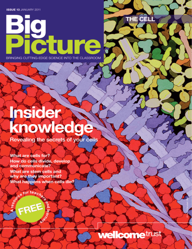
Insider
knowledge
Revealing the secrets of your cells
e
E
FRE
© David S Goodsell, 2000
fo r t e a c h
a n d le a r n e
rs
A res o
ce
rs
ur
What are cells for?
How do cells divide, develop
and communicate?
What are stem cells and
why are they important?
What happens when cells die?
Big
Picture
Contents
Big Picture
Introducing the cell
A close-up look at the structure of animal cells.
Beginnings and basics
Big Picture is a free resource for teachers,
school and college students, and learners
of any age. Published twice a year, each
issue comes as a printed magazine with
accompanying online articles and other
content. Here’s how to get the most from
your issue.
3
4–5
6–7
8–9
10–11
12–13
14–15
How do cells divide, develop and grow into complex organisms?
Cells and their surroundings
How do cells interact with their environment?
A matter of life and death
How long do cells live? What happens when they die?
Stem cells and development
What are stem cells and how are they used in medical research?
Stem cells and the future
What might developments in stem cell science mean for us?
Real voices
Three people’s stories about the roles of cells in their lives.
How to use Big Picture magazine
How to use the Big Picture website
Each issue of Big Picture focuses on a topic that is current,
relevant to the biology curriculum, rooted in science, and that has
debate-provoking aspects. The issues are divided into doublepage spreads, each dealing with a different aspect of the topic.
The spreads are a jigsaw of articles, images, diagrams and ‘Fast
Facts’, allowing you to dip into each issue, and each spread, as
you need. Every issue contains a series of ‘real voices’ – interviews
with people whose lives are affected and shaped by the topic in
question. Whether you want to stimulate debate, to provide up-todate, scientifically accurate examples around particular issues, or
to get across complex ideas to your students, Big Picture helps
you to bring cutting-edge science into your classroom.
Using stem cells
You seem different…
How are cells specialised
Stem cells
What do we mean by
2
stem cells?
be
all its descendants will
If a differentiated cell divides,
do more,
other. A stem cell can
identical to it and each
differentiated cells. They’re
producing stem cells and
on
means that they can go
also self-renewing, which
and on dividing.
can
potency – how much they
Stem cells vary in their
of
products
the
and
egg
differentiate. A newly fertilised totipotent cells. These
made of
its first few divisions are
cell type in the body, including
cells can give rise to any
produce a whole organism.
the placenta, and so can
egg in
a few steps beyond the
Embryonic stem cells,
– they can become any
development, are pluripotent
organism.
whole
a
not
but
type of specialised cell,
of
can make just a few types
Multipotent cells, which
red
marrow that can generate
cell, include those in bone
found
adult stem cells (those
or white blood cells. Most
and organs) are multipotent.
in differentiated tissues
just one
those in the skin, make
Unipotent cells, such as
usually where lots of new
fully differentiated cell type,
cells are needed regularly.
transitions between
the
on
focuses
A lot of research
is whether pluripotent
these states. A basic question
by default, or need some
cells carry on as they are
them to stop differentiating.
continuing signal to remind
cells
that embryonic stems
Research strongly suggests
cells, as
keep making more stem
are self-sustaining, and
protein
signals from a particular
long as they receive no
that triggers cell differentiation.
10 • Big Picture 13:
– including
for studying specific diseases
muscular dystrophy
Huntington’s, Parkinson’s,
have been made by
and type 1 diabetes –
from patients into a
reprogramming adult cells
stem cells would
pluripotent state. Pluripotent
to test drugs
give researchers the chance
if they can
only
but
on different types of cell,
how and when
develop ways of controlling
these stem cells differentiate.
found in
Adult stem cells (those
organs) are
differentiated tissues and
in
potentially useful too. Researchers single adult
using
are
Cambridge, for example,
the biological signals
skin stem cells to explore
Scanning electron micrograph
Many types of cell are
in the lab for research.
4
micrograph of epithelial
This way up
Direction is important
track
absorption. The cell keeps
so that
of which end is which,
way.
molecules go the right
is
A more complex example
a type
found in the ear, where
inner ear
of epithelial cell in the
into
turns vibrational signals
we can
electrical messages so
which
hear. These hair cells,
cilia, have
have a bundle of fine
have to
a top and bottom, but
direction
be arranged in the right
well. If they
along another axis as
planar
lose this orientation, or
hearing
polarity, the sense of
may be impaired.
3
© 2010 Nucleus Medical
Art, all rights reserved.
www.nucleusinc.
grown
1
4
2
3
FAST FACTare
Stem cell therapies
already in use in the
form of bone marrow
of
transplants – the first
which was performed
in 1956. Source:
Organs can grow by
getting more cells,
or larger ones.
two ways.
An organ can grow in
it
Adding more cells makes
bigger. But so can increasing
(called
the size of individual cells
both
‘hypertrophy’). We use
heart
methods. The embryonic
extra
increases in size by adding
hearts
cells, but after birth, our
grow by hypertrophy.
By exercising, we stimulate
hearts of
this process, and the
larger
athletes can grow much
still. This so-called physiological
when
hypertrophy is normal;
heart
you stop training, your
reduce
will adapt and slowly
cells.
be kept alive outside
Many kinds of cell can
dish. If they grow and
the body in a laboratory
cell culture. Many cell
reproduce, you have a
are used in research.
culture ‘lines’ (or types)
substitute for whole
To some extent, they can
for animal testing of
organisms, particularly
However, cultures
toxicity or drug effects.
(a layer one cell
usually consist of a monolayer the mix of cells
cell, not
thick) of a single type of
also lack the threefound in real tissues. They
of tissues and organs,
dimensional structure
and support their
which have defined shapes
arrangements.
cells in carefully ordered
is as much art
Successful tissue culture
carefully controlled
as science. Cells need
the right
conditions to grow, including
growth factors in
temperature, gas mix and
in. The longest-lived
the medium they grow
cancer cells, which
cultures often derive from
normal controls
have found ways to override
lines have to be
on cell division. Such cell
cancerous cells may
checked continually. The
do in a tumour that’s
go on changing, as they
easy for cultures to
still in the body. It is also
by other cells. If this
become contaminated
may not be
goes unnoticed, experiments
think.
testing what the investigators
More, and bigger
(skin) cells.
and function.
in cells’ development
– including
A cell’s development
its direction – is constantly
influenced by the cells
surrounding it. An epithelial
sheet, for example, is
– the
asymmetric. One face
to
apical surface – is exposed
the gut,
the watery contents of
The
or to the air in the lungs.
surface
opposite face – the basal
of
– sits on supporting layers
tissue.
collagen and connective
into
Cells that secrete molecules
membrane
the gut need different
bottom,
proteins at the top and
for
and so do those specialised
of embryonic kidney
. This approach
involved in cell differentiation
drugs for, in this
can also be used to screen
tissues.
instance, repairing damaged
Getting some culture
Medical illustration copyright
Transmission electron
Annie Cavanagh
www.biotechlearn.org.nz
Munich
– not
Stem cells are very special
s but
only can they renew themselve ted
differentia
they can also become an embryo
in
cells. Stem cells found
cells found
can become any of the hold great
in the body and, as such,
ent
replacem
g
promise for generatin
a number
tissues and cells to treat , including
of diseases and disorders
disease and
diabetes, Parkinson’s
multiple sclerosis.
potential for
control the
If we can learn how to
cells we might be able
differentiation of stem
of cell damage in the
to remedy many kinds
cells are pluripotent,
body. Embryonic stem
but their use
and so the most versatile,
(for more, see
is not without controversy
pages 12–13).
companies use
Some pharmaceutical
drugs. Stem cells
new
test
stem cells to
digestive
range of hormones and
of the
Each cell type is specialised
enzymes. Small regions
c
Islets of
for the role it plays; specifi
pancreas known as the
from
different
characteristics range
Langerhans contain four
general
make
making key proteins to
cell types, which each
Red blood
most
properties like shape.
different hormones. The
small
cells
beta
–
cells, for example, are
there
common cells
shape
biconcave discs. This
– make insulin and amylin.
area,
specialise?
gives a large surface
cells
How do
oxygen
a signal
helping the cells to ship
A stem cell will receive
tissues,
that
from the lungs to the
from its surroundings
dioxide
pattern
and a little of the carbon
triggers a change in the
on and
turned
the other way.
are
that
of genes
a
The shape also gives
off, directing the cell towards
cells
Having
flexibility, helping the
more specialised state.
smallest
g of
squeeze through the
a detailed understandin
red blood
they
capillaries. Developing
these changes and how
and
us to
cells begin with a nucleus
are triggered may allow
before
organelles, but lose them
control them.
in effect
they start work, which
full
reduces the cells to bags
EM Unit, UCL Medical
of haemoglobin, the protein
School, Royal Free Campus
carbon
that carries oxygen and
distorted,
dioxide. If the shape is
example,
disease can result. For
sicklein the inherited disease
blood
cell anaemia, some red
cells become sickle-shaped
owing to abnormal haemoglobin
molecules clumping together.
Organs often contain
The
blood cells.
sub-populations of cells.
Sickled and normal red
makes a
pancreas, for instance,
University of Edinburgh
cells and
1 Stem
development
Stem cells hold great
treating disease.
for their roles?
Also on this site, you can download PDFs of the current and all
13 past issues of Big Picture. There are also curriculum-matching
details, and info on how to order copies of the magazines.
Normal heart (cut section).
which
again. In heart failure,
things
can result from many
diet and
such as infection, poor
is an
high blood pressure, there
heart that
increased load on the
Medical Art Service,
Big
Picture
As well as twice-yearly magazines, there’s loads of other
useful material available on the Big Picture website
(www.wellcome.ac.uk/bigpicture). The exclusive online
content is created for each issue, and includes articles, videos,
games, image galleries, animations, lesson plans, activities and
more. Our selected links help you and your students to get the
most out of the web for each topic.
too thick
Heart muscle becomes
(hypertrophy).
In this
stimulates hypertrophy.
in
case, despite the increase
actually
size, heart performance
worsens.
Cross-section of bone.
JANUARY 2011 • 11
THE CELL
1
Title – Each spread deals with a particular aspect of the
title topic.
2
Intro – This sets the scene for the spread within the issue,
and the wider world.
Article – Each article can be used alongside others from the same spread or the rest of the issue, or as a standalone
component for a lesson.
4
Image/diagram – Illustrations and diagrams help you express complex ideas in a visual way, as well as helping to place the
issues in question into a real-life context.
3
2 • Big Picture 13: THE CELL
Left-hand menu – Browse Big Picture online by title, article, activity or resource.
1
2
Current issue heading – Explore the online content for
the latest issue.
3
Recent issues heading – Explore the online content
for past issues.
4
Sign-up link – Sign up to receive issues of Big Picture
and other Wellcome Trust publications.
Introducing the cell
Join us as we explore the building block of life.
The cell is the smallest unit of life. Some
simple organisms consist of just one cell,
whereas more complex beasts, like us, have
vast numbers of them. Humans are among
the organisms built up from eukaryotic
cells, which have their DNA parcelled
up in a nucleus, and lots of subcellular
compartments, called organelles. Prokaryotic
organisms (such as bacteria) are simpler:
cells still with DNA, but having no nucleus
or membrane-bound structures. The vast
majority of these are unicellular, while most
eukaryotic organisms are multicellular.
The intricately organised insides of
eukaryotic cells allow them to have different
things happening in different compartments.
Keeping a cell going depends on getting
the right molecules to the right place at the
right time. Having distinct spaces does half
the job, but it also requires sophisticated
machinery to ensure the right things get
into each section. Only material the cell has
finished with, for example, can be allowed
into a lysosome, where powerful enzymes
microtubules – small, tubular assemblies of
protein, made from repeating tubulin subunits,
which help maintain the cell’s internal structure
and move organelles and cytoplasm using
molecular motors. Part of the cytoskeleton
are poised to break down the material into
smaller molecules.
Cell theory was put forward in the
1830s, soon after the cell nucleus was first
identified in eukaryotes. It recognised that
living things are made of cells, that cells
are the basic units of life, and that new
cells are created by old ones dividing into
two. Viruses – simple entities of genes and
protein – need to get into a cell and hijack its
cytoplasmic machinery to copy themselves.
We describe these as acellular, and they are
not considered to be living.
In this issue, we’ll be focusing on animal
cells – how they reproduce, grow, move,
communicate and die. So join us to explore
what we know – as well as what we still don’t
understand – about the cells that are the
basis of all of us.
lysosomes – membrane-bound
organelles that are the cell’s rubbish
disposal and recycling units; contain
hydrolytic enzymes
vacuoles – internal bags,
surrounded by a membrane,
which cells use for storage
of food or waste
extracellular matrix – the
material in between cells that
holds tissues together, usually
made of scaffolding proteins
such as collagen
nucleus – the information centre
of the eukaryotic cell, where the
DNA is stored, replicated and
copied into RNA (transcribed)
microfilaments – smaller
than microtubules, these are
made from repeating actin
subunits. Responsible for
cell movement and changes
in shape, and make muscle
contraction possible. Part of
the cytoskeleton
nuclear envelope –
double membrane that
separates the contents
of the nucleus from the
cytoplasm
centrioles – a pair of
organelles that organises
microtubules into spindles
on which chromosomes
are separated when
cells divide
nuclear pores – gaps
in the nuclear envelope
that allow substances
to move in and out of
the nucleus
Golgi apparatus –
one of the wondrously
complex membrane
systems in the cytoplasm,
which modifies, packages
and directs newly made
proteins to where they
are needed
nucleolus – part of the
nucleus that produces
ribosomes
endoplasmic reticulum
(ER) – an extensive network
of membranes. Rough ER is
studded with ribosomes and
is a site where proteins are
made, modified and processed
for shipping. The roles of
smooth ER include lipid and
steroid synthesis and drug
detoxification
cytoplasm – everything in
the cell outside the nucleus;
a viscous fluid containing
proteins, other organic
and inorganic molecules,
membranes and organelles
mitochondria (singular:
mitochondrion) – rod-shaped
bodies in the cytoplasm that
supply chemical energy to the
rest of the cell
plasma membrane – a phospholipid
bilayer that contains cholesterol
and proteins. It surrounds the cell and
enables it to communicate with its
neighbours and detect and respond
to changes in the environment
ribosomes – molecular machines,
built from RNA and protein, that
make new proteins. They are found
in the cytoplasm and bound to the
rough endoplasmic reticulum
JANUARY 2011 • 3
Big
Picture
Beginnings and basics
Each of us has developed from a single egg and sperm to become a
complex, sophisticated organism made of trillions of cells. So, how
do cells divide, develop and work together to make this possible? And
if each of our cells contains all of our genes, then how do they turn into
different types of cell with distinct structures and functions? Why can’t
our cells go on dividing for ever?
Dividing we stand
The Golgi apparatus, which processes
and packages proteins in the cell.
Cell division is a tightly controlled process.
The most complex thing a cell can do is to split into two identical
cells. In actively reproducing cells, this begins with the copying of
the cell’s contents, including the chromosomes. The duplicates
separate into different halves of the cell and then, during mitosis,
the cell splits down the middle – and then the cycle starts again.
A more complex type of cell division, meiosis, which generates
eggs or sperm, has an extra stage. Each chromosome pair is
separated, so that each cell has a single chromosome.
Seen under a microscope, the dance of the chromosomes
looks magical, but it has a precise molecular mechanism.
The movements of the chromosomes, like those of the cell, are
controlled by microtubules. These are long tubes of protein
subunits, which can assemble, break down and reassemble
very quickly in a different arrangement.
Microtubules play a crucial role in mitosis, rearranging
themselves from their usual place in the cytoskeleton to build
a complicated machine called the mitotic spindle, whose other
parts include motor proteins. The spindle grabs hold of the
chromosomes and lines them up. It pulls the pair of chromatids
that make up each chromosome in opposite directions so they
end up in different halves of the cell.
The whole process is a carefully orchestrated sequence
of movements involving hundreds of different proteins. A key
component is the protein complex (two or more proteins that
associate with each other) that assembles on chromosomes and
provides a place for microtubules to latch on to, known as the
kinetochore. Cell division is a very active area of research because
it is one of the key cellular processes that goes wrong in cancer.
Paul J Smith and Rachel Errington
Human cancer (melanoma) cells dividing.
4 • Big Picture 13: THE CELL
Seeing is believing
Cells were first seen
over 300 years ago.
No one knew that cells existed
before the invention of the
microscope. Robert Hooke
in 1665 saw spaces in dead
sections of cork that he called
‘cells’, and the Dutch pioneer
Anthony van Leeuwenhoek
was astonished by living cells
20 years later. The insides of
cells were observed much
later, with more powerful light
microscopes. Even the best
light microscopes, though, can
only show objects larger than
0.2 µm (micrometres, millionths
of a metre).
Electron microscopes can
go smaller still. Today, they can
even show how single molecules
of newly made protein nestle
inside the ‘chaperone’ proteins
that help them fold up into their
proper shape. Analysing images
of hundreds of these protein
complexes has shown just how
they fit together. Understanding
how proteins fold properly is
important, because misfolded
proteins can accumulate
and cause diseases such as
Alzheimer’s.
Who’s in control?
Your cells contain the same genome but different
genes are in use.
The store of genes in the cell
nucleus, the genome, makes your
cells human. But every human has
around 200 different cell types,
each with an identical gene store.
The differences lie in which genes
are actually in use. Specific sets
of genes are switched on and off
as cells start to adopt specialised
functions during development,
a process called differentiation.
The genes, in turn, will
generate unique patterns of RNA
messages from reading the DNA
that is in use, and a signature
population of proteins and smaller
regulatory molecules. These
patterns change in response to
gene signals, the contents of
the cytoplasm and messages
from outside the cell. The result
is a complex developmental
conversation. At one level, the
nucleus is in charge of the cell.
But there is also a sense in which
the cell, with its surroundings, is in
charge of the information store in
the nucleus, and how it gets used.
Scientists have decoded
the entire human genome, but
that does not give a picture of
which information is active in
any cell. This is registered in the
transcriptome (the complete
catalogue of messenger RNA
molecules in a cell), and the
proteome (the list of all the
different proteins present). Unlike
the genome, these are constantly
shifting, so each is a snapshot
in the life of the cell. How the
transcriptome and proteome
are tweaked by small changes
in the cell’s circumstances is
one of the biggest topics in current
biological research.
Dr David Furness
One, two,
or many?
No limits?
Why don’t our cells go on dividing for ever?
Not all of your cells have
a single nucleus.
It is important to have just the right amount
of cell division, so the process has lots of
checks and balances. If the controls fail,
there is a final limit. Each time a chromosome
is copied, a repetitive stretch of DNA at its
end, the telomere, gets shorter. The telomere
is needed for the proteins that copy DNA to
work properly, so when it is all gone there is
no more chromosome copying, and no more
cell division. In effect, telomeres count cell
divisions. Human cells can normally manage
between 40 and 60 divisions – this is called
the Hayflick limit, named after Leonard
Hayflick’s 1965 discovery. Stem cells, and
many cancer cells, can get round
this limit using the enzyme
telomerase, which
rebuilds the ends
of chromosomes.
Snaprender/iStockphoto
How do things get around in the cell?
The inside of the cell is a scene of constant motion. Soluble molecules move around apparently
randomly in the cytoplasm, but many components are transported more precisely. Many proteins
are allowed only into one of the cellular compartments. Cells also have an elaborate network of
fine protein filaments (strands), an interior cytoskeleton, which helps them keep their shape and
provides the rails of a transport system. Small protein motors pull little bags, or vesicles, of cell
products up or down microtubules or actin filaments. Special labels ensure the right cargo is sent
to the right destination. These are usually proteins, or parts of proteins, sticking out of the vesicle.
The same system also moves organelles around or anchors them in place. The cell uses a lot
of energy for all this transport, but it needs to speed things along. A protein molecule might take
years to travel the length of the longest nerve cell by simple diffusion, but if it is bagged up and
dragged along a microtubule it can cover 10 cm in a day. This is vastly more than most distances
within cells, and means that even the ends of the long, drawn-out extensions of nerve cells, the
axons, in your fingers or toes can be reached in a few days.
Prof. Giorgio Gabella
Inside the cell
Most cells have the standard
single nucleus, but not all. Skeletal
muscles, such as your biceps, have
very long cells with many nuclei.
They form by fusion of cells with
one nucleus. Some cells in adult
heart muscle have two nuclei. This
happens because the cells take the
normal cell cycle through copying
and separation of chromosomes,
but do not go the whole way and
divide. Why does this happen?
We don’t know for sure, but one
recent idea is that one nucleus lies
dormant, but triggers further cell
division if the muscle gets damaged.
Red blood cells in mammals have
no nucleus. For more on these cells,
see page 10.
Transmission electron micrograph of
smooth muscle cells.
Under development
Controlled cell division is a
key part of development.
Tissues contain cells and material between
the cells. In connective tissues, like bone
or tendon, this material – the extracellular
matrix – predominates. In other tissues, it is
distributed more sparingly. In all tissues, the
cells and their matrix communicate, chemically
and physically. The physical link is via proteins
that cross the external membrane of the cell,
which are known as integrins. One end of an
integrin is tied into the cytoskeleton, the other
to collagen fibres in the extracellular matrix.
The matrix also contains chemical
messengers, often bound to gels made up of
proteoglycans, a complex mix of proteins and
the sugar-derived polysaccharides. These
C Merrifield and M Duchen
Cells communicate with the matrix
surrounding them.
The filaments, coloured red, show part of the
cytoskeleton.
include growth factors and signal molecules,
which affect many processes, including cell
migration. Some come from the cells that
make the matrix, some from other cell types
– and the cells, in turn, respond to the mix of
signals as it changes over time.
FAST FACT
Human eggs are made in
the embryo, so the egg
cell that fused with a
sperm to become you
was actually produced
around six months before
your mum was born.
JANUARY 2011 • 5
Yorgos Nikas
Exploring the matrix
Perhaps the most remarkable
fact in biology is that the several
tens of trillions (1 trillion =
1 000 000 000 000) of cells
that make you develop from
a single cell – a fertilised ovum,
or zygote. Controlled cell division
is crucial for development. The
dividing cells in the embryo are
also differentiating and forming
structures that fold, get reshaped,
or even migrate to different
locations. Disruption of one of the
many genes involved in controlling
all these subtle shifts increases the
risk of developmental defects. The
effects of these defects may be
felt much later on in adult life, not
just at birth.
Big
Picture
Cells within cells?
Cells and their
surroundings
In any multicellular organism, cells don’t
exist in isolation. A cell’s environment
has a profound effect on how it develops,
grows and functions, making effective
communication between a cell and its
surroundings – including neighbouring
cells – vitally important. How do cells
interact with their environment? What
happens when these signalling systems
are disrupted?
A society of cells
How do cells organise in an organism?
The human body is a closely organised community
of cells. Tissues, collections of similar cells, work
together in groups to form an organ. These organs
work with each other in organ systems. The liver,
for instance, is made up of 80 per cent of one kind
of cell, hepatocytes, which make, store or secrete
many proteins, fats and digestive enzymes. The
remaining 20 per cent consists of several other
specialised liver cell types, along with blood vessels,
nerve cells and others. All have to work together for
the liver to do its job. To keep the whole organism
functioning correctly, most organs or organ systems
also have to respond to the state of other organs,
which may be a long way away.
For a smooth-running community, cells need
good communication. They manage this in different
ways, but all use either chemical or electrical
signals. Nerve cells, for example, send electrical
signals down their long fibres, but communicate
with the next cell in the chain by
sending chemicals across a tiny gap
at the synapse. Heart muscle cells,
on the other hand, join to their
neighbours via gap junctions.
At these points, the cells are
close enough together
for protein complexes with
tiny channels inside to span
both cell membranes, and
allow small molecules and
ions to pass between
them. This allows
waves of
depolarisation
to pass along a
series of heart
cells and keep
the heart beating
regularly.
Heart, showing the
coronary arteries.
6 • Big Picture 13: THE CELL
Were some organelles
originally bacteria?
Some organelles inside eukaryotic cells look rather like cells
themselves. Mitochondria – and the energy-generating chloroplasts
in plant cells – even have a little DNA of their own. According to
endosymbiotic theory, eukaryotes originated when symbiotic
bacteria (which exist alongside cells in a mutually beneficial
relationship) began to live inside larger cells, giving them readymade compartments. Over time, these bacteria became permanent
additions – chloroplasts and mitochondria – to the cells we see today.
Researching membrane proteins
Why do researchers study membrane proteins?
Around one-third of all proteins
are linked to membranes in some
way. The ones that sit within the
membrane are hard to study
because pulling them out of their
fatty surrounding often wrecks
their normal structure. But
examination of these proteins’
structure and function shows
how their roles in signalling can
be exploited, either by viruses
trying to bypass cell defences or
by researchers developing drugs
to treat disease.
One important focus for
studies of membrane proteins is
the action of HIV. The virus enters
one kind of cell in the immune
system by first binding to a normal
cell surface receptor protein
called CD4. HIV has an outer
envelope that can fuse with the cell
membrane, allowing viral genes
and proteins inside. Several other
cell-surface receptors, and other
chemical messengers, can help
or hinder the fusion of membranes.
Work on understanding exactly
how they interact with each other
suggests new targets for antiHIV drugs.
R Dourmashkin
HIV particles budding from the surface
of a T cell.
Passing on the message
Cells pass on signals via cascades of events.
Cells have evolved efficient ways
of processing the many signals
they receive. Many cell-surface
receptors are linked to other
proteins inside the cell. When
the right molecule (the ‘first
messenger’) from outside the cell
binds to the receptor, it changes
shape, and triggers changes in the
internal protein as well. That in turn
may activate an enzyme, most
often one that makes the molecule
cyclic adenosine monophosphate
(cAMP), which is a messenger
inside many cells. As cAMP is
produced after this small but
significant cascade of events, it is
known as a second messenger.
The effects of cAMP depend
on both the first messenger (often
a hormone) and the target cell.
Several important hormones use
cAMP as a second messenger.
For example, adrenaline increases
heart rate and how strongly the
heart beats, as well as promoting
glycogen breakdown in muscle.
Different hormones whose
actions are mediated by cAMP
can cause the same response
in a given cell – for example,
adrenaline, glucagon and other
hormones trigger the breakdown
of triglycerides in fat cells.
Communication breakdown
Dr David Furness
Problems in cell signalling can be bad news.
When messages between cells
are blocked or scrambled, there
are usually harmful results.
Autoimmune diseases, in which
our own immune cells attack
body tissues, are partly due
to errors in identifying cells. In
multiple sclerosis, misdirected T
cells remove the insulating sheath
around nerve fibres, while tumours
begin when cells ignore messages
telling them not to replicate, or
when they misread signals to keep
dividing. Teratomas (tumours that
can contain hair, teeth and bone)
arise from germ cells (sperm and
eggs) that are triggered to begin
dividing inside the body.
Some diseases affect cell–cell
signalling directly. In Alzheimer’s
disease, toxic clumps of a protein
called amyloid appear in the brain.
They build up from fragments
of a precursor protein present
at synaptic junctions, which is
abnormally processed. Another
example, whose full details are still
being worked out, is diabetes. In
type 1 diabetes, sugar metabolism
gets out of control because the
cells that make the hormone
insulin die off. In the more
common type 2 diabetes, there
is insulin in the circulation, but the
cells that normally respond to it
are deaf to the signal.
Colour-enhanced image of two myelinated nerve fibres.
Don’t stop moving
How and why do some cells move?
Mind your membranes
Membranes are important inside and around the cell.
The cell’s plasma membrane
separates inside from out and
allows communication. Its
structure is based on a foamy
bilayer of phospholipid molecules,
but it is studded with proteins
that regulate the traffic back and
forth. Some just make pores that
allow small, soluble molecules to
get through. Some use energy to
move their chosen cargo more
actively from one side to the other.
Other membrane proteins bind
signalling molecules, such as
hormones, that float by outside,
and then pass a message to
the interior.
When larger quantities of some
substances need to be moved
from cell to cell, they are bagged
up into membrane-bound sacs
called vesicles that can fuse with
the cell membrane. As usual, the
vesicles are tagged with special
proteins that label the contents,
and interact with yet more
membrane proteins to make sure
the correct cargo is delivered.
The same system operates
for internal transport between the
cell’s compartments. Vesicles bud
out from one membrane, then
fuse with another. Recent work at
the University of Cambridge has
shown how a mutation in protein
complexes that regulate vesicle
transport can cause a rare
genetic disorder that leads to
albinism, impaired lung function
and chronic bruising.
Karin Hing
Maurizio De Angelis
Ion channels in the cell’s plasma membrane.
One simple response to a signal – for a few cell types in adults
at least – is to get moving. Cells can move slowly by crawling or
sliding along, aided by changes in the cytoskeleton, but faster
movements depend on specialised external organelles. Motile
cilia are small projections that wave or beat and either propel a
cell through a fluid or waft the fluid past a line of cells, as happens
when mucus is cleared from your windpipe. The larger flagella
are used only to move cells. The long, whip-like tail of a sperm
cell is an important example. Research on how its movement is
activated, and how it changes when a sperm gets close to the
surface of an egg, may help overcome some causes of infertility.
It could also pave the way to an effective male contraceptive.
Cell movement is also an important part of wound healing.
A gash in the skin triggers new mitosis around the edges and
also induces cells to move into the wound space to begin
covering the opening.
Two human bone-forming cells (osteoblasts) crawling over ceramic crystals.
FAST FACT
A man makes 1500
sperm per heartbeat
Source: Science
328(5974):15.
Joyce Harper, UCL
JANUARY 2011 • 7
Big
Picture
A matter of life and death
Cells have very different lifespans. While some cells stay with
us for our whole lifetime, others have fleeting, single-day roles
as part of our bodies. For example, while men produce sperm
throughout their adult lives, women are born with all their eggs –
how might the age of a woman and her eggs affect any potential
offspring? Proper recycling and breakdown of a cell’s unwanted
parts is necessary to keep things in working order. So how do
cells dispose of their waste and – when cells actually
die – themselves?
The life expectancy of the cell
The lifespans of different cells vary greatly.
Mark Lythgoe and Chloe Hutton
Cells can last a human lifetime, but not many do. Some, such as the
white blood cells that hunt down bacteria, are gone in less than a day,
while cells in the lining of the gut hang around for nearly a week. Most,
though, last a good deal longer – liver cells for a year or so, bone cells
for perhaps ten years.
Ten years, in fact, is about average. Just a few kinds of cell can
endure from birth to their owner’s death. They include cells inside the
Magnetic resonance imaging scan of the head.
Building your brain
Can we grow new nerve cells
in our brains?
Some organs, such as the liver, can regenerate
if they are damaged, and will, within limits, grow
to cope with demand. Exercise your muscles in
the gym, and they get larger. Sadly, this will not
work for your brain: although ‘exercising’ your
brain may alter cell–cell connections, the
number of neurons will not change.
Or will it? There is controversy about
whether the brain can grow new neurons,
especially in the cerebral cortex, home of
advanced thinking skills. If so, the numbers
New cells for old
The age of eggs and sperm can be very different.
Men produce sperm continually after puberty, and dispose of
old ones after a couple of months. Human eggs (ova), on the
other hand, are made in the embryo, and released from the ovaries
much later on. Actually, these are primary oocytes, which have not
completed meiosis. After puberty, one oocyte a month matures
into a secondary oocyte, which can then be fertilised.
The older a woman, the older her eggs, and the greater the
chance that she will have a baby with chromosomal abnormalities.
Serious chromosomal problems may lead to miscarriage, so older
women are at greater risk of this than younger women. Very recent
studies indicate that there are stem cells in mammalian ovaries
that can produce new egg cells in adults. This might help to treat
infertility, or to reduce risks of some birth defects.
8 • Big Picture 13: THE CELL
lens of the eye, which become inert once they are in place in the
embryo, cells in heart muscle and, perhaps most importantly, neurons
in the brain (see below).
Counting all the cells a person ever has would take several lifetimes.
The average turnover of all human cells in different tissues is seven to
ten years, so the lifetime cell count is perhaps ten times the adult total
(at least several tens of trillions of cells). That ignores a lot of other cells,
like the 180 or so types of bacteria and other microorganisms that live in
and on our bodies. It’s thought that each of us carries ten times as many
of these cells as we have our own, human cells.
are small. The vast majority of neurons in the
brain of the oldest man or woman have been
there for their entire lifetime.
It was discovered in the 1990s that the
hippocampus, where new memories form,
can produce new neurons late in life. Since
then, evidence has mounted that stem cells
that make extra neurons are found in the
cerebral cortex as well, at least in mice and
monkeys. If further work confirms this finding,
researchers will want to know whether the
cells can be activated, to help heal injuries
such as stroke or even, perhaps, improve
brain function.
FAST FACT
Chemotherapy can lead to hair loss because
the hair follicle epithelial cells – like cancer
cells – divide rapidly, and hence are also targets
of many anticancer drugs.
ker
Source: Scientific American, 5 January 2001.
Spike Wal
So long, cells
Cells die in a variety of ways.
Cells, like people, die if they are starved or poisoned.
Blockage of an artery leading to the heart causes death of
heart muscle cells in a heart attack, for instance. A similar
obstruction in the brain leads to the destruction of brain
cells in a stroke. This kind of death is called necrosis. But a
cell’s life can also end in other ways.
One, whose importance only became clear recently, is
programmed cell death, or apoptosis. Cells have an inbuilt
self-destruct routine that keeps them poised on the brink
of suicide. Normally, signals from neighbouring cells keep
them from doing themselves in, but if the signals change,
the cell pushes the suicide button.
This is a neat and tidy death. While necrotic cells
swell and burst, spilling lysosomal enzymes and
damaging surrounding cells, apoptosis involves the orderly
dismantling of organelles and proteins, while the cell shrinks
and ends up as a few vesicles, which are cleared up by
other cells.
Apoptosis happens in different places at different times.
The developing embryo uses it to sculpt fingers from a
web-shaped hand, by removing the cells in between the
fingers. Cells with damaged DNA
may sense the damage and sacrifice
themselves for the common good,
for example, when you get
sunburn. Viruses may also
set off apoptosis of infected
cells. Triggering apoptosis
is one way that HIV depletes
the immune system of vital
defensive cells.
Human chromosomes.
Getting on a bit
Ageing cells have a
number of tell-tale signs.
Over time, the waste disposal
can get clogged up. Old, tangled
proteins and other cellular junk
defy the lysosomal enzymes.
Remnants of fatty acids are a
particular problem and they make
up a big portion of the yellowish
pigment granules known as
lipofuscin, whose appearance
is a sure sign of an ageing cell.
Other signs of cell ageing, such as
shortening of the telomeres
on the ends of the chromosomes,
are related to how many times the
cell has divided. Most, though,
are the result of normal wear and
tear. Mitochondria age faster
than other organelles. Their job
of energy production exposes
them to reactive chemicals – free
radicals – that can damage DNA.
Mitochondria have some essential
genes in their own DNA, and
the proportion of them that have
serious defects increases as the
person, and their cells, grow older.
Waste disposal
How do cells dispose of their unwanted parts?
chemical, a ganglioside,
accumulates and eventually
destroys cells in the brain
and spinal cord, causing a
progressive loss of mental
and physical function that
usually results in death
before the age of five.
The right packaging is
also crucial for recycling and
disposal. Cells constantly pinch
off bits of outer membrane,
turning a small pit in the
membrane into a vesicle, which
is brought inside. Larger vesicles
import material from outside.
The vesicles then fuse with an
extensive network of tubes and
bags, known as endosomes,
which sort incoming material.
New vesicles bud off from here,
and shift designated contents
onward – to other parts of the
cell, back to the outer membrane
or to the lysosomes. Cell surface
receptors are recycled as part of
this process, too.
al Ge
netics
Centr
e
FAST FACT
The 2009 Nobel Prize in Physiology or
Medicine went to Elizabeth Blackburn,
Carol Greider and Jack Szostak for their
work on how chromosomes are protected
by telomeres and the enzyme telomerase.
Source: www.nobelprize.org
Wess
gion
ex R e
Cells are continually making new
molecules, while old ones are
broken down and recycled. The
main site for this is the lysosome,
which acts as a cellular stomach.
A typical human cell has about
a hundred lysosomes, each a
collection of potent hydrolytic
enzymes, which break down
substances, enclosed in a
membrane. Old organelles, other
cellular waste and, in immune
system cells, old red blood cells or
bacteria swallowed by the cell are
all wrapped in membranes of their
own. These then fuse with the
lysosome, where they are quickly
broken down into small molecules
for re-use.
In cases where one of the
lysosomal enzymes fails, the cell
cannot keep up with removing
waste. In the rare genetic
condition Tay–Sachs disease,
for example, an enzyme that
mops up a fatty chemical
in neurons is defective. The
JANUARY 2011 • 9
Stem cells and
development
Stem cells are very special – not
only can they renew themselves but
they can also become differentiated
cells. Stem cells found in an embryo
can become any of the cells found
in the body and, as such, hold great
promise for generating replacement
tissues and cells to treat a number
of diseases and disorders, including
diabetes, Parkinson’s disease and
multiple sclerosis.
Stem cells
What do we mean by stem cells?
If a differentiated cell divides, all its descendants will be
identical to it and each other. A stem cell can do more,
producing stem cells and differentiated cells. They’re
also self-renewing, which means that they can go on
and on dividing.
Stem cells vary in their potency – how much they can
differentiate. A newly fertilised egg and the products of
its first few divisions are made of totipotent cells. These
cells can give rise to any cell type in the body, including
the placenta, and so can produce a whole organism.
Embryonic stem cells, a few steps beyond the egg in
development, are pluripotent – they can become any
type of specialised cell, but not a whole organism.
Multipotent cells, which can make just a few types of
cell, include those in bone marrow that can generate red
or white blood cells. Most adult stem cells (those found
in differentiated tissues and organs) are multipotent.
Unipotent cells, such as those in the skin, make just one
fully differentiated cell type, usually where lots of new
cells are needed regularly.
A lot of research focuses on the transitions between
these states. A basic question is whether pluripotent
cells carry on as they are by default, or need some
continuing signal to remind them to stop differentiating.
Research strongly suggests that embryonic stems cells
are self-sustaining, and keep making more stem cells, as
long as they receive no signals from a particular protein
that triggers cell differentiation.
10 • Big Picture 13: THE CELL
You seem different…
How are cells specialised for their roles?
Each cell type is specialised
for the role it plays; specific
characteristics range from
making key proteins to general
properties like shape. Red blood
cells, for example, are small
biconcave discs. This shape
gives a large surface area,
helping the cells to ship oxygen
from the lungs to the tissues,
and a little of the carbon dioxide
the other way.
The shape also gives
flexibility, helping the cells
squeeze through the smallest
capillaries. Developing red blood
cells begin with a nucleus and
organelles, but lose them before
they start work, which in effect
reduces the cells to bags full
of haemoglobin, the protein
that carries oxygen and carbon
dioxide. If the shape is distorted,
disease can result. For example,
in the inherited disease sicklecell anaemia, some red blood
cells become sickle-shaped
owing to abnormal haemoglobin
molecules clumping together.
Organs often contain
sub-populations of cells. The
pancreas, for instance, makes a
range of hormones and digestive
enzymes. Small regions of the
pancreas known as the Islets of
Langerhans contain four different
cell types, which each make
different hormones. The most
common cells there – beta cells
– make insulin and amylin.
How do cells specialise?
A stem cell will receive a signal
from its surroundings that
triggers a change in the pattern
of genes that are turned on and
off, directing the cell towards a
more specialised state. Having
a detailed understanding of
these changes and how they
are triggered may allow us to
control them.
EM Unit, UCL Medical
School, Royal Free Campus
Sickled and normal red blood cells.
University of Edinburgh
Big
Picture
Transmission electron micrograph of epithelial (skin) cells.
This way up
Direction is important in cells’ development and function.
A cell’s development – including
its direction – is constantly
influenced by the cells
surrounding it. An epithelial
sheet, for example, is
asymmetric. One face – the
apical surface – is exposed to
the watery contents of the gut,
or to the air in the lungs. The
opposite face – the basal surface
– sits on supporting layers of
collagen and connective tissue.
Cells that secrete molecules into
the gut need different membrane
proteins at the top and bottom,
and so do those specialised for
absorption. The cell keeps track
of which end is which, so that
molecules go the right way.
A more complex example is
found in the ear, where a type
of epithelial cell in the inner ear
turns vibrational signals into
electrical messages so we can
hear. These hair cells, which
have a bundle of fine cilia, have
a top and bottom, but have to
be arranged in the right direction
along another axis as well. If they
lose this orientation, or planar
polarity, the sense of hearing
may be impaired.
Using stem cells
Stem cells hold great potential for
treating disease.
If we can learn how to control the
differentiation of stem cells we might be able
to remedy many kinds of cell damage in the
body. Embryonic stem cells are pluripotent,
and so the most versatile, but their use
is not without controversy (for more, see
pages 12–13).
Some pharmaceutical companies use
stem cells to test new drugs. Stem cells
for studying specific diseases – including
Huntington’s, Parkinson’s, muscular dystrophy
and type 1 diabetes – have been made by
reprogramming adult cells from patients into a
pluripotent state. Pluripotent stem cells would
give researchers the chance to test drugs
on different types of cell, but only if they can
develop ways of controlling how and when
these stem cells differentiate.
Adult stem cells (those found in
differentiated tissues and organs) are
potentially useful too. Researchers in
Cambridge, for example, are using single adult
skin stem cells to explore the biological signals
Annie Cavanagh
Scanning electron micrograph of embryonic kidney cells.
involved in cell differentiation. This approach
can also be used to screen drugs for, in this
instance, repairing damaged tissues.
Getting some culture
Many types of cell are grown
in the lab for research.
Many kinds of cell can be kept alive outside
the body in a laboratory dish. If they grow and
reproduce, you have a cell culture. Many cell
culture ‘lines’ (or types) are used in research.
To some extent, they can substitute for whole
organisms, particularly for animal testing of
toxicity or drug effects. However, cultures
usually consist of a monolayer (a layer one cell
thick) of a single type of cell, not the mix of cells
found in real tissues. They also lack the threedimensional structure of tissues and organs,
which have defined shapes and support their
cells in carefully ordered arrangements.
Successful tissue culture is as much art
as science. Cells need carefully controlled
conditions to grow, including the right
temperature, gas mix and growth factors in
the medium they grow in. The longest-lived
cultures often derive from cancer cells, which
have found ways to override normal controls
on cell division. Such cell lines have to be
checked continually. The cancerous cells may
go on changing, as they do in a tumour that’s
still in the body. It is also easy for cultures to
become contaminated by other cells. If this
goes unnoticed, experiments may not be
testing what the investigators think.
FAST FACT
Stem cell therapies are
already in use in the
form of bone marrow
transplants – the first of
which was performed
in 1956. Source:
Organs can grow by
getting more cells,
or larger ones.
An organ can grow in two ways.
Adding more cells makes it
bigger. But so can increasing
the size of individual cells (called
‘hypertrophy’). We use both
methods. The embryonic heart
increases in size by adding extra
cells, but after birth, our hearts
grow by hypertrophy.
By exercising, we stimulate
this process, and the hearts of
athletes can grow much larger
still. This so-called physiological
hypertrophy is normal; when
you stop training, your heart
will adapt and slowly reduce
www.biotechlearn.org.nz
Normal heart (cut section).
Heart muscle becomes
too thick (hypertrophy).
again. In heart failure, which
can result from many things
such as infection, poor diet and
high blood pressure, there is an
increased load on the heart that
stimulates hypertrophy. In this
case, despite the increase in
size, heart performance actually
worsens.
Medical Art Service, Munich
More, and bigger
Medical illustration copyright © 2010 Nucleus Medical Art, all rights reserved. www.nucleusinc.
Cross-section of bone.
JANUARY 2011 • 11
Big
Picture
Stem cells and
the future
Advances in stem cell technologies are
occurring rapidly, bringing with them different
ethical, moral and social questions to consider.
Take a look at the timeline and the three
scenarios below to explore your thoughts on
the sources and use of stem cells in medicine
and human enhancement.
Umbilical stem cells
Many of the embryonic stem cells used in research in the UK
come from fertilised eggs donated by people undergoing in vitro
fertilisation (IVF). They decide how any ‘excess’ embryos from these
procedures are used, and can choose to donate them to research.
The use of embryonic stem cells is controversial and opponents
argue that destroying embryos to remove stem cells is inhumane.
Using umbilical cord stem cells is an alternative source, potentially
with fewer ethical concerns.
Cord blood stem cell transplants have been used to help people
with diseases such as leukaemia and lymphoma. However, if the
donor and the recipient are not genetically similar enough then
donor cord blood can be rejected by the patient. This is less likely to
happen than during bone marrow transplants, though, and as cord
blood banks grow, there will be a greater chance of finding suitable
matches.
In 1978, blood stem cells were discovered in human umbilical
cord blood. At first it was thought that cord blood stem cells were
only able to differentiate into types of blood cell, but research now
suggests that they may have greater potency than this.
Scientists in the USA have used human umbilical cord stem cells
to treat mice with thin, cloudy corneas. The stem cells were shown
to take on the properties of cells in the cornea of the eye, showing
the potential of this research to help those with loss of vision.
“It was easy to donate my cord
blood and I hope that one day
the stem cells in my sample will
help someone who needs them.”
“I don’t feel comfortable donating
my umbilical cord blood – who
knows what they might be able to
do with it in the future?”
Question: Should it become obligatory for women
who give birth to donate their umbilical cord blood for research?
?
Snapshots of the stem cell story
•
•
1981: Scientists successfully culture (grow) pluripotent
mouse embryonic stem cells
12 • Big Picture 13: THE CELL
1996: ‘Dolly the sheep’ is the first mammal to be
cloned by somatic nuclear transfer – adding a nucleus from an adult sheep cell to an unfertilised egg with
the nucleus removed
•
1998: Scientists at the University of Wisconsin isolate and grow the first stem cells from
human embryos left
over from IVF
•
1990: Human Fertilisation and Embryology Act passed in the
UK, includes founding of Human
Fertilisation and Embryology
Authority, which regulates the
creation, use and storage of human embryos in treatment
and research
•
2004: Britain opens
the world’s first government-financed
stem cell bank, containing embryonic and stem cell lines
K Hardy
Ph Plailly/Eurelios/
SPL
1958: Leroy Stevens identifies pluripotency
of certain
mouse cells
Wellcome Trust
Sanger Institute
•
•
2001: UK Parliament rules embryonic stem cell research
can occur using government funding. Human embryos
can be created for research
purposes only, but not kept beyond 14 days
•
2006: Korean scientist Woo-suk Hwang found to
have fraudulently claimed the creation
of human embryonic cells through cloning
Admixed embryos
Induced pluripotent stem cells
Another way to avoid using human embryonic stem cells could be to create
hybrid embryos, by removing the nucleus from an animal’s egg and replacing
it with a human body cell nucleus. The egg is not fertilised, but is triggered to
develop with a jolt of electricity. If it works this could lead to early embryonic
cell division, and production of human stem cells. However, the absence of
the regulatory chemicals present in a human egg makes such work tricky
to accomplish.
Since 2008, three research groups in the UK have been granted licences by
the Human Fertilisation and Embryology Authority (HFEA) to create cytoplasmic
hybrid embryos for research. Human admixed embryos are not allowed
to develop beyond 14 days, it is prohibited to implant them into humans or
animals, and their use is regulated by the HFEA.
Now, consider a future where researchers have discovered
how to ‘reset’ adult stem cells and give them pluripotency,
without a danger of them becoming cancerous when
introduced into the body (so induced pluripotent stem
cells – IPS cells – could be readily produced in the lab).
See below for the type of advert that this could lead to.
PLURI−U
PUTTING THE 'I' INTO IPS CELLS!
Transplants from donors are a thing of the past, thanks
to Pluri-U. Simply attend your local clinic and have
a small skin sample taken. Then, technicians will treat
your cells with the right combination of chemicals and
produce whichever cells you need for your condition,
tailor-made to match your own immune system.
No rejection! No immune-suppressing drugs!
But don’t take our word for it!
Marion, 41, says: “A lifetime of type 1 diabetes, and my disease was
gone after one procedure. No more insulin, no more monitoring my
blood sugar! My only regret is that this wasn’t available 20 years ago!”
“I’m really excited about being able
to use hybrid embryos. Working
with stem cells created this way
will help me to research genetic
neurodegenerative disorders such
as Alzheimer’s and Parkinson’s.”
Mark, 35, says: “Our son was born with sickle-cell anaemia, but thanks
to Pluri-U’s cell reset and gene therapy package, his blood cells are
now normal and he’s disease-free!”
“It seems unnatural to be
mixing up animal and human
cells. How do we know the
long-term effects of this?”
Question: What about using stem cells to
enhance humans – do you think
it would be acceptable to use the
same technology to make you
stronger or able to run faster?
Question: Is creating a human admixed embryo from a
mature human cell and an animal egg cell
for research more acceptable ethically than
using a human embryonic stem cell?
?
?
2008: The second UK Human Fertilisation and Embryology Act passed, amending the 1990 Act, allowing researchers, under
tight controls, to create animal–human
hybrid embryos by replacing the nucleus from an animal egg with a nucleus from a human body cell
•
2007: A Japanese team including Yamanaka and Takahashi and a separate US team create the first induced pluripotent stem cells from human cells
•
2009: An international team of
researchers creates the first human induced pluripotent stem
cells without using viruses
•
2008: Scientists at the Harvard Stem Cell Institute create stem cells for ten genetic disorders, which will
allow researchers to understand better how diseases develop in cultured cells
Maurizio De Angelis
•
Domiwo/stock.xchng
2006: Dr Shinya Yamanaka (below) and Dr Kazutoshi Takahashi create and name the first ‘induced pluripotent stem cells’ by treating mouse skin cells so they become like embryonic stem cells
Rubenstein/flickr
•
•
2010: In the USA, a patient with
spinal cord damage becomes
the first person in the world to be injected with embryonic stem
cells in the first official clinical trial of this therapy, in humans, which will test if it’s safe and if it works
See www.
wellcome.ac.uk/
bigpicture/cell
for a lesson plan
to use with these
articles.
JANUARY 2011 • 13
Big
Picture
Real voices
Three people talk to us about the role of cells in their lives. Meet Spike
Walker, an award-winning micrographer, Olly Rofix, who was diagnosed
with a rare form of leukaemia in his early 20s, and Andrew Evered,
whose job as a cytologist means he screens cell samples for cancer.
Spike Walker
Olly Rofix
Micrographer
(takes photos through
microscopes)
When did you first get into
microscopy?
When I was 11, a friend of mine
told me there was a microscope
at his school. So I asked my father
for one. He was on about £2.50
a week but he bought me one for
£4.50. And it’s been an interest for
65 years now.
What has kept your interest
so long?
It’s an entirely different world.
And it’s accessible. People will
spend a lot of money going out
to Kenya to see lions in the wild.
I can go up the lane, and take a
tubeful of dirty water out of one of
the ditches and I’ve no idea what
I’m going to see, out of possibly
30 000 species. A lot of them are
single cells, and they’re absolutely
fascinating. The average cell in
your body does one thing. If it’s
a muscle cell it spends its time
contracting, for example. These
cells living on their own in water,
and do everything: they propel
themselves about, catch their
prey, digest it and excrete the
remains, and find a mate.
Which cell do you find
particularly fascinating?
There’s a one-celled animal called
a perenema. It’s a very elastic,
transparent sack with a stout
whip sticking out the front end
of it, which propels it around.
The very tip of it wiggles like a
nose. Sometimes I’m looking at
something else in a drop of water,
and suddenly one of these twitchy
little fingers appears in the corner
of your eye, followed by yards of
14 • Big Picture 13: THE CELL
nothing and then, unbelievably,
this sack-like body. There are
times I’ve nearly fallen off my
chair laughing.
What’s tricky about making
images with a microscope?
If you’re making a portrait of
someone, you can decide
exactly where your subject is
going to sit. And other people
hopefully don’t come rushing in
and out and getting involved in
the photography. But if you’re
photographing things in dirty
water, other things will be there
and they’ll swim in and out. Or
the dancer you’re photographing
won’t keep still and keeps moving
out of the frame.
What’s your favourite image?
There’s one of oxidised vitamin
C that got a Wellcome Special
Award of Excellence in 2008.
I made it by scratching boxes
on the slide with a needle. The
crystals grew in the boxes, and
the image looks like a Victorian
wall decoration – little boxes with
shells inside.
What awards have you won?
I’ve won 19 Wellcome Image
Awards since I started entering in
2002. This year I won the Royal
Photographic Society’s Combined
Royal Colleges Medal – probably
for being the oldest micrographer
still in existence!
For a video on Spike and his
work, see www.youtube.com/
watch?v=Xo7mr90GYLA
What do you do?
I’m 25, I used to be an engineer
at the port of Felixstowe, but I left
work last year to concentrate on
rebuilding my boat. I bought it as
an inspiration to get me through
the bone marrow transplant
I had for leukaemia five
years ago.
What are your plans next?
Next year I’m sailing around Britain
to raise money for the Anthony
Nolan charity. They run the UK’s
largest stem cell register. They
found me my donor and saved
my life. I also want to show other
young people with cancer that if
you’ve got a focus or a goal, you
can live longer, or you can beat it.
How did you find out you had
leukaemia?
I was first diagnosed in 2005 with
glandular fever because I started
to get very tired. Then a routine
blood test showed that I had
leukaemia. The doctors told me
I was only the third person in the
world to be diagnosed with this
type of leukaemia – and the other
two were dead. So I needed a
bone marrow transplant urgently.
Anthony Nolan scanned the
register and found me one with
a really good tissue match.
How did they give you the
transplant?
I had to have another lot of
chemotherapy and total-body
radiation – the harshest form they
Nick David
Sailor and bone
marrow recipient
ever give to anyone – to destroy
my own bone marrow. Then they
gave me the new bone marrow.
It just dripped in from a bag like
a blood transfusion, through a
tube that went directly through
my chest muscle and ribs into
my heart. It took about two hours
for the bone marrow to drip in.
As I watched it dripping in, I was
thinking, whose blood is that?
Who is this one person in the
world who’s saving my life?
How did you understand the
stem cell therapy?
I saw it in an engineering way, as
giving me an oil change: getting
rid of my bone marrow and putting
fresh in.
Did the doctors tell you what
your prognosis was?
Yes, the survival rates of the
transplant then weren’t fantastic,
10 to 15 per cent. It was quite
scary – your whole life is put into a
figure. Now, I’m in remission, and if
I get the all-clear next March, that
will be five years post-transplant.
Did you meet your donor?
Yes, earlier this year. I met him
and was able to thank him, as I
wouldn’t be here without him.
Find out more at www.oliverstravels.co.uk. If you want to
become a bone marrow donor,
join the Anthony Nolan register –
visit www.anthonynolan.org.
makeanotherme/iStockphoto
Extra online
resources
What cells do you look at?
Some 90 per cent of the screens
we do are cervical, for the UK’s
Cervical Screening Programme.
We decide if they are normal
and healthy, or pre-cancerous.
The other 10 per cent are nongynaecological screens of cells
in body fluids: sputum, urine and
chest drains and so on. We’re
looking for signs of other cancers,
such as lung or bladder cancer.
How can you spot a precancerous cell?
Through changes to the nucleus.
In pre-cancerous cells or cancers
– they’re all on a spectrum – the
nucleus loses its roundness,
becomes irregular in shape and
gets larger. It also increases its
uptake of the stains and dyes we
use to see it, so it looks darker.
It’s more complex than that – but
that’s it in a nutshell.
Do you find other things?
We find a lot of infections that
weren’t clinically suspected. The
cervix is very prone to the fungal
infection thrush, for example.
And two common viral infections
we find are the human papilloma
virus (HPV) and the herpes virus.
They’re too small to see but they
have an effect on the cells. HPV
•
•
•
•
•
an animation and lesson plan on hearing
a guide to the evolution of cells
the story of Henrietta Lacks and HeLa cells
a video on working with cells in the lab
image galleries on cell division, cell types
and the history of cell imaging.
The Wellcome Trust, the charity behind Big Picture, has a YouTube
channel packed with videos on the research it supports. Free to
use and share, the videos cover many different areas of biomedical
science, from appetite to MRI, Parkinson’s disease to chronic pain.
Many include interviews with people researching and living with these
issues. www.youtube.com/wellcometrust
Cytologist
(screens cells
for cancer)
If you go into my cytology lab
you’ll see lots of people looking
down microscopes. They don’t
look very busy, they’re not moving
around, but their brains are
working overtime. They’re looking
at human cells on glass slides that
have been stained with dyes so
we can see them better.
Go to www.wellcome.ac.uk/bigpicture/cell to find
more articles, videos, image galleries and lesson plans
around the cell. This issue’s resources include:
Free science videos and films
Andrew Evered
What do you do?
Get online for even more
cell-related material from
the Big Picture team.
takes over a cell’s machinery, and
uses it to make its own new DNA.
You can see that in the size and
shape changes in the nucleus.
How do you become a cytologist?
I did a degree in biomedical science
and gradually moved up through
the ranks to become a consultant.
Cytology isn’t the easiest of
healthcare disciplines to learn. You
have to develop the visual skills to
recognise cancer, and that takes a
lot of practice. So there’s a two-year
training programme. You have to
work in a cytology lab and screen a
minimum of 5000 cervical smears,
then pass a rigorous exam. After
that you’re certified to sign out
cervical specimens. It takes several
more years of practice to become
proficient at reporting non-cervical
specimens. You can also enter the
profession with four GSCEs. You do
the same two years’ training, screen
5000 samples and take the exam.
But that can limit your career as you
can only screen cervical samples.
What qualities do you need?
Your eyesight must be good or
possible to correct with spectacles,
and you must have passed a recent
standard eye test. You also need to
be able to sit still and concentrate
for long periods. It’s meticulous
work. And you must be able to
make sound judgements about
whether a sample is abnormal.
Has the technology changed?
Unlike other areas of biomedicine,
such as haematology and
biochemistry, cytology hasn’t been
automated. There’s no machine that
can adequately take the place of
the human eye.
The Wellcome Film YouTube channel is home to hours of archived
medical film, free to use. Browse the channel for all kinds of films,
from brain surgery to UK public information films on smoking.
www.youtube.com/wellcomefilm
Free science images
Wellcome Images is one of the world’s richest image collections,
with themes ranging from medical and social history to contemporary
healthcare. It includes over 40 000 biomedical images from the UK’s
leading teaching hospitals and research institutes. Images are free to
browse and use for educational purposes. images.wellcome.ac.uk
About the cover
The cover image is ‘Blood’ (© David S Goodsell,
2000) by David Goodsell, an Associate Professor
at the Scripps Research Institute, USA. This
illustration shows a cross-section through the
blood, with blood serum in the upper half and
a red blood cell below. There are Y-shaped
antibodies, long, thin fibrinogen molecules (in
light red) and many small albumin proteins in the
serum. The large UFO-shaped object is a low-density lipoprotein and
the six-armed protein is complement C1. The cell wall, in purple, is
braced on the inner surface by long spectrin chains connected at one
end to a small segment of actin filament.
mgl.scripps.edu/people/goodsell/illustration/public
Education editor: Stephanie Sinclair
Editor: Chrissie Giles
Writers: Jon Turney, Chrissie Giles, Penny Bailey
Graphic designer: Malcolm Chivers
Illustrator: Glen McBeth
Project manager: Ruth Paget
Assistant editor: Tom Freeman
Teachers’ advisory board: Peter Anderson, Paul Connell, Alison Davies, Helen English, Ian
Graham, Stephen Ham, Kim Hatfield, Jaswinder Kaur, Marjorie Smith
Advisory board: Frances Balkwill, Ilan Davies, Nan Davies, Andrew Greenfield, Margarete
Heck, Anthony Hollander, Jane Itzhaki, Ross MacFarlane, Tim Mohun, Giles Newton, Laura
Pastorelli, Emlyn Samuel, Fiona Watt, Stephen Wilkinson, Cathryn Wood, Emily Yeomans.
Wellcome Trust: We are a global charitable foundation dedicated to achieving
extraordinary improvements in human and animal health. We support the brightest
minds in biomedical research and the medical humanities. Our breadth of support
includes public engagement, education and the application of research to improve
health. We are independent of both political and commercial interests.
The future of science depends on the quality of science education today.
All images, unless otherwise indicated, are from Wellcome Images (images.wellcome.ac.uk).
Big Picture is © the Wellcome Trust 2011 and is licensed under Creative Commons
Attribution 2.0 UK. ISSN 1745-7777.
This is an open access publication and, with the exception of images and illustrations, the
content may unless otherwise stated be reproduced free of charge in any format or medium,
subject to the following conditions: content must be reproduced accurately; content must
not be used in a misleading context; the Wellcome Trust must be attributed as the original
author and the title of the document specified in the attribution. The Wellcome Trust is a charity registered in England and Wales, no. 210183. Its sole trustee
is The Wellcome Trust Limited, a company registered in England and Wales, no. 2711000
(whose registered office is at 215 Euston Road, London NW1 2BE, UK).
E-4886/12K/11–2010/MC
JANUARY 2011 • 15
Big
Picture
Big
Picture
THE CELL
• What are cells for?
• How do cells divide, develop and communicate?
• What are stem cells and why are they important?
• What happens when cells die?
We all share something amazing in common
– that we developed from a single sperm and
egg to become complicated, sophisticated
organisms made of trillions of cells. But
how can cells grow and develop to form
such complex creatures? In this issue of Big
Picture, we explore the secrets of the cell –
looking at both what scientists understand
so far, as well as what’s still to be uncovered
in this area.
Join us as we take a close-up look at
animal cells and get to grips with how
these cells develop, grow and specialise to
produce a vast array of tissues and organs
with distinct structures and roles. Explore
how cells communicate with each other
and their surroundings to keep everything
in working order, and what happens when
these processes goes wrong. Find out what
happens when cells die, and when they
become immortal. Finally, we ask if stem
cells might really hold the secret to treating
and curing many diseases, and – if so – at
what cost?
Big Picture subscriptions
Sign up to get regular copies
ISSUE
Big
Pictur
e
Sign up to receive regular copies of
Big Picture at www.wellcome.ac.uk/
bigpicture/order
bring
Here, you can also order more copies of
this issue of Big Picture, or any of the past
issues, which include Nanotechnology,
Evolution and Obesity, for free!
50%
This document was printed on material
made from 25 per cent post-consumer
waste & 25 per cent pre-consumer waste.
16 • Big Picture 13: THE CELL
enCe
inTo THe
CLASSr
ADDICT
ION
ooM
Gettin
hookeg
d?
ou
What is
addictio
How is
n?
the bra
in addic
in involv
tion?
ed
How is
ad
What mi diction treate
d?
of addic ght the future
tion ho
ld?
rc
e fo
r teac h
er
FREE
Sergey
Mirono
v/iStoc
kphoto
s
Are you a teacher in the UK?
If so, you can order class sets
of any of these issues – just
email publishing@wellcome.ac.uk
Dge SCi
n d le a r n e r
Big Picture series
Wellcome Trust
FREEPOST RLYJ-UJHU-EKHJ
Slough SL3 0BP
Ting-e
sa
T +44 (0)20 7611 8651
E publishing@wellcome.ac.uk
ing CuT
e 2010
Explorin
behind g the science
addictio
n
A re s
Or you can contact us:
12 jun
Feedback
Questions, comments, ideas?
Share your thoughts on Big Picture by emailing us:
bigpicture@wellcome.ac.uk


