Interactive Shoulder Part 1
Bones Structure
By Primal Pictures
EDITOR’S NOTE: The following is a small sample of the Interactive
Shoulder CD in the ground breaking Primal Pictures 3-D Anatomy CD ROM
series
Shoulder complex
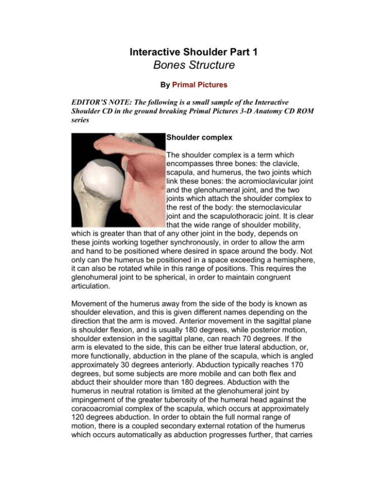
The shoulder complex is a term which
encompasses three bones: the clavicle,
scapula, and humerus, the two joints which
link these bones: the acromioclavicular joint
and the glenohumeral joint, and the two
joints which attach the shoulder complex to
the rest of the body: the sternoclavicular
joint and the scapulothoracic joint. It is clear
that the wide range of shoulder mobility,
which is greater than that of any other joint in the body, depends on
these joints working together synchronously, in order to allow the arm
and hand to be positioned where desired in space around the body. Not
only can the humerus be positioned in a space exceeding a hemisphere,
it can also be rotated while in this range of positions. This requires the
glenohumeral joint to be spherical, in order to maintain congruent
articulation.
Movement of the humerus away from the side of the body is known as
shoulder elevation, and this is given different names depending on the
direction that the arm is moved. Anterior movement in the sagittal plane
is shoulder flexion, and is usually 180 degrees, while posterior motion,
shoulder extension in the sagittal plane, can reach 70 degrees. If the
arm is elevated to the side, this can be either true lateral abduction, or,
more functionally, abduction in the plane of the scapula, which is angled
approximately 30 degrees anteriorly. Abduction typically reaches 170
degrees, but some subjects are more mobile and can both flex and
abduct their shoulder more than 180 degrees. Abduction with the
humerus in neutral rotation is limited at the glenohumeral joint by
impingement of the greater tuberosity of the humeral head against the
coracoacromial complex of the scapula, which occurs at approximately
120 degrees abduction. In order to obtain the full normal range of
motion, there is a coupled secondary external rotation of the humerus
which occurs automatically as abduction progresses further, that carries
the greater tuberosity posteriorly to avoid impingement. Ranges of
motion tend to decrease with advancing age.
If the shoulder is slightly flexed, so that the humerus is brought anterior
to the thorax, the shoulder can be adducted, bringing the arm towards
the centerline of the body, by approximately 60 degrees. The arm can
also be moved in a horizontal plane after abducting the shoulder 90
degrees, and this allows the arm to move across the front of the body to
approximately 140 degrees range of motion, and 45 degrees posteriorly
from the lateral direction. Thus, the shoulder complex allows an overall
range of motion of 180 degrees in almost all directions. This range of
motion of the humerus in space is accomplished by motion of the
scapula relative to the thorax, in addition to the dominant glenohumeral
motion. This sharing of mobility has been studied most extensively for
humeral abduction in the plane of the scapula. Different researchers
have found variations in motion, but overall it seems that 60% of this
motion arises at the glenohumeral joint and 40% at the scapulothoracic
joint. Similarly, shoulder motion with the humerus in a horizontal plane
includes scapular protraction and retraction movement. This scapular
mobility is essential for maintaining some reduced shoulder function if
arthritis with loss of bone requires an arthrodesis of the glenohumeral
joint. Scapular motion is linked to the clavicle, which controls the position
of the acromioclavicular joint in relation to the thorax at the
sternoclavicular joint.
Rotation of the humerus can also compound the mobility of the shoulder
complex. When the arm is alongside the thorax with the elbow flexed 90
degrees, internal rotation is limited by the hand reaching the body, while
external rotation is limited to 90 degrees by tightening of the soft tissues
crossing the anterior aspect of the shoulder, such as the anterior
glenohumeral ligaments and subscapularis muscle. Similar ranges of
humeral rotation are found when the humerus is rotated with the
shoulder abducted 90 degrees. If the elbow is extended when rotation
occurs, the hand rotates further than the humerus, due to the additional
mobility of the radioulnar joints in the forearm.
The great mobility of the shoulder complex allows the hand to be
positioned in space; it also allows the hand to reach to a particular point
via different paths of motion. This adaptability has led to some
apparently paradoxical observations on complex shoulder motion, most
notably that known as Codmans paradox. This paradox poses the
question of whether the humerus is in a position of internal or external
rotation when the forearm is held horizontally above the head. If the
humerus is returned to the side of the body by flexion of the
glenohumeral joint in the sagittal plane, the forearm ends up across the
front of the body, so the humerus is in internal rotation . Conversely, if
the humerus is lowered by rotating in an arc of adduction in the coronal
plane, the forearm ends up pointing away from the body the humerus
clearly in external rotation. This paradox arises because the path taken
through space, and the final destination, are affected by the sequence in
which rotations occur about perpendicular axes. This phenomenon is
well known among engineers, who refer to the three mutually
perpendicular rotations as pitch, yaw and roll, and conventions are used
to ensure uniformity of the sequence of application of the rotations and,
therefore, of the motion described.
Scapula
This bone acts primarily to provide extensive attachment areas for
muscles that contribute to the rotator cuff: supraspinatus, infraspinatus
and subscapularis. They focus their tensions across the glenohumeral
joint, pulling the head of the humerus medially, thus causing the
compressive joint force component that stabilizes the head of the
humerus into the relatively shallow articulation of the glenoid. The blade
of the scapula can be transparently thin, and it is protected from buckling
failure, under the actions of the muscles which attach across it, by the
spine of the scapula, that separates the attachments of the
supraspinatus and infraspinatus muscles. The scapula depends on the
clavicle to control its position, as the link to the thorax is somewhat
tenuous. The scapula is effectively suspended below the lateral end of
the clavicle by the coracoclavicular ligaments, and is also joined to it at
the acromioclavicular joint, which is surrounded by acromioclavicular
ligaments.
Glenohumeral joint
This joint has the largest range of motion in the body. It is essentially
part-spherical. In order to allow the wide range of motion, the concavity
of the glenoid has a shallow rim, to delay impingement by the humerus
as it approaches the limits of motion, that is augmented by a soft labrum.
The shallow articulation means that the joint has little inherent stability,
and so it depends on the coordinated actions of the muscles to maintain
stability, particularly those which constitute the rotator cuff. The
ligaments of the glenohumeral joint capsule only act to stabilize the joint
when tightened, which only occurs at the limits of the ranges of motion.
The lack of restraint in the glenohumeral joint means that although it has
a spherical geometry, there is usually some inherent laxity. The concave
surface of the glenoid often has a greater radius of curvature (i.e. it is
relatively flat) than does the articular surface of the head of the humerus.
This leads to a relatively small area of contact between them. This has
two side effects: firstly, it causes the joint contact pressure to rise;
secondly, it allows some joint mobility. This is seen as a linear translation
movement, that is a slight subluxation, of the humeral head relative to
the glenoid as the head rotates. If the head were located in a glenoid
with matching radius of curvature, then it would not be free to translate,
and so it would be forced to spin in one place as the humerus rotates,
and this causes a relative sliding motion between the articular surfaces.
This is the case for the hip joint. The glenohumeral joint has some rolling
motion mixed in with the sliding: as the humerus starts a movement of
external rotation , for example, the humeral head will initially roll
posteriorly over the surface of the glenoid, perhaps for 3 mm. As it
encounters increasing restraint from the slope of the labrum, so it tends
to stop rolling and start sliding in one place . Noting this arrangement for
natural joints, some joint replacements have been made with their metal
humeral head with a smaller radius than the polyethylene glenoid
surface, in order to allow these small natural secondary movements. The
downside to this, however, is the possibility of increased wear due to the
raised contact pressure. The glenoid is somewhat egg shaped, with the
narrower part superiorly, and it is typically 35 mm in superior-inferior and
25 mm in anterior-posterior directions. Sometimes, the face of the
glenoid is angled away from the normal orientation, and this can be
exaggerated by arthritic erosions, and leads to a tendency to instability,
as the head of the humerus tends to slip off.
The articular surface of the humeral head is nearly hemispherical and is
angled superiorly approximately 45 degrees and posteriorly
approximately 30 degrees. This allows greater ranges of abduction and
external rotation before impingement occurs. When transverse sections
of the proximal humerus are viewed, the articular surface is seen not to
be centered on the long axis of the shaft: it is offset posteriorly. The
combination of offset and angulation is variable, and this is allowed for
by some shoulder prosthesis designs.
There is little data available about forces acting across the glenohumeral
joint, but a relatively simple analysis can show that it will reach three
times body weight, and perhaps higher, during abduction to 90 degrees
while supporting a load of 12% body weight in the hand. The force
analysis shown in the diagram has been simplified greatly by assuming
that all of the shoulder elevation moment comes from the deltoid tension.
In reality, this moment is shared with other muscles which act to elevate
the humerus and also to stabilize the head of the humerus into the
glenoid, especially the supraspinatus. It is possible to apportion muscle
forces using schemes which allow for the number of muscle fibers (the
physiological cross-sectional area) and the degree of muscle activation,
that can be assessed by electromyography. However, all other muscles
have a humeral elevation moment arm which is smaller than that of the
deltoid, and this means that the simplification leads to a lower bound for
the muscle force acting across the glenohumeral joint. The force vector
diagram shows the relative magnitudes of the muscle and joint forces,
and also their directions in the plane of the scapula. It is clear that the
majority of the joint force is caused by the muscle action. The resultant
joint force shown will cause the head of the humerus to press against the
central superior rim of the glenoid. Activities such as shoulder flexion or
abduction while holding a weight of 2kg in the hand lead to joint forces of
approximately 1.5 body weight. These forces act centrally and superiorly
or superiorly and anteriorly onto the glenoid, giving a tendency for the
head of the humerus to sublux superiorly. When this occurs, the
subluxation is limited by the head of the humerus impinging against the
coracoacromial ligament, which passes in an anterior-posterior direction
above the head of the humerus.
Scapulothoracic joint
The scapulothoracic joint is not a true articular joint, but simply a plane of
separation of the scapula and the subscapularis muscle from the thorax,
that allows relative motion. The thoracic surface here is the superficial
aspect of the serratus anterior overlying the ribs. It allows the scapula to
slide anteriorly/posteriorly around the rib cage with shoulder
protraction/retraction. As it does so, the scapula rotates about a vertical
axis, since the ribs are curved, and so shoulder protraction causes the
glenoid to face more anteriorly, and more posteriorly with retraction. The
scapula also rotates in its own plane as the humerus is
abducted/adducted . In shoulder abduction, approximately 40% of the
humeral abduction motion occurs at the scapulothoracic articulation.
There is typically some 75 degrees motion here in 180 degrees
abduction. By rotating the scapula, the glenoid is angled superiorly as
the humerus is abducted, and so superior impingement is delayed.
Further, the superior angulation of the glenoid allows the compressive
joint force to remain within the articular concavity, which is stable. If the
scapula did not abduct with the humerus, then the muscles would pull
the head of the humerus into inferior (caudal) subluxation. Rotation of
the scapula in its own plane is caused by coordinated actions of the
serratus anterior and the upper and lower parts of the trapezius muscles:
their actions can create a rotation moment acting onto the scapula
without a resultant force tending to move it linearly. Scapular protraction
and retraction are due to actions of serratus anterior, and rhomboid plus
trapezius actions, respectively.
Movement at the scapulothoracic joint is essential to patients who have
their glenohumeral joint arthrodesed, as some upper limb mobility still
remains.
Acromioclavicular joint
This joint is loaded primarily in compression by the actions of the
muscles which pull the shoulder towards the centerline of the body, such
as the anterior pectoral muscles. There is also a superior-inferior
shearing load, due to muscles which tend to elevate the clavicle, but this
is counteracted by the inferior component of the tensions in pectoralis
major and anterior deltoid, which pass to the humerus from the clavicle.
Since the acromioclavicular joint surfaces are oblique, the loads applied
tend to cause the lateral end of the clavicle to sublux superiorly if the
joint is disrupted: the lateral end of the clavicle tends to slide uphill
across the slope of the joint surface on the acromion. This tendency is
resisted by the coracoclavicular ligaments, damage to which allows the
characteristic superior prominence of the end of the clavicle. The
acromioclavicular ligaments which surround the lateral end of the
clavicle are slack enough to allow motion between the clavicle and the
scapula, with a relative scapular rotation of approximately 20 degrees
during humeral abduction, and shearing actions at the joint during
shoulder protration/retraction, when the motion is centered at the conoid
part of the coracoclavicular ligaments. Similarly, shoulder abductionadduction motion causes the scapula to rotate about a horizontal
anterior-posterior axis relative to the clavicle (which is itself rotating
similarly about the sternoclavicular joint), that is centered amidst the
coracoclavicular ligaments. Since these attach away from the end of the
clavicle, this motion also causes the end of the clavicle to slide across
the surface of the acromion as the scapula rotates.
Clinical Pathology Text
The suprascapular nerve is located in the supraspinous or spinoglenoid
notch, at the superior border of the supraspinous fossa. In this location
the suprascapular nerve may be compressed by a ganglion or
entrapped, secondary to thickening of the suprascapular ligament.
The origin of the coracoid is superior and medial to the glenoid on the
scapular neck, and the tip of the coracoid projects anterior and lateral to
the glenoid. The coracoid is an important surgical landmark because
neurovascular structures travel along its inferomedial surface.
The acromion is classified as one of three types, depending on its
morphology. A type 1 acromion has a flat or straight undersurface with a
high angle of inclination. A type 2 acromion has a curved arc and
decreased angle of inclination. A type 3 acromion is hooked anteriorly,
with a decreased angle of inclination. The angle of inclination is formed
by the intersection of a line drawn from the posteroinferior aspect of the
acromion to the anterior margin of the acromion with a line formed by the
posteroinferior aspect of the acromion and the inferior tip of the coracoid
process.
At the lateral angle of the scapula is the glenoid cavity (glenoid fossa)
with its supra- and infraglenoid tuberosities. The glenoid version angle
varies, and may contribute to instability patterns of the shoulder.
Acromioclavicular Separations
There are three types of AC separations: type 1is a sprain or incomplete
tear of the AC joint capsule, type 2 is a complete tear of the AC joint
capsule with intact coracoclavicular ligaments, and type 3 involves
disruption of both the AC joint capsule and the coracoclavicular
ligaments. Widening of the AC joint space to 1.0 to 1.5 cm and a 25% to
50% increase in coracoclavicular distance is associated with tearing of
the AC joint capsule and sprain of the coracoclavicular ligament.
Widening of the AC joint to 1.5 cm or a 50% increase in the
coracoclavicular distance correlates with coracoclavicular ligament
disruption.
Arthritis
Degenerative osteoarthritis of the glenohumeral joint is relatively
common. It is characterized by cartilage-space narrowing, hypertrophic
bone formation, subchondral cysts, and associated soft-tissue
abnormalities of the rotator cuff. In rheumatoid disease, unlike
osteoarthritis, joint space narrowing is more uniform and symmetric,
without osteophytosis. Rheumatoid erosions occur at the margins of the
articular cartilage, including the greater tuberosity.
Avascular necrosis (AVN) of the humeral head ( Slide 1 , Slide 2 ) can
usually be differentiated from osteoarthritis on MR scans by the
restriction of subchondral low signal intensity ischemia to the humerus,
without associated glenoid involvement (i.e., sclerosis). Avascular
necrosis of the humeral head is associated with trauma, steroid use,
sickle-cell disease, and alcoholism
CLICK HERE for more information on the Primal Pictures 3-D Anatomy
series and for details on exclusive PTontheNET.com member discounts
Disclaimer
No warranty is given as to the accuracy of the information on any of the pages in this website. No responsibility is accepted for
any loss or damage suffered as a result of the use of that information or reliance on it. It is a matter for users to satisfy themselves
as to their or their client’s medical and physical condition to adopt the information or recommendations made. Notwithstanding a
users medical or physical condition, no responsibility or liability is accepted for any loss or damage suffered by any person as a
result of adopting the information or recommendations.
© Copyright Personal Training on the Net 1998 2003 All rights reserved

