Lab: Blood Vessels DUE: 11/27 Name
advertisement
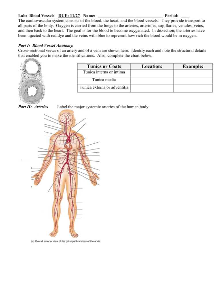
Lab: Blood Vessels DUE: 11/27 Name: ______________________________ Period: ____ The cardiovascular system consists of the blood, the heart, and the blood vessels. They provide transport to all parts of the body. Oxygen is carried from the lungs to the arteries, arterioles, capillaries, venules, veins, and then back to the heart. The goal is for the blood to become oxygenated. In dissection, the arteries have been injected with red dye and the veins with blue to represent how rich the blood would be in oxygen. Part I: Blood Vessel Anatomy. Cross-sectional views of an artery and of a vein are shown here. Identify each and note the structural details that enabled you to make the identifications. Also, complete the chart below. Tunics or Coats Location: Tunica interna or intima Tunica media Tunica externa or adventitia Part II: Arteries Label the major systemic arteries of the human body. Example: Part III: Veins Label the following major systemic veins of the human body. Part IV. Hepatic Portal System. Label the anatomy of the hepatic portal system. Compare the role of the hepatic vein versus the hepatic portal vein. Conclusion: 1. Fill in the blank with the correct artery or vein. o longest vein in the lower limb - __________ o supplies the diaphragm- __________ o artery serving the kidney- __________ o major artery serving the arm- __________ o artery generally used to take pulse on wrist- __________ o supplies massive amounts of blood to the neck- __________ o supplies the heart- __________ 2. Why are valves present in veins and not arteries? 3. What is the Circle of Willis? 4. How are pulmonary arteries and veins different from all other arteries and veins in the body?

