HLTH603: Gross Human Anatomy Lower Extremity Pre
advertisement
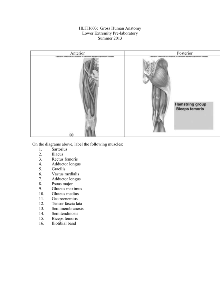
HLTH603: Gross Human Anatomy Lower Extremity Pre-laboratory Summer 2013 Anterior On the diagrams above, label the following muscles: 1. Sartorius 2. Iliacus 3. Rectus femoris 4. Adductor longus 5. Gracilis 6. Vastus medialis 7. Adductor longus 8. Psoas major 9. Gluteus maximus 10. Gluteus medius 11. Gastrocnemius 12. Tensor fascia lata 13. Semimembranosis 14. Semitendinosis 15. Biceps femoris 16. Iliotibial band Posterior Anterior Identify the following muscles on the diagram above: 1. Gastrocnemius 2. Soleus 3. Tibialis anterior 4. Peroneus longus 5. Extensor digitorium 6. Achilles tendon (yes, I know it is not a muscle) 7. Biceps femoris 8. Semitendinosis Posterior Anterior On the diagrams above, label the following muscles/structures: 1. Vastus medialis 2. Vastus lateralis 3. Rectus femoris 4. Patella 5. Gastrocnemius 6. Soleus 7. Peroneus longus 8. Achilles tendon 9. Patellar tendon 10. Tendon of peroneus longus/brevis Lateral Actions of the Lower Extremity List the actions associated with each of the following joints: Joint Actions Hip Knee Ankle Arteries of the Lower Extremity Palpate the following arteries on a partner: 1. Femoral 2. Popliteal 3. Posterior tibial 4. Dorsal pedal On the diagram above, label the following arteries: 1. Femoral 2. Profunda (Deep femoral) 3. Lateral circumflex 4. Medial circumflex 5. Popliteal 6. Posterior tibial 7. Anterior tibial In the following diagram, color the external iliac artery in red, femoral artery in green, profunda femoris artery in blue, medial femoral circumflex artery black, lateral femoral circumflex artery in orange, descending branch of lateral circumflex in brown, popliteal artery in purple and the anterior tibial artery in mauve. Of course, if you don’t have these colors, choose your favorites. Nerves of the Lower Extremity What the two main divisions of the sciatic nerve? What regions/muscles do these divisions innervate? Define dermatome. What is the dermatome distribution for L4? What is the dermatome distribution for S1? Define myotome. What test would you perform to test the L3 myotome? What test would you perform to test the L5 myotome? Using your text (or some other Human Anatomy text) and using general terminology, fill in the chart below with the origin, insertion and action of the following lower extremity muscles Muscle Iliopsoas Sartorius Adductor magnus Adductor longus Adductor brevis Pectineus Gracilis Rectus femoris Vastus lateralis, intermedius, medialis Tensor fascia lata Gluteus maximus Gluteus medius Piriformis Semimembranosus Semitendinosus Biceps femoris Gastrocnemius Soleus Extensor digitorum longus Plantaris Origin Insertion Action Popliteus Tibialis anterior Extensor hallucis longus Fibularis (peroneus) longus Fibularis (peroneus) brevis Flexor digitorum longus Flexor hallucis longus From the list of muscles above, list all muscles which complete the following joint movements: Joint movement Hip extension Knee extension Ankle dorsiflexion Hip abduction Ankle eversion Muscles Great toe flexion 2-4 toe flexion Great toe extension 2-4 toe extension

