Sheep Eye Dissection
advertisement
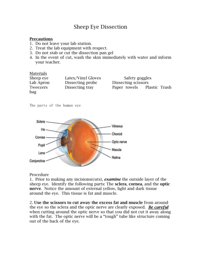
Sheep Eye Dissection Precautions 1. Do not leave your lab station. 2. Treat the lab equipment with respect. 3. Do not stab or cut the dissection pan gel 4. In the event of cut, wash the skin immediately with water and inform your teacher. Materials Sheep eye Lab Apron Tweezers bag Latex/Vinyl Gloves Dissecting probe Dissecting tray Safety goggles Dissecting scissors Paper towels Plastic Trash The parts of the human eye Procedure 1. Prior to making any incisions(cuts), examine the outside layer of the sheep eye. Identify the following parts: The sclera, cornea, and the optic nerve. Notice the amount of external yellow, light and dark tissue around the eye. This tissue is fat and muscle. 2. Use the scissors to cut away the excess fat and muscle from around the eye so the sclera and the optic nerve are clearly exposed. Be careful when cutting around the optic nerve so that you did not cut it away along with the fat. The optic nerve will be a “tough” tube like structure coming out of the back of the eye. 3. The sheep eye has 4 external attached muscles which control the eye movement. The human eye has six. The additional muscles allow humans the ability to “roll” and move their eyes in all directions. 4. Although the muscles of each eye work collectively as a team, the eyes themselves do not focus or work together until months after birth. Most people have one dominant eye. To find out which of your eyes is dominant, form a circle with your thumb and index finger. Hold that position and place your hand in front of you. With both eyes, look at an object through the circle. Continue to hold that position and close one eye then open it. Close the other eye, open it. The eye that you could still see the object with is your dominant eye. Which eye is your dominant eye? Left Right (circle one) 5. Use the probe to pierce the sclera, about half way between the pupil and the back of the eye. You will be cutting the eye in half, through the sclera. Be careful when you cut so that you do not destroy the pupil and lens. 6. Follow a circular pattern around the sclera, rotate the eye while continuing to carefully cut the eye in half. 7. A clear jelly like fluid will be inside the eye. This is the vitreous humor. Carefully lay the two sides of the eye in the dissection pan. 8. Observe the back half of the eye. You will see a dark reflective lining. This is called the tapetum and is not found in the human eye. This is why an animal’s eyes glow in the dark. This reflective covering reflects light back toward the lens and allows the animals to see better in limited light. 9. Attached to the optic nerve is a wrinkled saclike structure connected to the back of the eye. This is the retina and is considered the innermost layer of the eye. Just as the tapetum surrounds the inside of the sclera, the living tissue of the retina would have been smooth and would have rested against the tampetum. The point at which the retinal nerve tissue connects to the optic nerve is the eye’s blind spot. 10.Separate the retina from the back portion of the eye. 11.Remove the tapetum from the tough shell like outer layer of the sclera and see if you can find the choroid. 12.Now observe the front half of the eye. Place the eye with the cornea down. 13. In the front of the eye, or mixed with the vitreous humor will be a small lens. This lens is convex and will have a slightly gray color. In a live organism, the lens would be transparent. 14.Remove the lens from the vitreous humor. Dry it with a piece of paper towel. Feel and feel how flexible it is. Why is the lens flexible? 15.Hold the lens up in front of your eye, look through it. Describe how the lens changes your vision. 16. A ring of tiny ciliary muscles are located along the inner side of the iris. These muscles connect to the lens. 17. The pupil surrounded by the iris is on the front of the eye is the actual opening located in the center of the pupil. The pupil is covered and protected by the transparent living tissue of the cornea.+ 18.The cornea would be transparent in a living organism. Use the tweezers and hold up the front part of the eye. Look through the sheep pupil and cornea. How does your vision change? Why does the cornea and lens have to be transparent? 19.ASSESSMENT: When you are finished with the dissection, raise your hand. I will come by and ask you to identify one part of the eye. You will have 2 chances to place the probe on the correct part. I will record who answers correctly. NO TALKING during this time by any lab partners. 20. Follow clean up procedures: a) Wrap all remains in a paper towel and dispose of in the plastic trash bag b) All dissecting instruments, tray and gel must be thoroughly washed in water. c) The gel should be in the dissection pan, cover with a clean dry paper towel. d) All used gloves are to be disposed of the trash bag. e) All lab aprons and goggles returned to the designated place. f) Nothing is in the sinks at your lab station. If any material is in the sink, use a paper towel to remove and dispose of the paper towel in the trash bag. g) Lab stations are clean and free of water/waste h) Each person washes their hands with soap and water This lab is adapted from http://www.carver.org/middle_school/Teams/Ciarfella/Sci2.htm Analysis Questions: 1. Why does the retina have to be smooth? Why not wrinkled? (Think about reflecting light off of a wrinkled piece of aluminum foil and a smooth piece.) 2. Think about learning the parts of the eye. Teachers can use a diagram, a model, or dissection to show students the parts. In your opinion, use examples from your own experience, which method (diagram, model, dissection) do you think helped you to understand the parts of the eye the most? Why?


