respiratory sys questions
advertisement
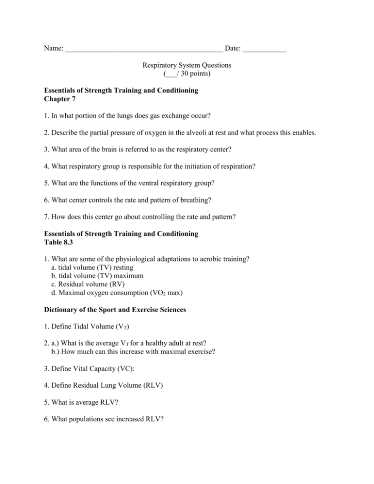
Name: ___________________________________________ Date: ____________ Respiratory System Questions (___/ 30 points) Essentials of Strength Training and Conditioning Chapter 7 1. In what portion of the lungs does gas exchange occur? 2. Describe the partial pressure of oxygen in the alveoli at rest and what process this enables. 3. What area of the brain is referred to as the respiratory center? 4. What respiratory group is responsible for the initiation of respiration? 5. What are the functions of the ventral respiratory group? 6. What center controls the rate and pattern of breathing? 7. How does this center go about controlling the rate and pattern? Essentials of Strength Training and Conditioning Table 8.3 1. What are some of the physiological adaptations to aerobic training? a. tidal volume (TV) resting b. tidal volume (TV) maximum c. Residual volume (RV) d. Maximal oxygen consumption (VO2 max) Dictionary of the Sport and Exercise Sciences 1. Define Tidal Volume (VT) 2. a.) What is the average VT for a healthy adult at rest? b.) How much can this increase with maximal exercise? 3. Define Vital Capacity (VC): 4. Define Residual Lung Volume (RLV) 5. What is average RLV? 6. What populations see increased RLV? 7. Define Total Lung Capacity (TLC) (Total Lung Volume (TLV)): 8. a.) Define VO2 (oxygen consumption) b.) What is the average VO2 for a young male at rest? c.) What can this value go up to during maximal exercise? 9. What is VO2 max? Essentials of Exercise Physiology (Paperback book): 1. Describe in your own words, the mechanics of inspiration and expiration 2. Using Figure 10.5, draw, define, and give male/female averages for tidal volume (TV), total lung capacity (TLC), residual lung volume (RLV), forced vital capacity (FVC), and functional residual capacity (FRC). Atlas of Human Anatomy: 1. a.) Define inspiration b.) Define expiration 2. What are the primary muscles of inspiration? 3. What are the muscles of expiration during quiet breathing? 4. What is the primary group of muscles responsible for forced or active expiration? 5. When the diaphragm contracts, what is the result? Clinically Oriented Anatomy: 1. What are the two main types of pleura? 2. Where are the two types of pleura found? 3. What is the purpose of the visceral pleura? 4. What is the pleural cavity? Where is it located? 5. What is contained within the pleural cavity? 6. What is the purpose of the substance found within the pleural cavity? Key Respiratory System Questions (___/ 30 points) Essentials of Strength Training and Conditioning Chapter 7 1. In what portion of the lungs does gas exchange occur? Alveoli 2. Describe the partial pressure of oxygen in the alveoli at rest and what process this enables. At rest, the partial pressure of oxygen in the alveoli is about 60 mmHg greater than that in the pulmonary capillaries. Thus, oxygen diffuses into the pulmonary capillaries. Carbon dioxide diffuses in the opposite direction of oxygen 3. What area of the brain is referred to as the respiratory center? The brainstem, containing the Pons and medulla oblongata 4. What respiratory group is responsible for the initiation of respiration? Dorsal respiratory group 5. What are the functions of the ventral respiratory group? Respiratory signals from these neurons contribute to the respiratory drive for increased pulmonary ventilation. Stimulation of some of the neurons in the ventral group causes inspiration or expiration depending on where the stimulus is located. (these neurons are essentially important in providing the expiratory signals to the powerful abdominal muscles during more forceful expiration.) 6. What center controls the rate and pattern of breathing? Pneumotaxic 7. How does this center go about controlling the rate and pattern? The center controls the duration of filling cycle of the lungs Essentials of Strength Training and Conditioning Table 8.3 1. What are some of the physiological adaptations to aerobic training? a. tidal volume (TV) resting – no change b. tidal volume (TV) maximum – 2.8-3.1 c. Residual volume (RV) – 5.8-6.7 d. Maximal oxygen consumption (VO2 max) – 47-55 mL/kg/min Dictionary of the Sport and Exercise Sciences 1. Define Tidal Volume (VT) amount of air inspired or expired per breath 2. a.) What is the average VT for a healthy adult at rest? 500mL b.) How much can this increase with maximal exercise? 3-4 times 3. Define Vital Capacity (VC): maximal volume of air forcefully expired after maximal inspiration 4. Define Residual Lung Volume (RLV) the volume of air that remains in the lungs after maximal expiration 5. What is average RLV? 1-2 L 6. What populations see increased RLV? Older peoples and smokers 7. Define Total Lung Capacity (TLC) (Total Lung Volume (TLV)): Maximum amount of air in the lungs; includes the sum of residual volume and vital capacity 8. a.) Define VO2 (oxygen consumption): The amount of oxygen taken up and used at the cellular level at rest, during submaximal exercise or in recovery b.) What is the average VO2 for a young male at rest? 250 mL/min c.) What can this value go up to during maximal exercise? 5,100 mL/min 9. What is VO2 max? Maximal oxygen consumption Highest amount of oxygen the body can consume during work for the aerobic production of ATP Essentials of Exercise Physiology (Paperback book): 1. Describe in your own words, the mechanics of inspiration and expiration Inspiration: diaphragm contracts, flattens toward the abdominal cavity. This elongates the chest cavity. The volume of the lungs increases, the air in the lungs expands decreasing intrapulmonic pressure to 5 mmHg below atmospheric pressure. The pressure differential between the lungs and ambient air literally suck air in through the nose and mouth and inflates the lungs Expiration: Passive process, occurs as air moves out of the lungs. It results from the recoil of stretched lung tissue and relaxation of inspiratory muscles. The diaphragm moves back toward the thoracic cavity. Cheat volume decreases increasing pressure and forcing air out of the respiratory tract 2. Using Figure 10.5, draw, define, and give male/female averages for tidal volume (TV), total lung capacity (TLC), residual lung volume (RLV), forced vital capacity (FVC), and functional residual capacity (FRC). TV- tidal volume: volume of air inspired or expired per breath (M 600 mL, F 500mL) TLC – total lung capacity: volume in lungs after maximal inspiration (M 6000mL, F 4200mL RLV – residual lung volume: volume of air lungs after maximal expiration (M 1200 mL, F 1000 mL) FVC – forced vital capacity: maximal volume of air expired after maximum inspiration (M 4800 mL, F 3200 mL) FRC – functional residual capacity: volume of air in lungs after tidal expiration (M 2400 mL, F 1800 mL) Atlas of Human Anatomy: 1. a.) Define inspiration Breathing in b.) Define expiration Breathing out 2. What are the primary muscles of inspiration? External Intercostals Internal Intercostals Diaphragm 3. What are the muscles of expiration during quiet breathing? This is a trick question, expiration results from passive recoil of the lungs 4. What is the primary group of muscles responsible for forced or active expiration? (Hint: put your hand on your stomach and breathe out or laugh) Abdominals 5. When the diaphragm contracts, what is the result? Domes descend, thus increasing the vertical dimension of the thoracic cavity Clinically Oriented Anatomy: 1. What are the two main types of pleura? Visceral and Parietal 2. Where are the two types of pleura found? A. Visceral surrounds the lungs and cannot be dissected from the lungs B. Parietal pleura lines the pulmonary cavities 3. What is the purpose of the visceral pleura? The visceral pleura provide the lung with a smooth, slippery surface enabling it to move freely on the parietal pleura 4. What is the pleural cavity? Where is it located? The pleural cavity is the potential space between the layers of the pleura 5. What is contained within the pleural cavity? Serous pleural fluid is contained within the pleural cavity 6. What is the purpose of the substance found within the pleural cavity? Serous pleural fluid lubricates the pleural surfaces and allow the layers of pleura smoothly over each other during respiration
