Control of Respiration
advertisement
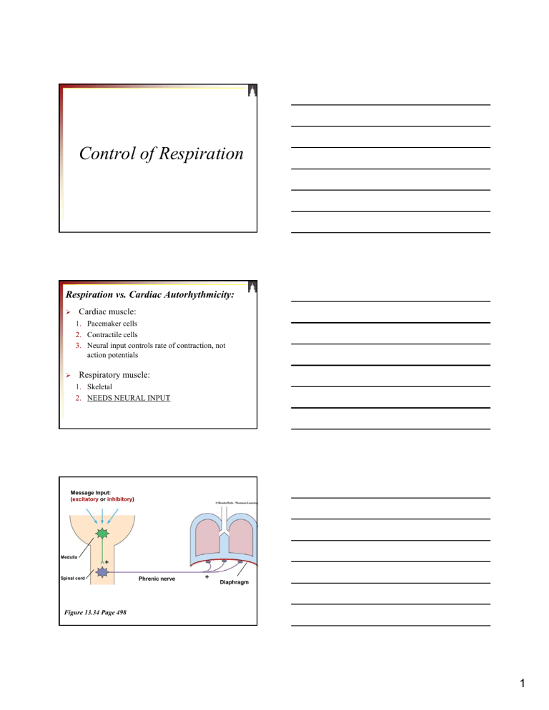
Control of Respiration Respiration vs. Cardiac Autorhythmicity: Cardiac muscle: ¾ 1. Pacemaker cells 2. Contractile cells 3. Neural input controls rate of contraction, not action potentials Respiratory muscle: ¾ 1. Skeletal 2. NEEDS NEURAL INPUT Message Input: (excitatory or inhibitory) Medulla Spinal cord Phrenic nerve Diaphragm Figure 13.34 Page 498 1 2 Neural Components: Figure 13.33 Page 497 Rhythmic inspiration/expiration 1. Respiratory control center Pons Pons respiratory centers Respiratory control centers in brain stem Pre-Bötzinger complex Medullary respiratory center Medullary respiratory center Pneumotaxic center Apneustic center Dorsal respiratory group Ventral respiratory group Medulla Dorsal respiratory group Ventral respiratory group Dorsal Respiratory Group (DRG) 1. • Inspiratory neurons 9 Firing causes inspiration (diaphragm & external intercostals) 9 Cease firing causes expiration Ventral Respiratory Group (VRG) 2. • Inspiratory & expiratory neurons 9 Inactive during normal quiet breathing 9 Work as “overdrive” for active breathing (muscles of expiration & inspiration) Pre-Bötzinger complex Pre-Botzinger complex 3. • • • Rhythm of ventilation Neuronal configuration similar to SA node Believed to control rate of DRG inspiratory neurons 2 Pons respiratory centers Pneumotaxic center Apneustic center “Fine-tuning” center Pneumotaxic center 1. • Neuronal input aiding in “turning off” inspiration Apneustic center 2. • Prevents inspiratory neurons from being “turned off” Hering-Breuer reflex & pulmonary stretch receptors 3. • Prevents overinflation (exercise) 2 Neural Components: Magnitude of ventilation 2. Sensory nerve fibers Three chemical factors: i. Carotid bodies PO2 • Monitored by peripheral chemoreceptors ii. PCO2 iii. H+ Aortic bodies PO2 fluctuation does NOT generally cause increased respiration i. • Hb-O2 saturation curves 3 Only if PO2 is dangerously LOW will it cause ventilation stimulation! Relieves Arterial PO2 <60 mm Hg Emergency life-saving mechanism Medullary respiratory center Peripheral chemoreceptors No effect on Central chemoreceptors Ventilation Arterial PO2 Figure 13.36 Page 498 PCO2 ii. • Major role in magnitude of ventilation Central chemoreceptors: 1. Sensitive to changes in CO2 induced H+ concentrations in the brain ECF 2. Increased brain H+ directly stimulates central chemoreceptors Arterial PCO2 Relieves Brain ECF PCO2 (when arterial PCO2 >70-80 mm Hg) Brain ECF H+ Weakly Peripheral chemoreceptors Medullary respiratory center Ventilation Arterial PCO2 Central chemoreceptors Figure 13.37 Page 501 4 ¾ Arterial H+ • Aortic and carotid bodies highly sensitive to changes in H+ Arterial H+ Acidosis Cannot penetrate blood-brain barrier Relieves Peripheral chemoreceptors Medullary respiratory center No effect on Central chemoreceptors Ventilation Arterial PCO2 Arterial -CO2 -H+ Figure 13.38 Page 502 Questions? 5


![Respiratory System Lecture:9 [PPT]](http://s2.studylib.net/store/data/010095677_1-807d395ef4decfdd1b95f3c6166ad4e0-300x300.png)
