Nervous System Lab Glial cells Neuron
advertisement
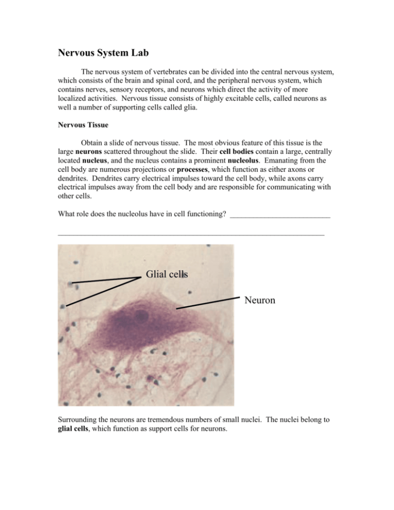
Nervous System Lab The nervous system of vertebrates can be divided into the central nervous system, which consists of the brain and spinal cord, and the peripheral nervous system, which contains nerves, sensory receptors, and neurons which direct the activity of more localized activities. Nervous tissue consists of highly excitable cells, called neurons as well a number of supporting cells called glia. Nervous Tissue Obtain a slide of nervous tissue. The most obvious feature of this tissue is the large neurons scattered throughout the slide. Their cell bodies contain a large, centrally located nucleus, and the nucleus contains a prominent nucleolus. Emanating from the cell body are numerous projections or processes, which function as either axons or dendrites. Dendrites carry electrical impulses toward the cell body, while axons carry electrical impulses away from the cell body and are responsible for communicating with other cells. What role does the nucleolus have in cell functioning? __________________________ _____________________________________________________________________ Glial cells Neuron Surrounding the neurons are tremendous numbers of small nuclei. The nuclei belong to glial cells, which function as support cells for neurons. Sheep Brain Dissection Again, the central nervous system is composed of the brain and the spinal cord. Obtain a sheep brain and a dissection pan from your instructor. The largest region of the sheep brain is the cerebrum, which is clearly divided into two hemispheres by a deep longitudinal fissure. The cerebrum contains regions that allow for such things as learning and memory, voluntary muscle control, and sensory perception. Just behind the cerebrum, is the cerebellum (“little brain”). The cerebellum coordinates voluntary movement and coordinates equilibrium. The surface of both the cerebrum and the cerebellum display folds (gyri) and grooves (sulci) which increase the surface area of these regions. Longitudinal fissure cerebrum cerebellum On the undersurface of the brain, the olfactory bulbs sit at the anterior end of the cerebrum. The olfactory nerves from the nasal cavity synapse with cells in this brain region. Is the sense of smell more important as a protective and food-getting sense in sheep or in humans? ______________________________________ Do you think the human olfactory bulbs would be larger or smaller (in proportion to the size of the brain) than those of the sheep brain? ________________________________________________ The optic nerves enter the brain just after a region called the optic chiasma, where some of the visual fibers “cross-over” to the opposite site of the body. This partial cross-over allows for increased depth perception, as compared to the visual system of an animal whose fibers completely cross to the opposite side. The pons and medulla oblongata form most of what is called the brain stem. The medulla oblongata is especially important as it houses neurons which control vital reflexes such as respiration and heart rate. A short segment of spinal cord may be attached to the posterior end of the medulla oblongata. Optic chiasma Medulla oblongata Pons Olfactory bulb If you use your scalpel, you may cut along the longitudinal fissure to cut the brain into sagittal sections. In the lower, central part of the cerebrum, you can clearly see the corpus callosum, which contains fibers that link the right and left cerebral hemispheres. Sensory Receptors An organism’s sensory receptors respond to stimuli from the internal or external environment. Receptors are specialized to respond to one type of stimulus, such as stretch, vibration, pain, or a chemical change in the environment. The structure of sensory receptors varies dramatically. The simplest receptors are free nerve endings. These typically respond to changes in pain and temperature and are widespread in the skin of humans and other animals. Some receptors are more complex, in that the nerve endings are encapsulated by connective tissue. One example of this type of encapsulated receptor is a Pacinian corpuscle, which responds to deep pressure. Look at the Pacinian corpuscle at the demonstration microscope. The thin nerve ending is in the center of the structure, and layers of connective tissue make up the bulk of the round or oval shape of this receptor. We will also look at a complex sensory receptor, the eye, in this lab. But first let’s test to see how numerous sensory receptors are in various locations over our body. Two-Point Discrimination Test. The density of touch receptors varies greatly in different body areas. Areas that have the highest densities of receptors are quite sensitive to touch, while areas with lower densities would be considered less sensitive. We are going to test the density of touch receptors in the areas of skin found in the table below. Which areas of the body would you predict might have the highest receptor density and thus be the most sensitive? ________________________________________________ ____________________________________________________________________ For the two-point discrimination test, you and a lab partner need one pair of calipers and a metric ruler. When testing each area of the skin, begin with the points of the caliper completely together. Touch your lab partner’s skin, and they should say whether they felt two points touching their skin or only one. If only one point was felt, move the calipers slightly apart, and touch their skin again. Repeat this procedure until your lab partner feels two points of contact. Measure this distance, which is called the two-point threshold, with your metric ruler. Body area tested Two-point threshold (in mm) Back of hand Palm of hand Fingertip Back of neck Ventral forearm Face Which area has the smallest two-point threshold? ________________________________ Which area has the largest two-point threshold? _________________________________ How well did your results compare with your prediction? _________________________ _______________________________________________________________________ Anatomy of the Eye In the last part of this lab, we are going to dissect the sheep eye as a representation of a complex sensory receptor. Obtain an eye and a dissecting pan from your instructor so that you can make the following observations. Your eye may be covered with a little bit of skin, fat or musculature which should be removed before continuing further. Anatomically, the wall of the eye consists of three layers. The outermost layer is made of fibrous connective tissue and is divided into two very different regions: the opaque sclera which covers most of the outer surface of the eye, and the clear cornea, which covers the most anterior surface of the eye. These two layers can be observed without any dissection. Extending out from the posterior side of the eye is a small optic nerve, which carries visual information into the central nervous system. To see the inner two layers of the eye, take your scalpel and cut the eye into two halves, cutting through the sclera, but not the cornea. As you open the eye, a thick jelly-like substance, the vitreous humor, may spill from the back part of the eye. If you remove the vitreous humor, be careful not to also remove a delicate, tancolored layer at the very back of the inside of the eye. This layer is the retina, and is where the photoreceptors are located. The vitreous humor holds the retina in place in the back of the eye, and the retina often falls away from the back of the eye when the vitreous has been removed. If you gently fold down some of the retina, you can observe the third layer of the eye, which rests under the retina. The middle layer of the eye is pigmented and carries most of the eye’s blood vessels. It too can be subdivided into different regions: the choroid, which is found in the more posterior part of the eye, and the ciliary body and iris which are in the anterior part of the eye, surrounding the lens. You may notice that the choroid is not uniformly dark, but is instead “painted” with bright, iridescent colors. This shiny, reflective layer is the tapetum lucidum, which helps animals see more acutely in dim light. Do you think the tapetum lucidum is found in human eyes? Why or why not? _________ _______________________________________________________________________ If you gently separate the lens from the ciliary body, you can look more closely at the anterior surface of the iris. It is the pigmented portion of the eye, and in its center is an opening called the pupil. Light enters the eye through the cornea and the pupil, and the cornea and lens help to direct the light so that it falls on the retina. Both the iris and the ciliary body contain smooth muscle fibers which aid in this process. Contraction and relaxation of muscle in the iris change the size of the pupil so that an appropriate amount of light is allowed to enter the eye, while contraction and relaxation of muscle in the ciliary body changes the shape of the lens for adequate focusing. Does the lens feel flexible? ____________ Is it clear or opaque? __________________ Are these features normal, or has the lens been modified by the preservatives? _____________________________________________________________________

