GRAM STAIN INTRODUCTION
advertisement
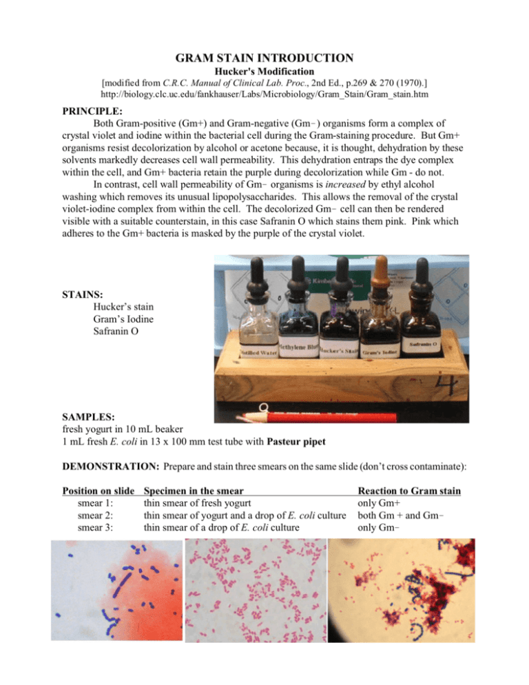
GRAM STAIN INTRODUCTION Hucker's Modification [modified from C.R.C. Manual of Clinical Lab. Proc., 2nd Ed., p.269 & 270 (1970).] http://biology.clc.uc.edu/fankhauser/Labs/Microbiology/Gram_Stain/Gram_stain.htm PRINCIPLE: Both Gram-positive (Gm+) and Gram-negative (Gm!) organisms form a complex of crystal violet and iodine within the bacterial cell during the Gram-staining procedure. But Gm+ organisms resist decolorization by alcohol or acetone because, it is thought, dehydration by these solvents markedly decreases cell wall permeability. This dehydration entraps the dye complex within the cell, and Gm+ bacteria retain the purple during decolorization while Gm - do not. In contrast, cell wall permeability of Gm! organisms is increased by ethyl alcohol washing which removes its unusual lipopolysaccharides. This allows the removal of the crystal violet-iodine complex from within the cell. The decolorized Gm! cell can then be rendered visible with a suitable counterstain, in this case Safranin O which stains them pink. Pink which adheres to the Gm+ bacteria is masked by the purple of the crystal violet. STAINS: Hucker’s stain Gram’s Iodine Safranin O SAMPLES: fresh yogurt in 10 mL beaker 1 mL fresh E. coli in 13 x 100 mm test tube with Pasteur pipet DEMONSTRATION: Prepare and stain three smears on the same slide (don’t cross contaminate): Position on slide smear 1: smear 2: smear 3: Specimen in the smear thin smear of fresh yogurt thin smear of yogurt and a drop of E. coli culture thin smear of a drop of E. coli culture Reaction to Gram stain only Gm+ both Gm + and Gm! only Gm! PROCEDURE: (Prepare thinly spread smears of fresh bacteria. See protocol on Smear & Staining, for smearing and fixing.) 1. PRIMARY STAIN: Stain with Hucker's Stain for 1 minute. (If over-staining results in improper decolorization of known Gram-negative organisms, use less crystal violet.) 2. Wash briefly in tap water (no longer than 2 seconds) to remove liquid Hucker's stain. 3. MORDANT: Flood the smear with Gram's iodine. Allow to remain for 1 minute. (You should see a metallic bronze on the surface of the iodine.) 4. DECOLORIZE with 95% ethyl alcohol: Rinse the specimen with EtOH over the sink, let sit, rinse with more EtOH. Repeat until the purple dye no longer flows from the smear. 5. Wash in tap water for 2 seconds to remove the EtOH. Blot gently (no rubbing.) 6. COUNTERSTAIN: Flood the smears with safranin 0. Allow to stain for 1 minute. 7. Wash with tap water. 8. Blot dry between sheets of bibulous paper or lint-free paper towel. Briefly flame to finish drying. 9. Examine under 1000x oil immersion lens. Illustrate morphologies and staining patterns observed. REAG ENTS: 1. Crystal violet (Hucker's Stain): Solution A crystal violet, certified 2.0 g ethyl alcohol, 95% 20.0 mL Solution B ammonium oxalate 0.8 g distilled water 80.0 mL Mix solutions A and B. Store for 24 hours before use. The resulting stain is stable. 2. Gram's iodine: Dissolve 0.33 g of iodine and 0.66 g of potassium iodide in 100 mL of distilled water. Alternately, dilute 0.1 N iodine 1:4. (Gram's Iodine solution which has weakened and appears tan will not work.) 3. Ethyl alcohol (95%) 4. Counterstain stock solution: Dissolve 2.5 g of certified safranin 0 in 100 mL of 95% ethyl alcohol. 5. Counterstain working solution Dilute stock solution 1:10 with dH2O.
