Action Potentials
advertisement
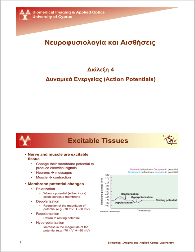
Biomedical Imaging & Applied Optics University of Cyprus Νευροφυσιολογία και Αισθήσεις Διάλεξη 4 Δυναμικά Ενεργείας (Action Potentials) Excitable Tissues • Nerve and muscle are excitable tissue • • • Change their membrane potential to produce electrical signals Neurons Æ messages Muscle Æ contraction • Membrane potential changes • Polarization • When a potential (either + or -) exists across a membrane • Depolarization • Reduction of the magnitude of potential (e.g. -70 mV Æ -50 mV) • Repolarization • Return to resting potential • Hyperpolarization • Increase in the magnitude of the potential (e.g. -70 mV Æ -90 mV) 2 Biomedical Imaging and Applied Optics Laboratory Excitable Tissues • Changes are triggered by • • • • Change of the local electrical field Interaction with chemical messenger and surface receptor Stimulus (e.g. sound, light, etc) Spontaneous change of potential by inherent ion leaks • Changes are caused by movement of ions • Leak channels • Gated channels • Open all the time • Can be open or closed (conformation change) • Types • • • • Voltage gated Chemically gated Mechanically gated Thermally gated • Electrical signals • • Graded Potentials Action Potentials Biomedical Imaging and Applied Optics Laboratory 3 Graded Potentials • Local changes in membrane potential • Confined to small area, the Active Area • Remaining cell is still at resting potential, the Inactive Area • Triggered by specific events • Gated channels (usually Na+ open) • Magnitude and duration proportional to triggering event Graded potential (change in membrane potential relative to resting potential) Resting potential Time Magnitude of stimulus Stimuli applied 4 Biomedical Imaging and Applied Optics Laboratory Graded Potentials • Propagate to adjacent areas • Movement of ions = current • Current spreads in the ECF and ICF (low resistance) but not through the membrane (high resistance) • Depolarizes adjacent regions • Graded potentials propagate Biomedical Imaging and Applied Optics Laboratory 5 Graded Potentials • Graded potentials die out over short distances • Loss of charge • Magnitude decreases as it moves away from the point of origin • Completely disappear with a few mm Portion of excitable cell Initial site of potential change Loss of charge Direction of current flow from initial site Loss of charge Direction of current flow from initial site * Numbers refer to the local potential in mV at various points along the membrane. • Grades potentials are important • • • • • 6 Postsynaptic potentials Receptor potentials End-plate potentials Pacemaker potentials Slow-wave potentials Biomedical Imaging and Applied Optics Laboratory Action Potentials • Large (~100 mV) changes in the membrane potential • • • • A.k.a spikes Can be initiated by graded potentials Unlike graded potentials action potentials propagate Transmit information Na+ equilibrium potential • Changes during an action potential • Gradual depolarization to threshold potential (-50 to -55 mV) • If not reached no action potential will occur • Rapid depolarization (+30 mV) • Portion between 0 and 30 mV is called an overshoot • • Rapid repolarization leading to hyperpolarization (-80 mV) Resting potential restored (-70 mV) K+ equilibrium potential • Constant duration for given cell type • E.g. Nerves Æ 1 msec Biomedical Imaging and Applied Optics Laboratory 7 Action Potentials • AP are a result of changes in ion permeability Voltage-gated Na+ channels • Voltage-gated channels • Proteins which change conformation depending on potential • Allow passage of ions • Voltage-gated Na+ channels • Activation (immediate) and inactivation gates (delayed) • Voltage-gated K+ channels • Activation gate (delayed) 8 Voltage-gated K+ channels Biomedical Imaging and Applied Optics Laboratory Action Potentials Time Event 0 msec 0.3 msec 0.5 msec 0.8 msec 1 msec Potential Resting state All channels are closed Graded potential arrives Begins depolarization - 70 mV Threshold reached Activation gates of Na+ channels open Activation gates of K+ channels begin to open slowly Inactivation gates of Na+ channels begin to close slowly - 50 mV Peak potential reached Inactivation gates of Na+ channels are now closed Activation gates of K+ channels are now open 30 mV Hyperpolarized state Activation gates of K+ channels close - 80 mV Resting state Na+-K+-pump restores resting potential Na+ channels are reset to close but active -70 mV Biomedical Imaging and Applied Optics Laboratory 9 Action Potentials • Neuron structure • Input Zone • Dendrites (up to 400 000) • Cell Body • Have receptors which receive chemical signals • Conduction zone • Axon or nerve fiber (axon hillock to axon terminals) <1 mm to >1m • Output zone • Axon terminal • Input • Synapse • Graded Potentials • Generated in the dendrites as a response to chemical signals • Can trigger action potentials in the axon • 10 Sensory nerve endings Biomedical Imaging and Applied Optics Laboratory Action Potentials • AP Propagation • APs initiated at the axon hilloc • More voltage-gated channels Æ lower threshold • Once initiated the AP travels the entire axon • Contiguous conduction • Saltatory conduction • Contiguous conduction • Flow of ions Æ depolarization of adjacent area to threshold • As AP is initiated in adjacent area, the original AP is ending with repolarization • The AP itself does not travel, it is regenerated at successive locations (like “wave” in a stadium) Biomedical Imaging and Applied Optics Laboratory 11 Action Potentials • Saltatory Propagation • Some neurons are myelinated • Covered with myelin (lipid barrier) • Formed by oligodendrocytes (CNS) and Schwann cells (PNS) • No ion movement across myelinated areas • Nodes of Ranvier • Areas between myelin sheaths • Ions can flow Æ APs can form • Local current can generate AP at the next node • APs “jump” from node to node Æ information travels 50x faster, less work by pumps to maintain ion balance • Loss of myelin can cause serious problems • E.g. multiple sclerosis 12 Biomedical Imaging and Applied Optics Laboratory Action Potentials • Refractory Period • APs do not travel backwards • Local currents do not regenerate an AP in the previously-activenow-inactive area • Certain time must pass before a second AP can be triggered Æ refractory period • Absolute refractory period • During an AP • No APs can be triggered Previous active New active area area returned toat peak of action resting potentialpotential New adjacent inactive area into which depolarization is spreading; will soon reach threshold “Forward” current flow excites new inactive area “Backward” current flow does not re-excite Direction of propagation of previously active area action potential because this area is in its refractory period Absolute Relative refractory refractory period period • Relative refractory period • Na+ channels are mostly inactive • K+ channels are slow to close • After an AP Æ second AP can be triggered only be exceedingly strong signals Action potential Na+ permeability K+ permeability • Refractory period sets an upper limit to the frequency of APs Æ~2.5 KHz Biomedical Imaging and Applied Optics Laboratory 13 Action Potentials • Characteristics of APs • How does strength vary? • Always the same! Æ All-or-None Law • Does not decrease during propagation • How are stronger stimuli recognized? • Faster generation of APs Æ ↑Frequency • More neurons fire simultaneously • What determines the speed of APs? • Myelination • Neuron diameter (↑ diameter Æ↓ Resistance to local current Æ ↑ Speed) • Large myelinated fibers: 120 m/sec (432 km/hr) Æ urgent information • Small unmyelinated fiber: 0.7 m/sec (2.5 km/hr) Æ slow-acting processes • Without myelin the diameter would have to be huge! (50 x larger) 14 Biomedical Imaging and Applied Optics Laboratory Regeneration of Nerve Fibers • Neurons in the PNS can regenerate • • • • Distal severed portion degenerates Schwann cells pick up the debris Schwann cells remain and form regeneration tube with nervegrowth-enhancing proteins Nerve grows through that tude • Neurons in the CNS can NOT regenerate • • Oligodendrocytes secrete nervegrowth-inhibiting hormones Necessary to keep a complex system such as the CNS stable (during the end of fetal development and later) Zebra fish axon induced to regenerate http://www.nbb.cornell.edu/neurobio/Fetcho/regeneration.htm • Many strategies to regenerate CNS neurons 15 Biomedical Imaging and Applied Optics Laboratory Επόμενη Διάλεξη … Διάλεξη 5 Συναπτική Μετάδοση (Synaptic Transmition) 16 Biomedical Imaging and Applied Optics Laboratory
