Meiotic telomeres: a matchmaker for homologous chromosomes
advertisement
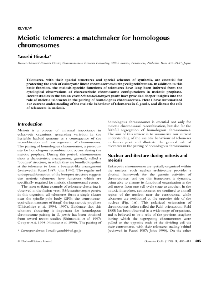
REVIEW Meiotic telomeres: a matchmaker for homologous chromosomes Yasushi Hiraoka* Kansai Advanced Research Center, Communications Research Laboratory, 588-2 Iwaoka, Iwaoka-cho, Nishi-ku, Kobe 651-2401, Japan Telomeres, with their special structures and special schemes of synthesis, are essential for protecting the ends of eukaryotic linear chromosomes during cell proliferation. In addition to this basic function, the meiosis-specific functions of telomeres have long been inferred from the cytological observations of characteristic chromosome configurations in meiotic prophase. Recent studies in the fission yeast Schizosaccharomyces pombe have provided deeper insights into the role of meiotic telomeres in the pairing of homologous chromosomes. Here I have summarized our current understanding of the meiotic behaviour of telomeres in S. pombe, and discuss the role of telomeres in meiosis. Introduction Meiosis is a process of universal importance in eukaryotic organisms, generating variation in the heritable haploid genome as a consequence of the recombination and rearrangement of chromosomes. The pairing of homologous chromosomes, a prerequisite for homologous recombination, occurs during the meiotic prophase. During this period, chromosomes show a characteristic arrangement, generally called a ‘bouquet’ structure, in which they are bundled together at the telomeres to form a bouquet-like arrangement (reviewed in Fussel 1987; John 1990). The regular and widespread formation of the bouquet structure suggests that meiotic telomeres have functions which are specifically required for meiotic chromosomal events. The most striking example of telomere clustering is observed in the fission yeast Schizosaccharomyces pombe; in this organism, all telomeres form a single cluster near the spindle-pole body (SPB; the centrosomeequivalent structure of fungi) during meiotic prophase (Chikashige et al. 1994, 1997). Evidence that this telomere clustering is important for homologous chromosome pairing in S. pombe has been obtained from several recent studies (Shimanuki et al. 1997; Cooper et al. 1998; Nimmo et al. 1998). The pairing of * Correspondence: E-mail: yasushi@crl.go.jp q Blackwell Science Limited homologous chromosomes is essential not only for meiotic chromosomal recombination, but also for the faithful segregation of homologous chromosomes. The aim of this review is to summarize our current understanding of the meiotic behaviour of telomeres in fission yeast and illustrate the general role of telomeres in the pairing of homologous chromosomes. Nuclear architecture during mitosis and meiosis Eukaryotic chromosomes are spatially organized within the nucleus; such nuclear architecture provides a physical framework for the genetic activities of chromosomes, and yet this framework is dynamic, being able to change its functional organization as the cell moves from one cell cycle stage to another. In the mitotic interphase, centromeres are confined to a small region of the nucleus near the centrosome, while telomeres are positioned at the opposite side of the nucleus (Fig. 1A). This polarized orientation of chromosomes (often called the Rabl orientation; Rabl 1885) has been observed in a wide range of organisms, and is believed to be a relic of the previous anaphase during which the segregating chromosomes were pulled to the opposite ends of the dividing cell via their centromeres, with their telomeres trailing behind (reviewed in Fussel 1987; John 1990). On the other Genes to Cells (1998) 3, 405–413 405 Y Hiraoka Figure 1 Nuclear architecture in mitosis and meiosis. (A) Rabl orientation of chromosomes in mitotic interphase (Rabl 1885). (B) The bouquet structure of chromosomes in meiotic prophase (e.g. Mohr 1916). hand, in meiotic prophase (Fig. 1B), telomeres are clustered in a small confinement near the centrosome (Mohr 1916; Hughes-Schrader 1943; reviewed in Fussel 1987; John 1990; Therman & Susman 1992; Dernburg et al. 1995). Thus, reorganization of the nuclear architecture could take place during meiosis by a rotation of the nuclear contents or a migration of the centrosome (Schreiner & Schreiner 1906; Gelei 1921, Janssens 1924; reviewed in Hughes-Schrader 1943). However, despite the large body of cytological observations of meiotic chromosomal events in a wide range of animals and plants which has accumulated over the course of this century, the underlying molecular mechanisms for meiotic nuclear reorganization are still not well understood. One model organism being studied is the fission yeast S. pombe. S. pombe, as illustrated in Fig. 2, shows distinct mitotic and meiotic configurations of chromosomes (Chikashige et al. 1994, 1997). With their distinct phenotype of telomere clustering and telomere-led nuclear movement, the fission yeast is providing a simple and unique experimental system for studying meiotic phenomena and the role of telomeres in these events. Search for homologous chromosome partners Pairing of homologous chromosomes is a requisite for meiotic recombination and faithful chromosome segregation. The pairing process is well characterized: chromosomes search for their homologous partners, the homologous chromosomes then become spatially aligned side by side, and finally a synapsis forms between the homologous chromosomes, stably holding the pair in register. The process by which the homologous chromosomes find each other, however, 406 Genes to Cells (1998) 3, 405–413 remains largely unknown. Several models have been proposed for mechanisms for homologous chromosome searching; these include a premeiotic alignment of homologous chromosomes, chance contact of homologous chromosomes, and homologous recombinationinitiated synapsis (Maguire 1984; Loidl 1990; Kleckner 1996; Roeder 1997). During meiotic prophase, chromosome movement is required to achieve an alignment of homologous chromosomes, and any heterologous chromosomes caught between homologous pairs need to be moved out of the way. The most extreme example of chromosome movement can be seen in zygotic meiosis in fungi in which the fusion of haploid nuclei is immediately followed by meiosis (Fussel 1987). In this case, homologous sets of chromosomes remain separate in their respective haploid nuclei until nuclear fusion; after fusion, the homologous chromosomes need to move about within the nucleus in order to align themselves alongside their homologous partners. This is best exemplified in the fission yeast S. pombe, in which the meiotic behaviours of the nucleus has been examined in living cells (Fig. 3). In the fission yeast, haploid cells of the opposite mating type conjugate upon nitrogen starvation and enter meiosis (zygotic meiosis); alternatively, diploid cells can be directly induced to undergo meiosis (azygotic meiosis) (Yamamoto et al. 1997). During meiotic prophase both in zygotic and azygotic meiosis, the whole nucleus migrates back and forth between the cell poles continuously for 2–3 h until the first meiotic division (Chikashige et al. 1994). A model has been proposed from observations of telomere-led nuclear movement in S. pombe (Kohli 1994; Ding et al. 1998; Chikashige et al. 1994, 1997); in this model, all of the chromosomes, both homologous and heterologous, are bundled into a small confined volume at the telomeres, then the linearly aligned chromosomes are shuffled around each other to search for a homologous partner during oscillatory nuclear movement. In azygotic meiosis in fungi, a diploid cell directly enters the meiotic process. Azygotic meiosis resembles the situation in higher eukaryotes in the sense that homologous chromosomes are retained within a diploid nucleus; in a diploid nucleus, homologous chromosomes may reside in close proximity to each other prior to meiosis, although the existence of such premeiotic alignment in many diploid organisms remains debatable (Fussel 1987). The arrangement of homologous chromosomes during azygotic meiosis has been examined in spread preparations of chromosomes in fission yeast: results show that a pair of q Blackwell Science Limited Telomeres in meiosis Figure 2 Nuclear architecture in the fission yeast S. pombe. (A) The genome of fission yeast S. pombe is composed of three chromosomes. Like those of other eukaryotes, fission yeast chromosomes have telomeric repeats of a short nucleotide sequence; the telomeric repeat is assigned as 50 -GGTTACA-30 with some variations in the number of G residues (Sugawara & Szostak 1986; reviewed in Henderson 1995; Hiraoka et al. 1998). Chromosomes I and II share a telomere-adjacent sequence; chromosome III does not have this telomere-adjacent sequence but instead the telomeric repeats are immediately flanked by repeats of ribosomal RNA genes (Yanagida et al. 1991). All three chromosomes share the centromeric repetitive sequence (Chikashige et al. 1989). Telomere repeats are indicated by the red box; centromeres are indicated by the green circle labelled ‘Cen’; the telomere-adjacent sequences are indicated by the yellow circle labelled ‘Telo’; and the repeats of ribosomal RNA genes are indicated by the light blue circle labelled ‘rRNA’. (B, left panel) The fission yeast nucleus in mitotic interphase is comprised of two hemispherical compartments, one hemisphere which is rich in RNA (a nucleolus) and a second hemisphere of chromatin with two protrusions of chromatin extending into the RNA-rich hemisphere (Yanagida et al. 1986); the protrusions of chromatin embedded in the nucleolus are repeats of ribosomal RNA genes located at both ends of chromosome III (Uzawa & Yanagida 1992). The three chromosomes of S. pombe are organized within the chromatin hemisphere and display a polarized configuration of centromeres and telomeres: the centromeres are clustered near the SPB which is located at the side away from the nucleolus, and telomeres are located proximal to the nucleolus (Funabiki et al. 1993). (B, right panel) In meiotic prophase, the fission yeast nucleus shows an elongated morphology and is generally referred to as the ‘horse tail’ nucleus because of its shape (Robinow 1977; Robinow & Hyams 1989). The horse-tail nucleus oscillates between the cell poles; throughout this oscillatory nuclear movement, the telomeres of all of the chromosomes remain clustered at the SPB located at the leading edge of nuclear movement and centromeres are trailing behind (Chikashige et al. 1994, 1997). Once meiotic chromosome division starts, chromosomes resume the mitotic configuration in which centromeres are again associated with the SPB. Reproduced from Chikashige et al. (1997). homologous chromosomes tends to occupy conjoinal territory prior to meiosis, and a pairing of homologous chromosomes is initiated at both the telomere and the centromere during meiotic prophase (Scherthan et al. 1994). It should be noted, however, that telomere clustering and telomere-led nuclear movement also take place during the prophase of azygotic meiosis (Chikashige et al. 1994). The joint territory occupied by homologous chromosomes may not be sufficient q Blackwell Science Limited for aligning the homologous chromosomes completely in register, and further shuffling of the chromosomes may be required. The dramatic nuclear movement exhibited by S. pombe has not been observed in any other organism. In diploid organisms, the nuclear movement, if any, may not be so striking; one of the rare examples of rotational movement of chromosomes within a stationary nucleus occurs in rat spermatocytes (Parvinen & Söderström Genes to Cells (1998) 3, 405–413 407 Y Hiraoka Figure 3 Nuclear reorganization during fission yeast meiosis. Haploid cells of the opposite mating type, hþ and h – , conjugate upon nitrogen starvation and enter zygotic meiosis. (A) At the time of induction to meiosis, centromeres form a single cluster near the SPB and telomeres are separated from the SPB. (B) In the haploid nuclei of the conjugated zygote, telomeres form a cluster close to the SPB and centromeres become separated from the SPB. (C) During nuclear fusion, the telomere is the site of initial contact between homologous chromosomes. After nuclear fusion, the entire nucleus and the chromosomes are pulled by the SPB; the movement distorts the nucleus into the horse-tail shape. (D) In the oscillating horse-tail nucleus, homologous sets of chromosomes attain the same orientation of polarized telomerecentromere configuration. 1976). It is likely that most commonly, the shuffling of chromosomes around the bouquet axis in meiotic prophase increases the chance of contact between homologous loci facilitating partner search. Once a pair of homologous chromosomes becomes spatially aligned side by side, an intimate physical contact, or synapsis, can be established between them. While the bouquet formation is well documented in a 408 Genes to Cells (1998) 3, 405–413 wide range of eukaryotic species, until fairly recently, it was not known whether the bouquet structure was simply carried over from the Rabl orientation or whether this structure formed de novo during meiotic prophase. Two reports have demonstrated that in mouse, human (Scherthan et al. 1996) and maize (Bass et al. 1997), the bouquet structure forms de novo after the cell enters meiotic prophase and is transient, observable only during a limited period of meiotic prophase. In S. pombe, it has been demonstrated that telomere clustering occurs de novo in response to the mating pheromone (Chikashige et al. 1997). Thus, the universality of directed telomere movement as the cell moves into meiotic prophase has been confirmed in such evolutionarily distant organisms as fission yeast, higher plants and mammals, supporting a model in which telomere movement is a fundamental aspect of meiosis, most likely being involved in homologous chromosome pairing via telomere-led nuclear movement in S. pombe, and by bouquet formation in most other eukaryotes. In the budding yeast Saccharomyces cerevisiae, there is actually no direct cytological evidence that telomere movement is involved in meiosis: in this organism, telomeres do not form a single cluster at any stage of mitosis or meiosis, but instead form multiple clusters near the nuclear membrane throughout the process of meiosis (Hayashi et al. 1998). However, mutation of the tam1/ndj1 protein, a meiosis-specific telomere-binding protein, affects chromosome synapsis in meiosis (Chua & Roeder 1997; Conrad et al. 1997), suggesting that in budding yeast also, telomeres are involved in meiotic events. Cytological observation of the centromeres in S. cerevisiae, unlike that of the telomeres, has shown that the centromeres undergo a dramatic repositioning as the cell enters meiotic prophase: interphase centromeres are clustered at the SPB, but become scattered within the nucleus in meiotic prophase (Jin et al. 1998; Hayashi et al. 1998). This situation is reminiscent of the separation of centromeres from the SPB which has been observed in S. pombe meiosis (Chikashige et al. 1994, 1997). The functional significance of centromere repositioning is not currently known, however, considerable chromosome movement is indicated. Driving forces for homologous chromosome pairing Cytological studies, mostly in higher plants, have shown that homologous chromosome synapsis is affected in pollen mother cells treated with a microtubule polymerization inhibitor, colchicine (reviewed in Fussel q Blackwell Science Limited Telomeres in meiosis that microtubules are involved in homologous chromosome searching but not in synapsis formation. In the fission yeast, dramatic reorganization of microtubules takes place during meiosis. In mitotic interphase, arrays of cytoplasmic microtubules extend along the length of the cell, but astral microtubules radiating from the SPB are absent (Hagan & Hyams 1988). When fission yeast cells are induced to undergo meiosis, the microtubules change their organization and originate exclusively from the SPB (Hagan & Yanagida 1995; Svoboda et al. 1995; Ding et al. 1998). Thus, the SPB acquires microtubule-nucleating activity upon entering meiosis. It has been demonstrated that telomere-led oscillatory nuclear movement is mediated by astral microtubules radiating from the SPB (Ding et al. 1998). Therefore, in both wheat and fission yeast, microtubules are implicated in the process of homologous chromosome searching. Because telomeres are attached to the nuclear membrane in many organisms (reviewed in Dernburg et al. 1995), it may be possible that telomeres interact with microtubules via telomerebinding nuclear membrane components. The driving forces that actually induce the clustering of the telomeres, however, have not yet been identified; there is no direct evidence that telomere clustering is driven by microtubules in fission yeast. The role of telomeres in homologous chromosome pairing Figure 4 Nuclear location of centromeres and telomeres in mutants. Position of centromeres and telomeres in a horse-tail nucleus in a wild-type cell (A); kms1 mutant (B); taz1 and lot2 mutants (C). In the kms1 mutant, the smaller squares represent fragments of the SPB as detected by anti-sad1 antibody (Shimanuki et al. 1997; also see Fig. 5). 1987). An interesting implication for the involvement of microtubules in homologous chromosome searching has been obtained from experiments in wheat pollen mother cells bearing an isochromosome. An isochromosome is a metacentric chromosome composed of two homologous chromosome arms joined to a common centromere; during the meiotic prophase, the homologous arms of an isochromosome are paired to each other. Colchicine inhibits the formation of synapsis in ordinary homologous chromosomes, but not in an isochromosome (Driscoll & Darvey 1970). Since the homologous arms of an isochromosome are relieved from searching for pairing partners, these results suggest q Blackwell Science Limited Insights into the molecular role of telomeres in meiosis have so far been provided only in the fission yeast S. pombe. In higher eukaryotes, while cytological evidence supports the role of telomeres in meiosis, the requirement of telomeric function for meiotic processes has not been demonstrated at the molecular level. In fission yeast, the role of telomeres in aligning homologous chromosomes has been indicated by studies on the localizations of an artificial linear minichromosome in the nucleus. The minichromosome contains an < 400 kbp centromeric portion of chromosome III directly flanked by telomeric repeats (Matsumoto et al. 1987; Niwa et al. 1989). In the meiotic prophase nucleus, the minichromosome is aligned with authentic telomeres and separated from authentic centromeres, indicating that telomere clustering, rather than DNA sequence homology, is a predominant step in aligning homologous chromosomes (Chikashige et al. 1997). These results also show that the telomeric repeats of 50 -GGTTACA-30 at the very ends of a chromosome—since they are the only sequences shared by the ends of the three authentic Genes to Cells (1998) 3, 405–413 409 Y Hiraoka chromosomes and the minichromosome—are sufficient to induce telomere clustering in meiotic prophase. More recently, it has been reported that the pairing of homologous chromosomes is impaired in several mutants that are defective in telomere clustering at the SPB (Fig. 4). The kms1 mutant fails to form a telomere cluster due to the disintegration of the SPB structure; intriguingly, a kms1 mutant strain exhibited a reduced rate of meiotic recombination (Shimanuki et al. 1997). The taz1 protein was identified as a telomere-binding protein in S. pombe (Cooper et al. 1997); in the taz1 mutant, telomeres fail to connect to the SPB in meiotic prophase, and in this mutant, recombination frequency is markedly reduced, indicating improper pairing of homologous chromosomes (Cooper et al. 1998). The lot2 mutant affects telomere length and transcriptional silencing at the telomere; here also, telomeres fail to cluster at the SPB in meiotic prophase and, similarly to the taz1 mutant, recombination frequency is markedly reduced (Nimmo et al. 1998). In addition, a mutant in the dynein heavy chain (designated dhc1), in which meiotic prophase nuclear movement is impaired, also exhibited a phenotype of reduced meiotic recombination frequency and improper positioning of homologous loci (A. Yamamoto, R.R. West, J.R. McIntosh & Y. Hiraoka, manuscript in preparation). Taken together, these results strongly support the idea that telomere clustering at the SPB and telomere-led nuclear movement are necessary for the proper pairing of homologous chromosomes. It should be pointed out that while recombination is reduced by about 2–10-fold, a significant level of recombination is retained in all of the above mentioned mutants, kms1, taz1, lot2 and dhc1. In these cells, homologous loci can presumably align, at a low level of efficiency, by chance contact even without telomere-led nuclear movement; recombination then takes place because the molecular machines required for recombination are operative. Do the ends justify the means?’ When the special scheme of telomere synthesis was first worked out, researchers thought that ‘the ends justified the means’ (Blackburn 1984). We have just begun asking again whether ‘the ends justify the means’ in meiosis. A universal goal of meiosis is to produce recombined sets of haploid genomes to be inherited by the parent’s offspring. Present-day organisms have evolved diverse strategies to ensure proper pairing, recombination and segregation of 410 Genes to Cells (1998) 3, 405–413 homologous chromosomes. Search and pairing of homologous chromosomes is a mechanistic process, and strategies for homologous partner search may depend on mechanistic systems which may be diverse among species. The oscillatory movement of an entire nucleus has not been observed in any organisms other than S. pombe. S. pombe appears to rely primarily on a chance contact between telomere-aligned homologous chromosomes, this chance being increased by the telomere-led chromosome movement. This choice of strategy may be related to one or more mechanistic features in S. pombe: this organism has a small number of chromosomes, exists primarily in the haploid state, and meiosis is predominantly zygotic (Yamamoto et al. 1997); unlike many other organisms, chromosomes in S. pombe show no detectable chromosome condensation during meiotic prophase (Chikashige et al. 1994; Scherthan et al. 1994); and S. pombe chromosomes do not form the typical tripartite structure of the synaptonemal complex (SC), although linear elements resembling axial elements of the SC are formed (Olson et al. 1978; Bähler et al. 1993; Scherthan et al. 1994). Lack of the SC, together with lack of chromosome condensation, results in a flexible chromosome structure, and the highly mobile meiotic prophase chromosomes may be reflective of this condition. S. pombe may thus have evolved a strategy of telomere-led nuclear movement for homologous chromosome searching due to—perhaps to compensate for—the lack of a stable SC structure. S. cerevisiae has provided a different view of homologous chromosome pairing. This organism, unlike S. pombe, exists primarily in the diploid state, and thus meiosis is routinely azygotic (Kupiec et al. 1997). During azygotic meiosis in this organism, it has been suggested that initial pairing of homologous loci occurs prior to meiotic S phase and results in the colocalization of homologous chromosomes to conjoined areas, the pairing interactions are disrupted as the cell replicates its DNA but are re-established and stabilized during meiotic prophase (Weiner & Kleckner 1994; Kleckner 1996). Higher eukaryotes, bearing a large number of chromosomes within a diploid nucleus, may have evolved different strategies from those in fungi (Loidl 1990; Roeder 1997; McKim et al. 1998). Nevertheless, considering the universality of the mitotic Rabl orientation and the meiotic bouquet structure, it is likely that, in general, telomere clustering is important in facilitating homologous chromosome searching by increasing the chance of contact between homologous q Blackwell Science Limited Telomeres in meiosis Figure 5 Meiotic telomere-SPB complex in fission yeast. The meiotic SPB acts as a nucleation centre of astral microtubules; these astral microtubules mediate oscillatory nuclear movement during meiotic prophase (Ding et al. 1998). Only a small number of components have been identified in the meiotic telomere–SPB complex in fission yeast. The sad1 protein was identified as a constitutive SPB component which is essential for mitotic growth (Hagan & Yanagida 1995). The kms1 protein is another constitutive component of the SPB (Shimanuki & Niwa, personal communication); the kms1 protein is nonessential in mitotic growth, and functions specifically in the progression of meiosis (Shimanuki et al. 1997). The S. pombe telomeric repeat is assigned as 50 -GGTTACA-30 (Sugawara & Szostak 1986; reviewed in Henderson 1995; Hiraoka et al. 1998). The taz1 protein is a constitutive telomere binding protein that specifically binds to the S. pombe telomeric repeat (Cooper et al. 1997). loci of chromosomes that are aligned in a defined spatial orientation. Our current understanding of the meiotic telomere– SPB complex in the fission yeast is summarized in Fig. 5. Meiosis-specific components or modification of constitutive components that bring telomeres to the SPB remain to be identified in the telomere, the SPB, or nuclear membrane. Because telomeric repeats are well conserved among species (Henderson 1995; Hiraoka et al. 1998) and telomere binding proteins, taz1 in fission yeast, Rap1 in budding yeast, and TRF1 in mammals, share a structural motif (Cooper et al. 1997; König & Rhodes 1997; van Steensel & de Lange 1997), we may expect that molecular machines for telomere-mediated search for homologous chromosomes are mechanistically conserved in eukaryotes. Further molecular and structural studies of chromosome ends—their activities, their clustering, the consequences of their justification—in diverse organisms will lead to a better understanding of universal means by which eukaryotes accomplish meiosis. Acknowledgements The author would like to thank Mizuki Shimanuki and Osami Niwa for communicating their observations prior to publication; q Blackwell Science Limited and David Alexander, Tokuko Haraguchi and Hubert Renauld for a critical reading of the manuscript. References Bähler, J., Wyler, T., Loidl, J. & Kohli, J. (1993) Unusual nuclear structures in meiotic prophase of fission yeast: a cytological analysis. J. Cell. Biol. 121, 241–256. Bass, H.W., Marshall, W.F., Sedat, J.W., Agard, D.A. & Cande, W.Z. (1997) Telomeres cluster de novo before the initiation of synapsis: a three-dimensional spatial analysis of telomere positions before and during meiotic prophase. J. Cell. Biol. 137, 5–18. Blackburn, E.H. (1984) Telomeres: do the ends justify the means? Cell 37, 7–8. Chikashige, Y., Ding, D.Q., Funabiki, H., et al. (1994) Telomereled premeiotic chromosome movement in fission yeast. Science 264, 270–273. Chikashige, Y., Ding, D.-Q., Imai, Y., Yamamoto, M., Haraguchi, T. & Hiraoka, Y. (1997) Meiotic nuclear reorganization: switching the position of centromeres and telomeres in fission yeast Schizosaccharomyces pombe. EMBO J. 16, 193–202. Chikashige, Y., Kinoshita, N., Nakaseko, Y., et al. (1989) Composite motifs and repeat symmetry in S. pombe centromeres: direct analysis by integration of NotI restriction sites. Cell 57, 739–751. Chua, P.R. & Roeder, G.S. (1997) Tam1, a telomere-associated meiotic protein, functions in chromosome synapsis and crossover interference. Genes Dev. 11, 1786–1800. Genes to Cells (1998) 3, 405–413 411 Y Hiraoka Conrad, M.N., Dominguez, A.M. & Dresser, M.E. (1997) Ndj1, a meiotic telomere protein required for normal chromosome synapsis and segregation in yeast. Science 276, 1252–1255. Cooper, J.P., Nimmo, E.R., Allshire, R.C. & Cech, T.R. (1997) Regulation of telomere length and function by a Myb-domain protein in fission yeast. Nature 385, 744–747. Cooper, J.P., Watanabe, Y. & Nurse, P. (1998) Fission yeast Taz1 protein is required for meiotic telomere clustering and recombination. Nature 392, 828–831. Dernburg, A.F., Sedat, J.W., Cande, W.Z. & Bass, H.W. (1995) Cytology of telomeres. In: Telomeres (eds E.H. Blackburn & C.W. Greider), pp. 295–338. Cold Spring Harbor, New York: Cold Spring Harbor Laboratory Press. Ding, D.-Q., Chikashige, Y., Haraguchi, T. & Hiraoka, Y. (1998) Oscillatory nuclear movement in fission yeast meiotic prophase is driven by astral microtubules as revealed by continuous observation of chromosomes and microtubules in living cells. J. Cell Sci. 111, 701–712. Driscoll, C.J. & Darvey, N.L. (1970) Chromosome pairing: effect of colchicine on an isochromosome. Science 169, 290–291. Funabiki, H., Hagan, I.M., Uzawa, S. & Yanagida, M. (1993) Cell cycle-dependent specific positioning and clustering of centromeres and telomeres in fission yeast. J. Cell. Biol. 121, 961–976. Fussel, C.P. (1987) The Rabl orientation: a prelude to synapsis. In: Meiosis (ed. P.B. Moens), pp. 275–299. San Diego, CA: Academic Press. Gelei, J. (1921) Weitere Studien über die Oogenese des Dedrocoelum lacteum. II Die Längskonjugation der Chromosomen. Arch. Zellforsch. 16, 88–169. Hagan, I.M. & Hyams, J.S. (1988) The use of cell division cycle mutants to investigate the control. of microtubule distribution in the fission yeast Schizosaccharomyces pombe. J. Cell Sci. 89, 343–357. Hagan, I. & Yanagida, M. (1995) The product of the spindle formation gene sad1þ associates with the fission yeast spindle pole body and is essential for viability. J. Cell. Biol. 129, 1033– 1047. Hayashi, A., Ogawa, H., Kohno, K., Gasser, S.M. & Hiraoka, H. (1998) Meiotic behaviours of chromosomes and microtubules in budding yeast: relocalization of centromeres and telomeres during meiotic prophase. Genes Cells, in press. Henderson, E. (1995) Telomere DNA structure. In: Telomeres (eds E.H. Blackburn & C.W. Greider), pp. 11–34. Cold Spring Harbor, New York: Cold Spring Harbor Laboratory Press. Hiraoka, Y., Henderson, E. & Blackburn, E.H. (1998) Not so peculiar: fission yeast telomere repeats. Trends Biochem. Sci. 23, 126. Hughes-Schrader, S. (1943) Polarization, kinetochore movements, and bivalent structure in the meiosis of male mantids. Biol. Bull. Woods Hole 85, 265–300. Janssens, F.A. (1924) La chiasmatypie dans les insectes. La Cellule 34, 134–359. Jin, Q., Trelles-Sticken, E., Scherthan, H. & Loidl, J. (1998) Yeast nuclei display prominent centromere clustering that is reduced in nondividing cells and in meiotic prophase. J. Cell. Biol. 41, 21–29. John, B. (1990) Meiosis. Cambridge, UK: Cambridge University Press. Kleckner, N. (1996) Meiosis: how could it work.? Proc. Natl. Acad. Sci. USA 6, 8167–8174. 412 Genes to Cells (1998) 3, 405–413 Kohli, J. (1994) Meiosis. Telomeres lead chromosome movement. Curr. Biol. 1, 724–727. König, P. & Rhodes, D. (1997) Recognition of telomeric DNA. Trends Biochem. Sci. 22, 43–47. Kupiec, M., Byers, B., Esposito, R.E. & Mitchell, A.P. (1997) Meiosis and sporulation in Saccharomyces cerevisiae. In: The Molecular and Cellular Biology of the Yeast Saccharomyces, Vol. 3. Cell Cycle and Cell Biology (eds J. R. Pringle, J.R. Broach & E.W. Jones), pp. 889–1036. Cold Spring Harbor, New York: Cold Spring Harbor Laboratory Press. Loidl, J. (1990) The initiation of meiotic chromosome pairing: the cytological view. Genome 33, 759–778. Maguire, M.P. (1984) The mechanism of meiotic homologue pairing. J. Theor. Biol. 106, 605–615. Matsumoto, T., Fukui, K., Niwa, O., Sugawara, N., Szostak, J.W. & Yanagida, M. (1987) Identification of healed terminal DNA fragments in linear minichromosomes of Schizosaccharomyces pombe. Mol. Cell. Biol. 7, 4424–4430. McKim, K., Green-Marroquin, B.L., Sekelsky, J.J., et al. (1998) Meiotic synapsis in the absence of recombination. Science 279, 876–878. Mohr, O.L. (1916) Studien über die Chromatinreifung der männlichen Geschlechtszellen bei Locusta viridissima. Arch. Biol. 29, 579–752. Nimmo, E.R., Pidoux, A.L., Perry, P.E. & Allshire, R.C. (1998) Defective meiosis in telomere-silencing mutants of Schizosaccharomyces pombe. Nature 23, 825–8. Niwa, O., Matsumoto, T., Chikashige, Y. & Yanagida, M. (1989) Characterization of Schizosaccharomyces pombe minichromosome deletion derivatives and a functional allocation of their centromere. EMBO J. 8, 3045–3052. Olson, L.W., Eden, U., Egel-Mitani, M. & Egel, R. (1978) Asynaptic meiosis in fission yeast? Hereditas 89, 189–199. Parvinen, M. & Söderström, K.O. (1976) Chromosome rotation and formation of synapsis. Nature 260, 534–535. Rabl, C. (1885) Über zelltheilung. Morph. Jahrb. 10, 214–330. Robinow, C.F. (1977) The number of chromosomes in Schizosaccharomyces pombe: light microscopy of stained preparations. Genetics 87, 491–197. Robinow, C.F. & Hyams, J.S. (1989) General cytology of fission yeasts. In: Molecular Biology of the Fission Yeast. (eds A. Nasim, P. Young & B. Johnson), pp. 273–330. San Diego, CA: Academic Press. Roeder, G.S. (1997) Meiotic chromosomes: it takes two to tango. Genes Dev. 11, 2600–2621. Scherthan, H., Bähler, J. & Kohli, J. (1994) Dynamics of chromosome organization and pairing during meiotic prophase in fission yeast. J. Cell. Biol. 127, 273–285. Scherthan, H., Weich, S., Schwegler, H., Heyting, C., Harle, M. & Cremer, T. (1996) Centromere and telomere movements during early meiotic prophase of mouse and man are associated with the onset of chromosome pairing. J. Cell. Biol. 134, 1109–1125. Schreiner, A. & Schreiner, K.E. (1906) Über die Entwickelung der männlichen Geschlechtszellen von Myxine glutonisa. II. Die Centriolen und ihre Vermehrungsweise. Arch. Biol. 21, 183–314. Shimanuki, M., Miki, F., Ding, D.Q., et al. (1997) A novel fission yeast gene, kms1þ, is required for the formation of meiotic prophase-specific nuclear architecture. Mol. Gen. Genet. 254, 238–249. van Steensel, B. & de Lange, T. (1997) Control. of telomere q Blackwell Science Limited Telomeres in meiosis length by the human telomeric protein TRF1. Nature 385, 740–743. Sugawara, N. & Szostak, J.W. (1986) Telomeres of Schizosaccharomyces pombe. Yeast 2(Suppl.), S372. Svoboda, A., Bähler, J. & Kohli, J. (1995) Microtubuledriven nuclear movements and linear elements as meiosisspecific characteristics of the fission yeasts Schizosaccharomyces versatilis and Schizosaccharomyces pombe. Chromosoma 104, 203– 214. Therman, E. & Susman, M. (1992) Human Chromosomes. Berlin: Spinger-Verlag. Uzawa, S. & Yanagida, M. (1992) Visualization of centromeric and nucleolar DNA in fission yeast by fluorescence in situ hybridization. J. Cell Sci. 101, 267–275. Weiner, B.M. & Kleckner, N. (1994) Chromosome pairing via multiple interstitial interactions before and during meiosis in yeast. Cell 77, 977–991. q Blackwell Science Limited Yamamoto, M., Imai, Y. & Watanabe, Y. (1997) Mating and sporulation in Schizosaccaromyces pombe. In: The Molecular and Cellular Biology of the Yeast Saccharomyces, Vol. 3. Cell Cycle and Cell Biology (eds J.R. Pringle, J.R. Broach & E.W. Jones), pp. 1037–1106. Cold Spring Harbor, New York: Cold Spring Harbor Laboratory Press. Yanagida, M., Morikawa, K., Hiraoka, Y., Matsumoto, S., Uemura, T. & Okada, S. (1986) Video-connected fluorescence microscopy of large DNA molecules, chromatin, and chromosomes. In: Applications of Fluorescence in the Biomedical Sciences (ed. D.L. Taylor), pp. 321–345. New York, NY: Alan R. Liss, Inc. Yanagida, M., Niwa, O., Chikashige, Y., et al. (1991) Genome analysis of Schizosaccharomyces pombe. In: Control of Cell Growth and Division (eds A. Ishikawa & H. Yoshikawa), pp. 255–262. Tokyo: Japan Science Society Press, and Berlin: SpringerVerlag. Genes to Cells (1998) 3, 405–413 413

