Advanced X-ray Analysis of Balloons Pigments
advertisement
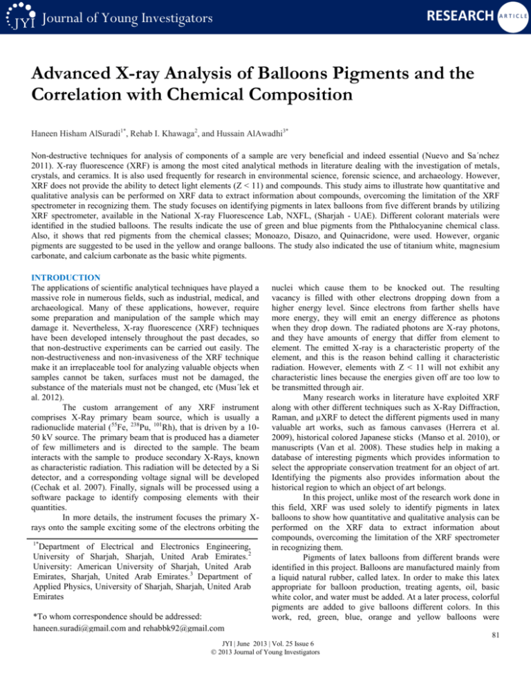
RESEARCH Journal of Young Investigators ARTICLE Advanced X-ray Analysis of Balloons Pigments and the Correlation with Chemical Composition Haneen Hisham AlSuradi1*, Rehab I. Khawaga2, and Hussain AlAwadhi3* Non-destructive techniques for analysis of components of a sample are very beneficial and indeed essential (Nuevo and Sa´nchez 2011). X-ray fluorescence (XRF) is among the most cited analytical methods in literature dealing with the investigation of metals, crystals, and ceramics. It is also used frequently for research in environmental science, forensic science, and archaeology. However, XRF does not provide the ability to detect light elements (Z < 11) and compounds. This study aims to illustrate how quantitative and qualitative analysis can be performed on XRF data to extract information about compounds, overcoming the limitation of the XRF spectrometer in recognizing them. The study focuses on identifying pigments in latex balloons from five different brands by utilizing XRF spectrometer, available in the National X-ray Fluorescence Lab, NXFL, (Sharjah - UAE). Different colorant materials were identified in the studied balloons. The results indicate the use of green and blue pigments from the Phthalocyanine chemical class. Also, it shows that red pigments from the chemical classes; Monoazo, Disazo, and Quinacridone, were used. However, organic pigments are suggested to be used in the yellow and orange balloons. The study also indicated the use of titanium white, magnesium carbonate, and calcium carbonate as the basic white pigments. INTRODUCTION The applications of scientific analytical techniques have played a massive role in numerous fields, such as industrial, medical, and archaeological. Many of these applications, however, require some preparation and manipulation of the sample which may damage it. Nevertheless, X-ray fluorescence (XRF) techniques have been developed intensely throughout the past decades, so that non-destructive experiments can be carried out easily. The non-destructiveness and non-invasiveness of the XRF technique make it an irreplaceable tool for analyzing valuable objects when samples cannot be taken, surfaces must not be damaged, the substance of the materials must not be changed, etc (Musı´lek et al. 2012). The custom arrangement of any XRF instrument comprises X-Ray primary beam source, which is usually a radionuclide material (55Fe, 238Pu, 101Rh), that is driven by a 1050 kV source. The primary beam that is produced has a diameter of few millimeters and is directed to the sample. The beam interacts with the sample to produce secondary X-Rays, known as characteristic radiation. This radiation will be detected by a Si detector, and a corresponding voltage signal will be developed (Cechak et al. 2007). Finally, signals will be processed using a software package to identify composing elements with their quantities. In more details, the instrument focuses the primary Xrays onto the sample exciting some of the electrons orbiting the 1* Department of Electrical and Electronics Engineering, University of Sharjah, Sharjah, United Arab Emirates. 2 University: American University of Sharjah, United Arab Emirates, Sharjah, United Arab Emirates.3 Department of Applied Physics, University of Sharjah, Sharjah, United Arab Emirates *To whom correspondence should be addressed: haneen.suradi@gmail.com and rehabbk92@gmail.com nuclei which cause them to be knocked out. The resulting vacancy is filled with other electrons dropping down from a higher energy level. Since electrons from farther shells have more energy, they will emit an energy difference as photons when they drop down. The radiated photons are X-ray photons, and they have amounts of energy that differ from element to element. The emitted X-ray is a characteristic property of the element, and this is the reason behind calling it characteristic radiation. However, elements with Z < 11 will not exhibit any characteristic lines because the energies given off are too low to be transmitted through air. Many research works in literature have exploited XRF along with other different techniques such as X-Ray Diffraction, Raman, and µXRF to detect the different pigments used in many valuable art works, such as famous canvases (Herrera et al. 2009), historical colored Japanese sticks (Manso et al. 2010), or manuscripts (Van et al. 2008). These studies help in making a database of interesting pigments which provides information to select the appropriate conservation treatment for an object of art. Identifying the pigments also provides information about the historical region to which an object of art belongs. In this project, unlike most of the research work done in this field, XRF was used solely to identify pigments in latex balloons to show how quantitative and qualitative analysis can be performed on the XRF data to extract information about compounds, overcoming the limitation of the XRF spectrometer in recognizing them. Pigments of latex balloons from different brands were identified in this project. Balloons are manufactured mainly from a liquid natural rubber, called latex. In order to make this latex appropriate for balloon production, treating agents, oil, basic white color, and water must be added. At a later process, colorful pigments are added to give balloons different colors. In this work, red, green, blue, orange and yellow balloons were JYI | June 2013 | Vol. 25 Issue 6 2013 Journal of Young Investigators 81 RESEARCH Journal of Young Investigators considered for analysis. The balloons were classified according to the manufacturing company. Five companies from different origins with different balloons' prices were selected in the analysis. ARTICLE set, blue, green, yellow, orange, and red. Samples were first cut by scissors into small pieces assuring that the sample area did not go below the area of the x-ray spot; 1.13 mm2. Then, they were fixed onto a plastic plate to be placed inside the XRF instrument. . Both quantitative and qualitative comparisons were made between elements of different balloons. Three measurements were considered per sample of balloon with a data acquisition time of 100 seconds for each, which yielded 75 readings in total. Prior to the measuring process, the instrument was calibrated using a metallic piece of copper. This instrument was designed to be calibrated by copper material. In the calibration process, the instrument assigned the dominant peak for Cu at 8.040 keV and each time a new measurement was made, peaks would be assigned to their corresponding elements relative to the copper peak. Statistical Analysis The statistical analysis was performed by the statistical software OriginPro 9. All graphical representations of data were also constructed using the same software. Fig. 1: XRF spectrum of the blue balloon from Hallmark. METHODS AND MATERIALS Instruments The instrument used to carry out this project is XGT-7200V XRay Analytical Microscope, which is designed to simultaneously measure and analyze the emitted X-ray image, fluorescent X-ray image, and X-ray spectrum of a sample (Horiba 2009). This analytical instrument available in the NXFL has been used for the analysis of the balloons. The bias source is operated at 50kV in voltage and 0.5mA in current intensity, and generates an X-ray beam from Rh anode that was collimated in order to produce a spot of 1.2mm in diameter under a total vacuum environment. In addition to XRF, Raman spectrometer was used in one case as an example of verifying the use of a pigment that was previously predicted based on the XRF data. Raman spectroscopy is a spectroscopic technique used to study vibrational frequency modes in a system. It relies on the scattering of monochromatic light, usually from a laser in the visible, near infrared, or near ultraviolet range. The laser light interacts with molecular vibrations, resulting in the energy of the laser photons being shifted up or down. The shift in energy gives information about the vibrational modes in the system and hence about the chemical composition of the material (Alison 2009). Experimental method In this project, five sets of colored latex balloons, categorized based on their manufacturer company, were analyzed. The selected manufacturing companies belonged to different origins and had different balloon prices. Details and balloon prices for each company are shown in Table 1 Five samples with different colors were taken from each RESULTS Examples of X-ray spectra obtained with the instrument are shown in (Fig.1 and Fig.2). Each spectrum represents the intensity versus the energy of the detected radiation, where each energy peak is a characteristic property of a certain element. The intensity of each characteristic radiation is related to the amount of each element in the material. The Rh Compton shift was detected at 18.8 keV as shown in (Fig. 3 ). It is worth mentioning that the Compton peak is produced from incoherent backscattering of X-ray radiation from the excitation source and is present in the spectrum of every sample. The constituting elements of the latex balloons were identified by their X-ray emission, as summarized in Table 2. No light elements were detected by the instrument since their energies were too weak to be detected. Nevertheless, the presence or absence of some heavy elements can be, to some extent, a good indicator of the compounds used in the balloons Fig. 2: XRF spectrum of the green balloon from Hallmark. 82 JYI | June 2013 | Vol. 25 Issue 6 2013 Journal of Young Investigators RESEARCH Journal of Young Investigators ARTICLE agent. In PAR, WR, & PBR, S was also present with higher intensity than in other colors. However, the red pigment used was not (Red 48:3), since both Cl and Sr were absent. Spectra shown in (Fig.9) illustrate the disappearance of both Cl and Sr peaks in WR in comparison with HR. (Red 41) and (Red 122) were possible candidate pigments, and other complementary techniques should be employed to correctly identify the red pigment used like: X-ray diffraction (XRD) which reveals information about the crystal structure of materials and hence their chemical composition (Hochleitner et al. 2003). XRF results also showed that Chlorine was a common element found in all yellow balloons from all companies. Despite the existence of Chlorine in all yellow balloons, this did not guarantee the usage of the same pigment, since there were many possible yellow pigments comprising of Chlorine. Chlorine was also found in all orange balloons from all companies except PAO. Like the yellow pigment, there were many possible orange organic pigments entailing Chlorine, so we could neither specify the pigment used nor insist it was consistent in all companies. In PAO, however, other organic pigments, which consisted of elements below the detection level of XRF instrument, might be used. In regards to other non-coloring additives, they could either form the basic layer of the balloons, as earlier mentioned, or affect the mechanical and the physical properties. Dominant elements, which had the higher weighted percentage, were most likely to be involved in comprising the basic pigment for the liquid latex. Fig. 3: Rh Compton shift. Table 1: Details of the selected companies and the notation used for each. The subscript letter added to the notation indicates the color of the balloon. (R: red, Y: yellow, O: orange, G: green, B: blue, e.g.: H R means the Red balloon of Hallmark company). DISCUSSION The different colors of the balloons were ascribed to the presence of different organic and inorganic coloring agents. Copper was exclusively detected in the blue and green balloons regardless of the company as shown in Table 3. In the blue balloons, the existence of copper strongly suggested that (Blue 15) was used as the blue coloring agent (Fig.4). In all green balloons, however, copper was not the only distinguished element, but Chlorine was also present with a higher weighted percent than in other colors (Fig.5). This result indicated that (Green 7) was used in all green balloons (Fig.4). Pigments, (Blue15) and (Green7), fall under the same chemical class, Copper Phthalocyanine. The derivatives of this chemical class were of major industrial importance as green and blue pigments. It yields brilliant turquoise and green shades not available from any other pigment category (Arzu et al. 2003). . The existence of Sr in only, HR & DR, suggested the use of the pigment (Red 48:3) (Fig.7). This assumption was further verified with the detection of Cl, in addition to the existence of S with higher intensity in HR & DR balloons. This might also explain the disappearance of Cl in PAR , WR, & PBR, where compounds other than (Red 48:3) were used as a red coloring Table 2: All elements detected in the studied samples. BDL stands o for: below detection level. 83 JYI | June 2013 | Vol. 25 Issue 6 2013 Journal of Young Investigators RESEARCH Journal of Young Investigators Table 3: Cu is exclusively detected in green and blue balloons from all companies. Both companies, H and PA, had mainly used titanium dioxide, TiO2, as an elementary white pigment. This assumption had been verified by the Raman analysis (Fig.7).Magnesium carbonate, MgCO3, was used to dye the latex in PB and W. Dingo Company, conversely, combined two white pigments, TiO 2 and CaCO3, with a total percentage of about 60% in all colors as shown in (Fig.8). However, the percentage of each of the two pigments was not the same in all colors. In warm colors (DR, DY, & DO), it was noticed that CaCO3 existed in higher percentage than TiO2. In contrast, cool colors (DG & DB) had a higher Fig. 4: Pigments involved in the analysis. (a) Blue 15 (b) Green 7 (c) Red 48:3 (d) Red 41 (e) Red 122. ARTICLE percentage of TiO2 than CaCO3. This variance in the partial percentages might be due to the difference in brilliance of CaCO3 and TiO2.The compound CaCO3 had an off-white look which might be compatible with the warm colors (Subramanian et al. 2012). On the other hand, the white pigment, TiO2, was more congruent for cool colors because of its high brilliance (Valandro et al. 1997). Finally, fillers, which ensured stability and high quality of rubbers, were recognized in the analysis as well. The element Silicon particularly was used as a major filler with different percentages in all companies. The surface properties of the filler Silicon guarantee the durability of the rubber material when exposed to the atmosphere (Kopylov 2011). Also, a small percentage of the most significant filler nowadays, Calcium, was used in the studied balloons. It was used in many polymer applications in order to ameliorate the mechanical, rheological and optical properties (Miao 2003). CONCLUSION The study carried out in Sharjah-UAE at the NXFL included the analysis of different balloons samples from different manufacturing using an XRF spectrometer to study the elemental composition of the pigments used in their manufacturing and obtain information about the employed materials. The choice of XRF as an analytical tool has proven itself to be very useful for the identification of pigments. The method is sensitive to most of the elements in the periodic table and it enabled more than one element to be identified quickly from a single measurement (Musı´lek et al. 2012). Also, XRF provides non-destructive analytical method, the feature that is of a tremendous importance in other fields like archeology and arts where an object is valuable and should be analyzed in-situ without any destruction. However, due to the energy threshold of detection by Xray spectrometers, light elements (Z <11) cannot be detected. Also, this technique would not have been efficient in a case of analyzing thick or multiple layers, since only a thin layer on the surface of an artwork can be analyzed. In addition, XRF is not sensitive to chemical compounds in which the elements are bonded (Musı´lek et al. 2012).Yet, as the study showed, a conclusion of the pigments used can still be reached by the presence or absence of elements within the detection limit. It was also shown that in some cases, XRF only allows one to establish uncertain hypotheses and narrow down the number of probable pigments. For example the red pigments used in PA, W, & PB are not uniquely determined. Other complementary technique is essential to determine whether (Red 41) or (Red 122) was used in PAR, WR, & PBR balloons to resolve doubts about the chemical compounds. However, in other cases, XRF cannot provide any clue about the pigments used as the case of the yellow and orange balloons, which arises the need of other analytical techniques. 84 JYI | June 2013 | Vol. 25 Issue 6 2013 Journal of Young Investigators RESEARCH Journal of Young Investigators ARTICLE Fig. 8: The percentages of Ca and Ti in all Dingo balloons. Fig. 5: Percentage of Cl in all balloons samples. ACKNOWLEDGEMENT We would like to express our sincere gratitude to Dr.Hussain Al Awadhi, Physics professor at University of Sharjah, for his continuous help, guidance and support. Also, special thanks go to the International Atomic Energy Agency (IAEA) for providing funding for the XRF instrument. REFERENCES Abel, A. (1998) The new colour index international of pigments and solvent dyes. Surface Coatings International Part B: Coatings Transactions 81, 2, 77-85. Fig. 6: XRF spectra of the red balloons from: (a) Hallmark "HR" and (b) Water Balloons "WR". Alison, J. et al. (2009) Stand-off Raman spectroscopy. Trends in Analytical Chemistry 28, 11. Arzu, Y. et al. (2003) Use of carbonised beet pulp carbon for removal of Remazol Turquoise Blue-G 133 from aqueous solution. Environ Sci Pollut Res 1-12. Bruni, S. et al. (1999) Spectrochemical characterization by micro-FTIR spectroscopy of blue pigments in different polychrome works of art. Vib. Spectrosc. 20, 15–25. Cechak, T. et al. (2007) X-ray fluorescence in investigations of archaeological finds. Nuclear Instruments and Methods in Physics Research B 263, 54–57. Fig. 7: Raman spectrum shows the compound TiO2 at 612 cm-1. Gil, M. et al. (2007) Yellow and red ochre pigments from southern Portugal: elemental composition and characterization by WDXRF and XRD, Nucl. Instrum. Methods Phys. Res. Sect. A 580, 728–731. 85 JYI | June 2013 | Vol. 25 Issue 6 2013 Journal of Young Investigators RESEARCH Journal of Young Investigators Herrera, L. et al. (2009) Advanced combined application of -Xray diffraction/ -X-ray fluorescence with conventional techniques for the identification of pictorial materials from Baroque Andalusia paintings. Talanta 80, 71–83. ARTICLE Van, G. et al. (2008) µ-XRF/µ-RS vs. SR µ-XRD for pigment identification in illuminated manuscripts. Appl. Phys. A 92, 59–68. Hochleitnera, B. et al. (2003) Historical pigments: a collection analyzed with X-ray diffraction analysis and X-ray fluorescence analysis in order to create a database. Spectrochimica Acta Part B 58, 641–649. Horiba (2009) XGT-7200V X-Ray Analytical Microscope Instruction Manual.Japan: Horiba,Ltd. Kopylov, V. et al. (2011) Silica fillers for silicone rubber. International Polymer Science and Technology 38. 4 , T35T47. Manso, M. et al. (2010) Characterization of Japanese color sticks by energy dispersive X-ray fluorescence, X-ray diffraction and Fourier transform infrared analysis. Spectrochimica Acta Part B 65, 321–327. Miao, S. (2003) Polymer surface modification and characterization of particulate calcium carbonate fillers. Applied Surface Science 220, 359–366. Mohd, Z. et al. (2011) Optimization of energy dispersive X-ray fluorescence spectrometer to analyze heavy metals in moss samples. American J. of Engineering and Applied Sciences 4 (3),355-362. Musı´lek, L. et al. (2012) X-ray fluorescence in investigations of cultural relics and archaeological finds. Applied Radiation and Isotopes 70, 1193–1202. Nuevo, M. et al. (2010). Application of XRF spectrometry to the study of pigments in glazed ceramic pots. Applied Radiation and Isotopes, 69, 574-579. Roine, J. et al. (2007) X-ray texture analysis of paper coating pigments and the correlation with chemical composition analysis. J. Appl. Phys. 102, 8. Sivasubramanian et al. (2012) Algal technology for effective reduction of total hardness in wastewater and industrial effluents. Phykos 42 (1), 51– 58. SzÖkefalvi-Nagy, Z. et al. (2004) Non-destructive XRF analysis of paintings. Nuclear Instruments and Methods in Physics Research B 226, 53–59. Valandro,V. et al. (1997) Environmental restoration of a TiO2 plant. J. Environ Pathol Toxicol Oncol16, 163-9. 86 JYI | June 2013 | Vol. 25 Issue 6 2013 Journal of Young Investigators
