Chapter 14 Topics - Defense Mechanisms - Non
advertisement
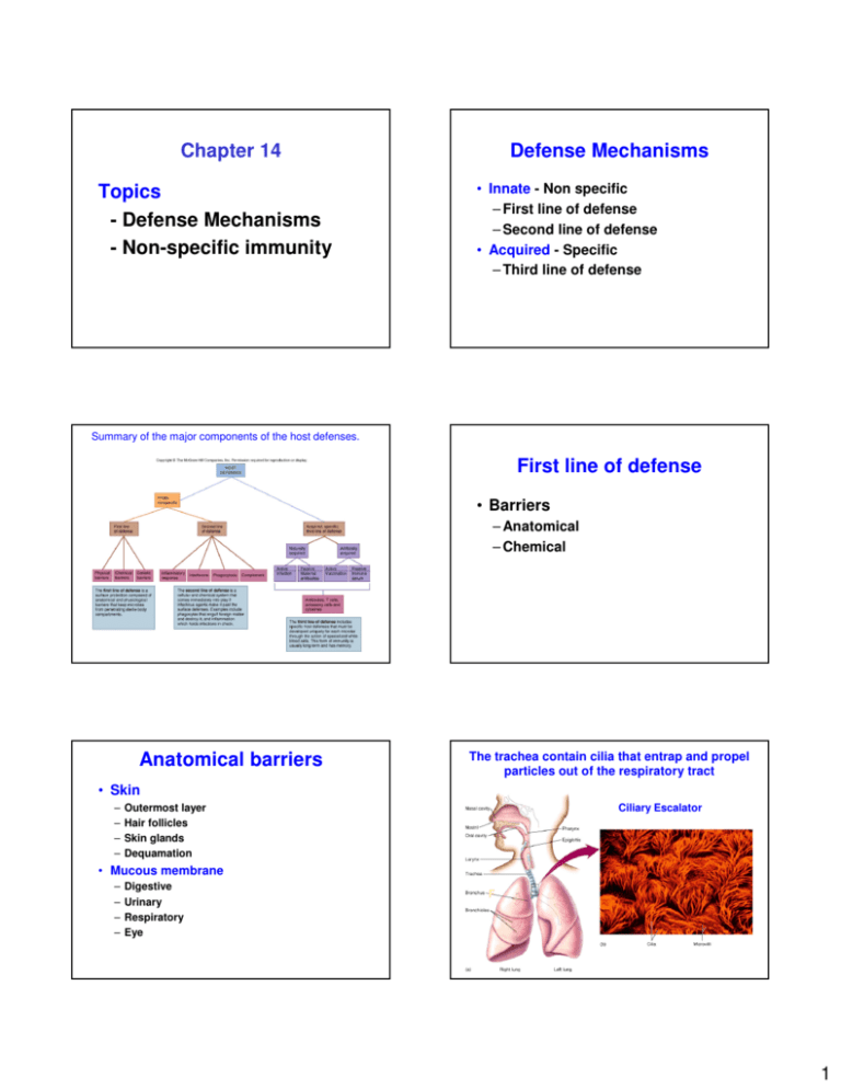
Chapter 14 Topics - Defense Mechanisms - Non-specific immunity Defense Mechanisms • Innate - Non specific – First line of defense – Second line of defense • Acquired - Specific – Third line of defense Summary of the major components of the host defenses. First line of defense • Barriers – Anatomical – Chemical Anatomical barriers The trachea contain cilia that entrap and propel particles out of the respiratory tract • Skin – – – – Outermost layer Hair follicles Skin glands Dequamation Ciliary Escalator • Mucous membrane – – – – Digestive Urinary Respiratory Eye F 1 Chemical barriers • Sebaceous secretions • Tears and saliva – lysozyme • Acidic pH – Sweat – Stomach – Skin – Semen – Vagina - mediated by presence of Lactobacillus WBC Immunology • Study of the development of resistance to infectious agents by the body – Surveillance of the body – Recognition of foreign material – Destruction of foreign material or agent • Involve nonspecific (Second line) and specific (Third line) immune defense systems • White blood cells (WBC) or leukocytes are involved Search, recognize, and destroy is the mandate of the immune system • WBC recognize "self" markers on the host cell – Do not attack or do not respond to host cell • WBC recognize non-self markers on the invading microbe –Attack or respond to microbe Blood Hemopoiesis • Stem cells precursors • Hemopoiesis • Components • Production of blood – Starts at the embryonic stage • Yolk sac and liver – Continues during adult stage – Hematopoietic stem cells in bone marrow 2 Lymphoid Myeloid White blood cells • Leukocytes – Granulocytes (large cytoplasmic granules) • Neutrophils • Basophils • Eosinophils The macroscopic composition of whole blood Neutrophils • Present in high numbers in blood and tissue • Phagocytizes bacteria – granules contain digestive enzymes • First to arrive during an immune response (inflammation) – Agranulocytes • T cells • B cells • Monocytes Eosinophils • Contain granules with hydrolytic enzymes • Attach and destroy large eucaryotic pathogens (worms) • Associated with inflammation and allergies Basophils • Present in low in number in the body • Function is similar to eosinophils. Involved in allergic reactions due to cytoplasmic granules • Localized basophils are called mast cells 3 Lymphocytes • Specific immunity – T cells cellular immunity – B cells humoral/antibody immunity • Third line of defense • Present throughout the body Monocytes • Agranulocyte • Differentiate into macrophages (circulation and lymphatics) and dendritic cells (tissue associated) • Phagocytosis Lymphatic system • Network of vessels, cells, and tissues that extend to most body areas • Connected to the blood system • Provides an auxiliary route for the return of extracellular fluid to the circulatory system • “Drain off” system for inflammatory response • Contains lymphocytes, phagocytes and antibodies Representation of the lymphatic system. Lymphatic system • • • • • • Fluids Vessels Nodes Spleen Thymus Miscellaneous (GALT Gut-associated lymphoid tissue (GALT) • Recognized incoming microbes from food • Supply lymphocytes for antibody response • Ex. Appendix, lacteals, Peyer’s patches 4 Non-specific Immunity Second Line of Defense • • • • Inflammation Phagocytosis Interferon Complement Inflammation - 1 Inflammation • Four major symptoms – Redness – Warmth – Swelling – Pain That result in Cellular Damage Causes • Trauma • Tissue injury due to physical or chemical agents • Reaction to foreign pathogens or bodies (ie medical implants) Inflammation - 2 Function • Mobilize and attract immune components to the site of injury • Localized and remove harmful substances • Destroy microbes and block their invasion • Aid in the repair of tissue damage 5 Chemical mediators during inflammation. 1. Vascular changes • Blood cells, tissue cells, and platelets release chemical mediators and cytokines • Chemical mediators – Vasoactive • Affect endothelial cells, smooth muscles of blood vessels – Chemotactic (chemokines) • Affect WBC The transmigration of WBCs is followed by chemotaxis. 2. Edema • Leakage of vascular fluid (exudate) into tissue • Exudate - plasma proteins, blood cells (WBC), debris, and pus • Migration of WBC is called diapedesis or transmigration – Chemotaxis 3. Fever • Caused by pyrogens – reset the hypothalamic thermostat (increase temperature) • Pyrogens – Microbes and their products (ex. LPS) – Leukocyte products (ex. lnterleukins) – IL-1 resets the thermostat • Inhibits microbe and viral multiplication, reduces nutrient availability, increases immune reactions Phagocytosis Neutrophils and monocytes/macrophages (and dendritic cells) are called professional phagocytes Eosinophils 6 Phagocytosis Neutrophils - First to arrive during an immune response (inflammation) • Neutrophils are primary components of pus Monocytes/Macrophages Differentiate into macrophages (circulation and lymphatics) and dendritic cells (tissue associated) Mechanism of Phagocytosis • • • • Chemotaxis Ingestion Phagolysosome Destruction Macrophages • Monocytes/macrophages motile • Specialized/Residents: – Alveolar lungs – Langerhan cells skin – Kupffer cells liver • 1) Responsible for phagocytosis • 2) Interact with B and T cells 1. Chemotaxis & binding • Directed by – Pathogen-associated molecular patterns (PAMPs) • Peptidoglycan • LPS – Foreign debris 2. Ingestion • Pseudopods enclose the pathogen or foreign material • Form a phagosome or phagocytic vacuole 7 3. Phagolysosome • Lysosomes fuse with the phagosome • Other antimicrobials chemicals are released into the phagolysosome 4. Destruction • Within the phagolysosome – A) Oxygen-dependent mechanisms – Similar to byproducts of respiration – B) Oxygen-independent mechanisms – due to numerous hydrolytic enzymes • Undigestible debris are released Interferon • Produced due to viral infections, microbe infections, RNA, immune products, and antigens 8 Activity Classes • Interferon alpha – Product of lymphocytes and macrophages • Interferon beta – Product of fibroblasts and epithelial cells • Interferon gamma – Product of T cells • Ex. Virus - binds to host cell • A signal is sent to the nucleus to synthesized (transcription and translation) interferon • Interferon is secreted • Binds to other host cells • Host cells produce antiviral proteins – inhibit viral multiplication or translation • Not virus-specific Interferon is produced, released, and taken-up by a near-by cell, where by original cell is not protected but the recipient cell is protected. Other Roles of Interferon • Activates and instructs T and B cell development • Inhibits cancer cells • Activates macrophages Fig. 14.20 The antiviral activity of interferon. Pathways Complement • Classical • Consist of ~26 blood proteins • Produced by liver hepatocytes, lymphocytes, and monocytes • Pathways • Cascade reaction • Stages – Activated by the presence of antibody bound to microbes • Lectin – Activated when a host serum protein binds a sugar (mannan) in the wall of fungi and other microbes • Alternative – Activated when complement proteins bind to cell wall or surface components of microbes 9 The three complement pathways, their activators, and the complement proteins involved. Stages • • • • Initiation Amplification and cascade Polymerization Membrane attack Table 14.1 Complement pathways Fig. 14.21a Fig. 14.21b Fig. 14.21d 10 Complement does 3 things • Inflammation C3a, C4a, C5a • Opsonization C3b • MAC killing C5-C9 11
