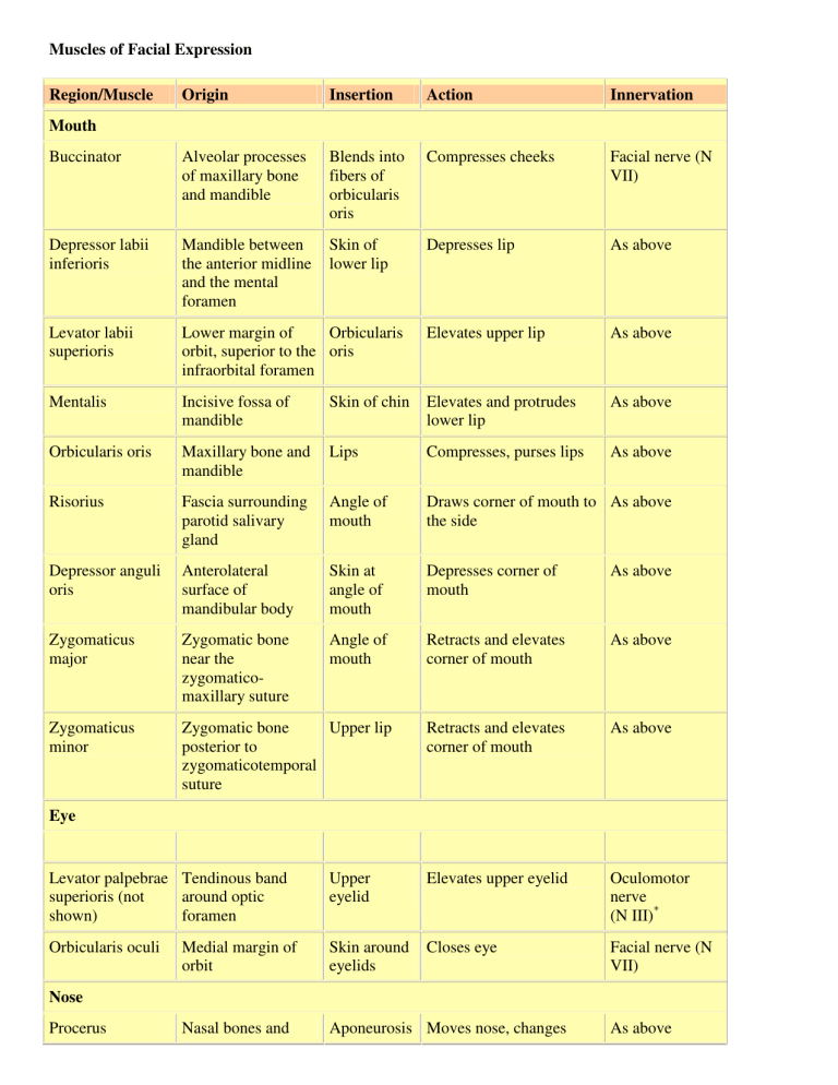
Muscles of Facial Expression Region/Muscle Origin Insertion Action
advertisement

Muscles of Facial Expression Region/Muscle Origin Insertion Action Innervation Buccinator Alveolar processes of maxillary bone and mandible Blends into fibers of orbicularis oris Compresses cheeks Facial nerve (N VII) Depressor labii inferioris Mandible between the anterior midline and the mental foramen Skin of lower lip Depresses lip As above Levator labii superioris Lower margin of Orbicularis orbit, superior to the oris infraorbital foramen Elevates upper lip As above Mentalis Incisive fossa of mandible Skin of chin Elevates and protrudes lower lip As above Orbicularis oris Maxillary bone and mandible Lips Compresses, purses lips As above Risorius Fascia surrounding parotid salivary gland Angle of mouth Draws corner of mouth to As above the side Depressor anguli oris Anterolateral surface of mandibular body Skin at angle of mouth Depresses corner of mouth As above Zygomaticus major Zygomatic bone near the zygomaticomaxillary suture Angle of mouth Retracts and elevates corner of mouth As above Zygomaticus minor Zygomatic bone Upper lip posterior to zygomaticotemporal suture Retracts and elevates corner of mouth As above Mouth Eye Levator palpebrae Tendinous band superioris (not around optic shown) foramen Upper eyelid Elevates upper eyelid Oculomotor nerve (N III)* Orbicularis oculi Medial margin of orbit Skin around eyelids Closes eye Facial nerve (N VII) Nasal bones and Aponeurosis Moves nose, changes Nose Procerus As above Nasalis lateral nasal cartilages at bridge of nose and skin of forehead position and shape of nostrils Maxillary bone and alar cartilage of nose Bridge of nose Compresses bridge, depresses tip of nose; elevates corners of nostrils As above Ear (extrinsic) Temporoparietalis Fascia around external ear Galea Tenses scalp, moves aponeurotica pinna of ear As above Skin of Raises eyebrows, eyebrow and wrinkles forehead bridge of nose As above Scalp (Epicranius) Frontalis Galea aponeurotica Occipitalis Superior nuchal line Galea Tenses and retracts scalp aponeurotica As above Upper thorax Mandible between cartilage of and skin of second rib and cheek acromion of scapula As above Neck Platysma Tenses skin of neck, depresses mandible Muscles of Mastication Muscle Origin Masseter Insertion Action Innervation Zygomatic arch Lateral surface of mandibular ramus Elevates mandible Trigeminal nerve (N V), mandibular branch Temporalis Along temporal Coronoid lines of skull process of mandible Elevates mandible As above Pterygoids (medial and lateral) Lateral pterygoid plate Medial: Elevates As above the mandible and closes the jaws, or moves mandible side to side Medial surface of mandibular ramus Lateral: Opens jaws, protrudes mandible, or As above moves mandible side to side Anterior Muscles of the Neck Muscle Origin Insertion Action Innervation Digastric Two Hyoid bone bellies: posterior from mastoid region of temporal; anterior from inferior surface of mandible at chin Depresses Trigeminal nerve mandible and/or (N V), mandibular elevates larynx branch, to anterior belly Geniohyoid Medial Hyoid bone surface of mandible at chin As above and Cervical nerve C1 pulls hyoid bone via hypoglossal anteriorally nerve (N XII) Mylohyoid Mylohyoid line of mandible Median connective tissue band (raphe) that runs to hyoid bone Elevates floor of Trigeminal nerve mouth, elevates (N V), mandibular hyoid bone, branch and/or depresses mandible Omohyoid Central tendon attaches to clavicle and 1st rib Two bellies: superior attaches to hyoid bone; inferior to superior margin of scapula Depresses hyoid Cervical spinal bone and larynx nerves C2-C3 Sternohyoid Clavicle Hyoid bone and manubrium As above Cervical spinal nerves C1-C3 Sternothyroid Dorsal Thyroid surface of cartilage of manubrium larynx and 1st costal cartilage As above As above Facial nerve (N VII) to posterior belly Stylohyoid Styloid process of temporal bone Hyoid bone Elevates larynx Facial nerve (N VII) Thyrohyoid Thyroid cartilage of larynx Hyoid bone Elevates thyroid, depresses hyoid bone Cervical spinal nerves C1-C2 via hypoglossal nerve (N XII) Together, they flex the neck; alone, one side bends head toward shoulder and turns face to opposite side Accessory nerve (N XI) and cervical spinal nerves (C2-C3) of cervical plexus Sternocleidomastoid Two Mastoid region bellies: of skull clavicular head attaches to sternal end of clavicle; sternal head attaches to manubrium Oblique and Rectus Muscles* Group/Muscl e Origin Insertion Action Innervation OBLIQUE GROUP Cervical region Scalenes Transverse and costal processes Superior surfaces of cervical vertebrae of first two ribs Elevates ribs Cervical and/or flexes neck spinal nerves Thoracic region External intercostals Inferior border of each rib Superior Elevate ribs border of the next rib Intercostal nerves (branches of thoracic spinal nerves) Internal intercostals Superior border of each rib Inferior Depress ribs border of the previous rib As above Transversus thoracis Medial surface of sternum Cartilages of As above ribs As above Abdominal region External oblique Lower eight ribs Linea alba and iliac Compresses abdomen, depresses ribs, flexes Intercostal, iliohypogastric, and ilioinguinal nerves crest or bends spine Internal oblique Lumbodorsal fascia and iliac crest Lower As above ribs, xiphoi d process , and linea alba As above Transversus abdominis Cartilages of lower ribs, iliac crest, and lumbodorsal fascia Linea alba and pubis As above Compresses abdomen RECTUS GROUP Thoracic region Diaphragm Xiphoid process, cartilages of ribs 4-10, and anterior surfaces of lumbar vertebrae Central Contraction Phrenic nerves (C3-C5) tendinous expands sheet thoracic cavity, compresses abdominopelvic cavity Abdominal region Rectus abdominis Superior surface of pubis around symphysis Inferior surfaces Depresses ribs, of costal flexes vertebral cartilages (ribs column 5-7) and xiphoid process Intercostal nerves (T7-T12) Muscles That Move the Pectoral Girdle Muscle Origin Insertion Action Innervation Levator scapulae Transverse processes of first 4 cervical vertebrae Vertebral border of scapula near superior angle Elevates scapula Cervical nerves C3-C4 and dorsal scapular nerve (C5) Pectoralis minor Ventral surfaces of ribs 3-5 Coracoid process of scapula Depresses and protracts shoulder; rotates scapula so glenoid cavity moves Medial pectoral nerve (C8, T1 ) inferiorly (downward rotation); elevates ribs if scapula is stationary Rhomboideus Spinous major processes of upper thoracic vertebrae Vertebral Adducts and performs Dorsal scapular nerve (C5) border of downward rotation scapula from spine to inferior angle Rhomboideus Spinous minor processes of vertebrae C7T1 Vertebral border of scapula near spine As above As above Serratus anterior Anterior and superior margins of ribs 1-9 Anterior surface of vertebral border of scapula Protracts shoulder, rotates scapula so glenoid cavity moves superiorly (upward rotation) Long thoracic nerve (C5C7 ) Subclavius First rib Clavicle (inferior border) Depresses and protracts shoulder Subclavian nerve Trapezius Occipital bone, ligamentum nuchae, and spinous processes of thoracic vertebrae Clavicle and scapula (acromion and scapular spine) Depends on active region and state of other muscles; may elevate, retract, depress, or rotate scapula upward and/or elevate clavicle; can also extend head and neck Accessory nerve (N XI) and cervical spinal nerves (C3-C4) Muscles That Move the Arm Muscle Origin Insertion Action Innervation Coracobrachialis Coracoid process Medial margin of shaft of humerus Adducts Musculocutaneous and nerve (C5-C7) flexes humerus Deltoid Clavicle and scapula (acromion and adjacent scapular spine) Deltoid tuberosity of humerus Abducts Axillary nerve humerus (C5-C6) Supraspinatus Supraspinous Greater Abducts Suprascapular fossa of scapula tubercle of humerus humerus nerve (C5) Infraspinatus Infraspinous fossa of scapula Greater tubercle of humerus Lateral Suprascapular rotation nerve (C5-C6) of humerus Subscapularis Subscapular fossa of scapula Lesser tubercle of humerus Medial Subscapular rotation nerves (C5-C6) of humerus Teres major Inferior angle of scapula Medial lip of the intertubercular groove of the humerus Extends, Lower adducts, subscapular nerve and (C5-C6) medially rotates humerus Teres minor Lateral border of scapula Greater tubercle of humerus Lateral Axillary nerve rotation (C5) of humerus Triceps brachii (long head) See Table 11-13 Latissimus dorsi Spinous processes of lower thoracic and all lumbar vertebrae, ribs 8-12, and the lumbodorsal fascia Floor of the intertubercular groove of the humerus Extends, Thoracodorsal adducts, nerve (C6-C8) and medially rotates humerus Pectoralis major Crest of greater tubercle and lateral lip of intertubercular groove of humerus Flexes, Pectoral nerves adducts, (C5-T1) and medially rotates humerus Cartilages of ribs 2-6, body of sternum, and inferior, medial portion of clavicle Muscles That Move the Forearm and Hand Muscle Origin Insertion PRIMARY ACTION AT THE ELBOW Action Innervation Flexors Biceps brachii Short head Tuberosity of from the radius coracoid process; long head from the supraglenoid tubercle (both on the scapula) Flexes elbow and supinates forearm; flexes arm Musculocutaneous nerve (C5C6 ) Brachialis Anterior, distal surface of Flexes elbow As above and radial nerve (C7-C8) Brachioradiali s Lateral Lateral aspect epicondyle of of styloid humerus process of radius As above Radial nerve (C5-C6) Extends elbow; moves ulna laterally during pronation Radial nerve (C7-C8) Tuberosity of ulna Extensors Anconeus Posterior surface of lateral epicondyle of humerus Lateral margin of olecranon on ulna Triceps brachii . lateral head Superior, lateral Olecranon process of margin of humerus ulna Extends elbow As above long head Infraglenoid As above tubercle of scapula As above As above medial head Posterior surface of humerus inferior to radial groove As above As above As above PRONATORS / SUPINATORS Pronator quadratus Medial surface of distal portion of ulna Anterolateral Pronates surface of forearm distal portion of radius Median nerve (C8-T1) Pronator teres Medial Distal lateral As above Median nerve (C6-C7) epicondyle of surface of humerus and radius coronoid process of ulna Supinator Lateral epicondyle of humerus and ridge of proximal ulna Anterolateral Supinates Deep radial nerve (C6) surface of forearm radius distal to the radial tuberosity PRIMARY ACTION AT THE HAND Flexors Flexor carpi radialis Medial epicondyle of humerus Bases of 2nd and 3rd metacarpal bones Flexes and abducts wrist Median nerve (C6C7 ) Flexor carpi ulnaris Medial epicondyle of humerus; adjacent medial surface of olecranon and anteromedial portion of ulna Pisiform bone, hamate bone, and base of 5th metacarpal bone Flexes and adducts wrist Ulnar nerve (C8T1 ) Palmaris longus Medial epicondyle of humerus Palmar aponeurosis and flexor retinaculum Flexes wrist Median nerve (C6C7 ) Extensor carpi radialis longus Lateral supracondylar ridge of humerus Base of 2nd metacarpal bone Extends and abducts wrist Radial nerve (C6C7 ) Extensor carpi radialis brevis Lateral epicondyle of humerus Base of 3rd metacarpal bone As above As above Extensor carpi ulnaris Lateral epicondyle of humerus; adjacent dorsal surface of ulna Base of 5th metacarpal bone Extends and adducts wrist Deep radial nerve (C6-C8) Extensors Muscles That Move the Fingers and Hand Muscle Origin Insertion Action Innervation Abductor pollicis longus Proximal dorsal surfaces of ulna and radius Lateral margin of 1st metacarpal bone Abducts thumb Deep radial nerve (C6-C7) Extensor digitorum Lateral epicondyle of humerus Posterior surfaces of the Extends fingers phalanges, fingers 2-5 and hand Deep radial nerve (C6-C8) Extensor pollicis brevis Shaft of radius distal to Base of proximal pollicis longus phalanx of thumb Extends thumb, abducts hand Deep radial nerve (C6-C7) Extensor Posterior and lateral Extends thumb, Deep radial nerve Base of distal phalanx pollicis longus surfaces of ulna and of thumb interosseous membrane abducts hand (C6-C8) Extensor indicis Posterior surface of ulna and interosseous membrane Posterior surface of phalanges of little (5th) finger, with tendon of extensor digitorum Extends and adducts little finger As above Extensor digiti minimi Via extensor tendon to lateral epicondyle of humerus, and from intermuscular septa Posterior surface of proximal phalanx of little finger Extends little finger As above Midlateral surfaces of middle phalanges of fingers 2-5 Flexes fingers, specifically middle phalanx or proximal phalanx; flexes wrist Median nerve (C7T1 ) Flexor Medial epicondyle of digitorum humerus; adjacent superficialis anterior surfaces of ulna and radius Flexor digitorum profundus Medial and posterior Bases of distal surfaces of ulna, medial phalanges of fingers 2-5 surface of coronoid process, and interosseus membrane Flexes distal phalanges and, to a lesser degree, the other phalanges and hand Palmar interosseous nerve, from median nerve and ulnar nerve (C8-T1) Flexor pollicis longus Anterior shaft of radius Base of distal phalanx and interosseous of thumb membrane Flexes thumb Median nerve (C8T1 ) Muscles That Move the Thigh Group/Muscle Origin Insertion Action Innervation Gluteal group Gluteus maximus Iliac crest of ilium, sacrum, coccyx, and lumbodorsal fascia Gluteus medius Gluteus minimus Iliotibial tract and gluteal tuberosity of femur Extends and laterally rotates thigh Inferior gluteal nerve (L5S2) Anterior iliac crest Greater of ilium, lateral trochanter of surface between femur posterior and anterior gluteal lines Abducts and medially rotates thigh Superior gluteal nerve (L4-S1) Lateral surface of ilium between inferior and anterior gluteal lines Abducts and medially rotates thigh As above Greater trochanter of femur Tensor fasciae latae Iliac crest and lateral surface of anterior superior iliac spine Iliotibial tract Flexes, abducts, and medially rotates thigh; tenses fascia lata, which laterally supports the knee As above Lateral rotator group Obturators (externus and internus) Lateral and medial margins of obturator foramen Trochanteric fossa of femur (externus); medial surface of greater trochanter (internus) Laterally Obturator nerve (externus: L3-L4) rotate and special nerve from sacral plexus thigh (internus: L5-S2) Piriformis Anterolateral Greater surface of trochanter of sacrum femur Laterally Branches of sacral nerves (S1-S2) rotates and abducts thigh Gemelli (superior and inferior) Ischial spine and tuberosity Medial surface of greater trochanter Laterally Nerves to obturator internus and rotates quadratus femoris thigh Quadratus femoris Lateral border of ischial tuberosity Intertrochanteric As above Special nerve from sacral plexus crest of femur (L4-S1) Adductor brevis Inferior ramus of pubis Linea aspera of femur Adducts, medially Obturator nerve (L3-L4) rotates, and flexes thigh Adductor longus Inferior ramus of pubis anterior to adductor brevis As above Adducts, flexes, and medially rotates thigh As above Adductor magnus Inferior ramus of pubis posterior to adductor brevis and ischial tuberosity Linear aspera and adductor tubercle of femur Adducts thigh; superior portion flexes and medially rotates thigh; inferior portion extends and laterally rotates thigh Obturator and sciatic nerves Adductor group Pectineus Superior ramus of pubis Pectineal line inferior to lesser trochanter of femur Flexes, medially rotates, and adducts thigh Femoral nerve (L2-L4) Gracilis Inferior ramus of pubis Medial surface of tibia inferior to medial condyle Flexes leg, adducts, and medially rotates thigh Obturator nerve (L3-L4) Iliacus Iliac fossa of ilium Femur distal to lesser trochanter; tendon fused with that of psoas Flexes hip and/or lumbar spine Femoral nerve (L2-L3) Psoas major Anterior surfaces and transverse processes of vertebrae T12-L5 Lesser trochanter in company with iliacus As above Branches of the lumbar plexus (L2-L3) Iliopsoas group Muscles That Move the Leg Muscle Origin Insertion Action Innervation Ischial tuberosity and linea aspera of femur Head of fibula, lateral condyle of tibia Flexes knee, extends and laterally rotates thigh Sciatic nerve; tibial portion (S1-S3; to long head) and common peroneal branch (L5S2; to short head) Semimembranosus Ischial tuberosity Posterior surface of medial condyle of tibia Flexes knee, medially rotates leg, and extends hip Sciatic nerve (tibial portion; L5-S2) Semitendinosus Ischial tuberosity Proximal, medial surface As above of tibia near insertion of gracilis Sartorius Anterior Medial surface of tibia superior iliac near tibial tuberosity spine Flexes knee, flexes hip, and laterally rotates thigh Femoral nerve (L2-L3) Popliteus Lateral condyle of femur Medially rotates tibia (or laterally rotates femur) Tibial nerve (L4-S1) Flexors of the Knee Biceps femoris Posterior surface of proximal tibial shaft As above Extensors of the Knee Rectus femoris Anterior inferior iliac spine and superior acetabular rim of ilium Vastus intermedius Tibial tuberosity via patellar ligament Extends knee, flexes thigh Femoral nerve (L2-L4) Anterolateral As above surface of femur and linea aspera (distal half) Extends knee As above Vastus lateralis Anterior and As above inferior to greater trochanter of femur and along linea aspera (proximal half) As above As above Vastus medialis Entire length As above of linea aspera of femur As above As above Extrinsic Muscles That Move the Foot and Toes Muscle Origin Insertion Action Innervation PRIMARY ACTION AT THE ANKLE Dorsiflexors Tibialis anterior Lateral condyle and proximal shaft of tibia Base of 1st metatarsal bone and medial cuneiform bone Dorsiflexes and inverts foot Deep peroneal nerve (L4S1) Plantar flexors Gastrocnemiu s Femoral condyles Calcaneus via calcaneal tendon Plantar flexes, inverts, and adducts foot; flexes knee Tibial nerve (S1-S2) Peroneus brevis Midlateral margin of fibula Base of 5th metatarsal bone Everts and plantar flexes foot Superficial peroneal nerve (L4-S1) Peroneus longus Lateral condyle Base of 1st of tibia, head and metatarsal bone proximal shaft of and medial Everts and plantar As above flexes foot; supports longitudinal arch fibula cuneiform bone Plantaris Lateral supracondylar ridge Posterior portion of calcaneus Plantar flexes foot; flexes knee Tibial nerve (L4-S1) Soleus Head and proximal shaft of fibula, and adjacent posteromedial shaft of tibia Calcaneus via calcaneal tendon (with gastrocnemius) Plantar flexes, inverts, and adducts foot Sciatic nerve, tibial branch (S1-S2) Tibialis posterior Interosseous Tarsal and membrane and metatarsal bones adjacent shafts of tibia and fibula Adducts, inverts, and plantar flexes foot As above PRIMARY ACTION AT THE TOES Digital Flexors Flexor digitorum longus Posteromedial Inferior Plantar surface of surfaces of flexes toes tibia distal 2-5 phalanges, toes 2-5 Flexor hallucis Posterior longus surface of fibula Inferior surface, distal phalanx of great toe Sciatic nerve, tibial branch (L5-S1) Plantar Sciatic nerve, tibial branch (L5-S1) flexes great toe Digital Extensors Extensor digitorum longus Lateral condyle of tibia, anterior surface of fibula Superior Extends surfaces of toes 2-5 phalanges, toes 2-5 Deep peroneal nerve (L4-S1) Extensor hallucis longus Anterior surface of fibula Superior surface, distal phalanx of great toe As above Extends great toe


