Unit 1: EPITHELIUM
advertisement
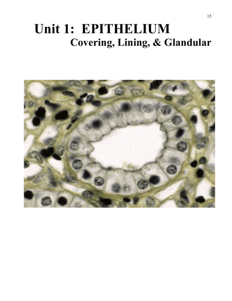
15 Unit 1: EPITHELIUM Covering, Lining, & Glandular 16 17 Epithelial Tissue Epithelial tissue is composed of cells physically close together that are connected by one or all three types of junctions (zonula occludens, zonula adherens, macula adherens) with very little intervening intercellular substance. Epithelial tissue is found lining internal surfaces and covering external surfaces of the body. Thus it is classified in general terms as covering and lining epithelium. Epithelial functions include protecting the body from the environment, transporting of molecules, establishing boundaries between compartments and creating fluid environments. The outer layer of tissue that makes up the skin is an epithelial tissue. The entire GI Tract is lined with epithelial tissue. The entire vascular system is lined with epithelial tissue. The other kind of epithelial tissue is glandular, ranging in composition from as small as a single cell secreting mucus, to a large gland like the liver consisting of millions of cells secreting multiple products. Your job in this unit is to learn to distinguish between the various types of epithelial tissues. Please be assured that, at this stage in learning histology, you are not responsible for identification of organs or their components, only the types of epithelia. Focus on the diagnostic features of epithelia. Covering and lining epithelium is classified according to the number of cell layers and the shape of the most superficial cells. Epithelia with one layer of cells are called simple and epithelia with two or more layers are called stratified. When naming a stratified epithelium you call it squamous, cuboidal, or columnar if the superficial layer of cells is flat, cuboidal or columnar shaped, respectively (the cells below the superficial layer are usually a different shape. You are responsible for learning to distinguish between the following types of covering/lining epithelia: Simple squamous epithelium (simple = one layer; squamous = flat) Simple cuboidal epithelium (shape = cube in 3D, but square in tissue sections) Simple columnar epithelium (shape = cylinder in 3D, but rectangular in tissue sections) Stratified squamous epithelium (more than one layer and superficial cells are flat) -Stratified squamous non-keratinized epithelium (if no keratin is present) -Stratified squamous keratinized epithelium (if keratin is present) Stratified cuboidal epithelium (more than one layer and superficial cells are cuboidal) Stratified columnar epithelium (more than one layer and superficial cells are columnar) Pseudostratified columnar epithelium (one layer and cells reaching surface are columnar) Transitional epithelium (more than one layer and cells at surface are flat or dome-shaped depending on whether the epithelium is stretched or relaxed) 18 Glandular epithelium is composed of epithelial cells that have differentiated and are equipped with the organelles to synthesize and secrete a given product. The product can be secreted directly into the lumen of the respiratory or GI tract by single cells, the so-called unicellular gland. The goblet cell of the intestinal and respiratory tracts is an example of a unicellular gland. The other main type of gland is multi-cellular. This means that the gland is composed not only of epithelial cells differentiated to synthesize and secrete, but also epithelial cells that are specialized to line ducts through which the secretions are conducted to the lumen of the GI tract, respiratory tract, or to the surface of the skin. Glandular epithelial cells may secrete mucus, protein, or lipid. Your text reading and lectures will help you learn the various types of classification and secretory methods of glandular epithelium. In this unit your job is to learn to distinguish between the following glandular epithelial cell types: Sebaceous glandular cell – secretes lipid (sebaceous gland cells of skin) Serous cell – secretes proteins like enzymes (salivary amylase by salivary gland, for example) Mucous cell – secretes mucus (salivary gland cells) Goblet cell – secretes mucus (unicellular gland ) 19 Learning the types of covering and lining epithelium Simple Columnar and Simple Squamous Epithelium Simple squamous epithelium lines all blood vessels forming a smooth surface which reduces the friction of flowing blood. In capillaries this epithelium functions to efficiently transport oxygen and carbon dioxide. Simple columnar epithelium lines the intestine where it moves the products of digestion from the lumen to the interstitium. In these two examples transportion of molecules is a common function. As you progress in your study of histology you will learn even more functions for these epithelia. To begin, go to the general histology section and under epithelium choose: Simple Columnar Epithelium (Renal Papilla) – Hematoxylin-Eosin. Scan the 5x specimen with the labels turned on. As you scan the specimen by moving the pale circle on the 5x (rectangular image lower left) you can observe and distinguish between epithelial tissue, connective tissue, blood vessels and a lumen of a kidney tubule in the 20x magnified view in the microscope field (circular image). (recall that when a structure is colored to highlight it you can click on the color and remove it – then clicking in the same area returns the colored highlighting). Remember, covering and lining epithelium always has to border on a space, either internal or external. Now change to the 20x image (by clicking on the 20x tab above the rectangular image) and scan this image with the microscope where the specimen is magnified 80x. With the labels turned on you can confirm the distinction between connective tissue, epithelial tissue and a lumen. The epithelial tissue labeled is simple columnar. Observe the rectangular shape of the cells. Now change to the 40x image and scan it observing all the labeled structures. You are now viewing in the microscope the specimen magnified 160x. Compare and contrast the appearance of the simple columnar and simple squamous epithelium in this field. The simple squamous epithelium is lining both the smaller tubules in the kidney (labeled) and the blood vessels (labeled). 20 Simple Cuboidal Epithelium This kind of epithelium functions in two distinctive ways depending on its location. It provides a lining for ducts that transport substances secreted by a gland (sweat gland or salivary gland) to a surface (of the skin or of the oral cavity). Simple cuboidal epithelium can also act as a semipermeable membrane that it transports some molecules but restricts others as exemplified in kidney tubules. To study simple cuboidal epithelium go to the organ section and under urinary system choose: Kidney – Cortex – Hematoxylin-Eosin. The kidney is an organ that is mostly constructed of epithelial tissues. There are many tubules in the kidney lined with all three types of simple epithelia. Select the second 20x image and scan it with the labels turned on until you find a tubule labeled ‘distal straight tubule’. This tubule, which is sectioned longitudinally, is lined with simple cuboidal epithelium. Observe the short cells with round nuclei. Now select the second 40x image and scan it until you find a structure labeled ‘distal tubule’. Here you can observe simple cuboidal epithelium at a higher magnification. You are probably already making the observation that the appearance of the cuboidal cells in this specimen differs from their depictions in textbooks. In real specimens, the cell borders are not always readily visible due to the interdigitation of the membranes of adjacent cells. To determine the specific classification of an epithelium lining a lumen, check to see that it is one layer of cells and then look at the nucleus. Squamous cells have flat nuclei, cuboidal cells have round nuclei, and columnar cells have ovoid nuclei. Before we study stratified ( 2 or more layers of cells) epithelia let’s look at one more specimen. Under epithelium Choose: Simple Columnar Epithelium (Renal Papilla) – Iron Hematoxylin After you have scanned at the 5x and 20x images with the labels turned on, choose the 100x image and scan it. The iron hematoxylin stain used on this specimen accentuates the cell borders, cell junctions and the basement membrane. Note the cell margins that are darkly stained, and the small round dark areas at the apical end (bordering the lumen) of the epithelial cells. These small round darkly stained areas represent the location of the junctional complexes (consisting of zonula occludens, zonula adherens, and macular adherens) between the cells. Click on the label and read the explanation. Also, observe the basement membrane. As a general rule, all covering or lining epithelial are attached to a basement membrane. 21 Stratified Epithelia Recall the epithelial naming rule. More than one layer of cells makes an epithelium stratified. The shape of the surface cells, usually bordering the lumen, determines whether the stratified epithelium is squamous, columnar, or cuboidal. Now let’s study stratified squamous epithelium. Stratified squamous non-keratinizing epithelium You will study two places in the body in which SSNK epithelium resides. First you will examine the lining of the esophagus that needs to withstand friction while food is swallowed. The mulitple layers of cells provides a buffer between the lumen and the tissue underneath the epithelium. Superficial cells can be lost and replaced by subsequent generations of cells. Mucus is secreted into the lumen by glands providing a slippery and we surface. Next, you will examine the lining of the vagina that is designed to withstand friction (sexual intercourse) with again the multiple layers of cells providing a buffer with cell loss an renewal playing a major role while also lubricated with mucus secreted by glands in the wall of the cervix. Desmosomes (macular adherens) provide strong connections between the cells and hemidesmosomes bind the epithelium to the underlying connective tissue via hemidesmosomes. Go to organ histology and under the gastrointestinal tract choose: Esophagus-Hematoxylin-Eosin The esophagus transports food from the oral cavity to the stomach. In this specimen you will study stratified squamous non-keratinized epithelium. Scan the 5x image and find the label epithelium to orient yourself. Now select the first 20x image and observe how the cells gradually become flattened from the basal to the superficial layer. By counting the nuclei from the basal to the superficial layer you can estimate the number of layers of cells in this epithelium (about 20 layers present). The cells of stratified epithelia are continually renewed by mitosis of stem cells in the basal (bottom) layer. Daughter cells generated here move through the layers to the surface where they are eventually shed. Turnover (movement of a daughter cell from basal to superficial layer), takes only a few days in the esophagus. The multiple layers provide a lining 22 that is substantial, and the renewal of cells insures replacement of cells removed by friction. Now select the first 40x image and carefully study the cell shape from basal to superficial layers. Note how the superficial cells still retain their nuclei. This is important to note because, in stratified squamous keratinizing epithelium the superficial cells lack nuclei and are full of the protein keratin. To study another example of stratified squamous non-keratinizing epithelium go to general histology and under epithelium choose: Stratified Squamous Epithelium (Vagina) – Goldner By studying this specimen you will learn how the multiple layers of cells are held together. This specimen is stained with Goldner’s stain. This is just a special name for a stain that is routinely known as a Trichrome Stain. The nuclei stain dark red, the cytoplasm stains light red, and the collagen in the connective tissue stains green. Scan this specimen first at 5x and observe the epithelium and connective tissue. Note the dark green staining reaction in the connective tissue indicative of a large amount of collagen. Click on the epithelium label and read about its appearance and the layers. Now choose the 20x image to scan and note that there are three regions defined in this stratified squamous epithelium. Click on the label for each of the three regions and learn the difference. Observe that the superficial cells are squamous and that they all have nuclei. The multiple layers of cells that comprise this kind of epithelium are fastened together by desmosomes, and the basal layer of cells are anchored into the connective tissue via the connecting hemidesmosomes at their basal surface. Select the first 100x image (note change in image orientation 90 degrees clockwise) and find the label ‘intercellular bridges’. This is an illustration of the multiple connecting sites between the cells. At each dark line there is a desmosome. Click on the label to learn about the ‘intercelluar bridges’. Now under Epithelium choose: Stratified Squamous Epithelium (Vagina) – Hematoxylin-Eosin. Scan the specimen at all magnifications. Try to observe the features of this epithelium as they appear with the H&E stain. Click on all of the labeled structures and read the comments. 23 Stratified Squamous Keratinizing Epithelium You will study one example of this epithelium in thin skin. The outer most component of skin is epidermis that is entirely composed of this kind of epithelium. In WEBMIC go to the organ section and look under skin and appendages for a specimen named: Thin Skin – Hematoxylin-Eosin First scan the 5x image to orient yourself and to find the label epidermis. Read the text related to that label. Next select and scan the second 20x image while observing the layers. Read the text related to all the labels, especially the label ‘stratum corneum’. The main point here is that the superficial cells of this kind of epithelium do not have nuclei and are full of the protein keratin. The outer layer of our skin, the epidermis, must also withstand frictional forces, but, rather than being slippery with mucus, it is slippery with oil produced by sebaceous glands located in the dermis, the layer of skin beneath the epidermis. To observe the layers of the epidermis in more detail select the first 40x image and note the histological difference between the stratum basale (find label: stratum basale cell), stratum spinosum, and stratum corneum. Focus on knowing the distinguishing characteristics of these regions. At this point, you are not responsible for the other terms. Stratified Cuboidal Epithelium The next epithelium we will study is stratified cuboidal epithelium. The ducts of sweat glands in the dermis of the skin are made up of this kind of epithelium. In WEBMIC go to the organ section and look under skin and appendages for a specimen named: Thin Skin – Hematoxylin-Eosin With the labels turned on scan the 5x and the two 20x images of the specimen in search for the sweat glands and their ducts. When you are comfortable with the context in which you will take a closer look at a sweat gland duct, click on the second 40x image. Now scan this image until you find the labeled sweat gland duct and the sweat gland body. Carefully observe the sweat 24 gland duct and read the text related to the label. You should be able to observe two layers of nuclei in the duct which indicates there are two layers of cells, thus named stratified cuboidal epithelium. Now observe the labeled sweat gland body and note the sweat gland secretory cell that is columnar shaped. The entire body (also known as the secretory portion of the sweat gland) is composed of simple columnar epithelium. If you click on the body of the sweat gland anywhere but on top of the labeled columnar cell, the coloring delineating the extent of the body will disappear allowing you to observe the mix of simple cuboidal and columnar epithelium. Comparing this epithelium with the sweat gland duct epithelium provides you with a demonstration of the difference between stratified and simple epithelium. Stratified Columnar Epithelium This kind of epithelium functions to compartmentalize the lumen of large ducts from the interstitium as in the large excretory duct of the parotid gland which carries substances secreted by the parotid gland into the oral cavity (Stenson’s duct) or the large duct which carries the secretions of the pancreas to the duodenum (duct of Wirsung). Larger ducts consist of more layers of cells than do small ducts. To study this kind of epithelium go to the organ section and under the gastrointestinal tract choose: Submandibular Gland-Hematoxylin-Eosin With the labels turned on scan the 5x image of this specimen and locate the duct in the upper left portion of the specimen. This is a rather large duct in the submandibular gland. It is draining secretions from a large collection of secretory cells. The lining of this duct begins as stratified cuboidal and changes to stratified columnar, as the ducts get larger. Now select the first 40x image and find the label: interlobular duct – epithelium. First observe that there are two layers of cells which make up the wall of this duct. Note that the cell next to the lumen has an ovoid shaped nucleus and the cell away from the lumen has a round nucleus suggesting stratified columnar epithelium. If you carefully examine the entire wall of the duct you will find portions that are stratified cuboidal evidenced by shorter cells with rounded nuclei at the surface. This is the case when one is observing a duct at it transition point between the two types of epithelia. Now you see the difference between stratified cuboidal and stratified columnar epithelium. 25 Transitional Epithelium We now have two remaining types of epithelium to study, pseudostratified columnar and transitional. First let’s look at transitional epithelium. Go to the organ section and look under the urinary system for a specimen named: Urinary Bladder-Hematoxylin-Eosin Transitional epithelium is a type of stratified epithelium that resembles stratified squamous epithelium. It is a unique epithelium that lines the urinary bladder and ureter & has important features that you will learn more about when you study urinary organ histology. Begin your observations by scanning the 5x image of this specimen with the labels turned on. Look in the 5x image for the epithelium label and read the text related to it by clicking on the label. Now select the first 20x image and scan the epithelium observing that it is composed of more than one layer of cells and that the surface cells are not flat. In some regions they may appear columnar and in others they may appear dome shaped. Now select the 40x image and study the epithelium more carefully. Note that that surface cells are neither columnar nor cuboidal in shape. The most common descriptive term for the shape you are observing is dome-shaped. In a distended bladder this epithelium will be stretched to the extent that the surface cells will be flattened. While stretched, transitional epithelium resembles stratified squamous epithelium. In stretched transitional epithelium the number of cellular layers is reduced to three. In actuality stratified squamous epithelium that has fewer than four layers of cells does not exist. Pseudostratified Columnar Epithelium The next and last type of covering and lining epithelium we will study in this unit is pseudostratified columnar epithelium. It is present lining the nasal cavity, the nasal pharynx, parts of the larynx, the trachea, and the extra- and intra-pulmonary bronchioles. In all of these sites this epithelium possesses cilia. Pseudostratified columnar epithelium may be found in parts of the male reproductive tract where, in some sites, the cells have stereocilia. Pseudostratified epithelium has only one layer of cells. All cells are connected to the basal lamina (basement membrane), but not all cells reach the surface. The way the nuclei are staggered in the 26 epithelium gives the impression that it is stratified. To study pseudostratifed columnar epithelium go to the epithelium section and look for a specimen named: Pseudostratified Columnar Epithelium (Trachea)-Hematoxylin-Eosin Scan the 5x image in its entirety and find the labeled epithelium to orient yourself. Note that this image includes the entire thickness of the trachea. Observe the lumen on the right and the connective tissue wrapping of the trachea on the left with the bulk of its structure being hyaline cartilage in the center. Click on the epithelium label and read the text explanation. Now select the 40x image and scan the entire length of the epithelium. Click on the epithelium label and read the text explanation. The best part of the epithelium in this image to look for the features of pseudostratified columnar epithelium is at the extreme left part of the epithelium. There you can see the outline of the lighter cells where they appear to almost reach the basement membrane. Even in this region it appears as if there are two layers of cells. In order to demonstrate this better you would need to look at a thinner specimen (which does not exist in this program), or examine an electron micrographic image of this kind of epithelium using an atlas or text. What you see here is the appearance of this kind of epithelium in routine specimens in the light microscope. Now you can probably appreciate that the cells appear columnar but due to the fact that not all cells reach the surface even though all cells attach to the basement membrane makes this epithelium appear stratified, but it is not, hence the name. Glandular Epithelium We will conclude our study of epithelial tissue with glandular epithelium. You were introduced to glandular epithelium previously in this unit when you studied stratified cuboidal epithelium. In that specimen, organs-skin & appendages-thin skin-hematoxylin-eosin, you observed the difference between the lining of the sweat gland duct and the secretory cells of the sweat gland. Although the epithelium that makes up the wall of the ducts of glands can be considered a part of glandular epithelium it is thought of as lining the duct and therefore classified as lining epithelium. The secretory cells in the glands are truly glandular epithelium and they consist of three main types, namely, serous cells (secreting a watery protein substance), mucous cells (secreting mucus) and sebaceous cells (secreting an oil based substance). Let’s now observe first 27 had the three types of glandular epithelial cells. First we will look at serous and mucous cells. In the organ section under the gastrointestinal tract choose: Submandibular Gland-Hematoxylin-Eosin With the labels turned on scan the 5x image finding the labeled ducts, the connective tissue septa, the partitioning of the gland into regions called lobules, and the appearance of the tissue within the lobules. Now select the 20x image and scan it to locate and observe the difference in the appearance of mucous and serous cells at low magnification. The mucous cells stain lighter because their content is water soluble mucous which is dissolved and washed away when samples are routinely prepared. In contrast the serous cells stain darker because their content is mostly protein that is not dissolved and washed away. Next scan the second 40x image and look for the mucous and serous cells (serous demilune label – look up this term in your text). Read the text explanations for the labeled mucous cells and serous demilune. Note that the nuclei of serous and mucous cells are shaped differently. Next scan the second 100x image and look for the labeled mucous cell and serous demilune reading the text explanation for each. In summary if you are looking for serous cells you should look for cells with round nuclei that contain some secretory granules that will stain with hematoxylin and eosin. If you are looking for mucous cells you should look for cells with dark and flattened nuclei with a cytoplasm that stains very lightly with hematoxylin and eosin. Serous and mucous cells are located in the salivary glands. Mucous secreting cells also are present in the lining of the intestine where they are called goblet cells due to their unique goblet shape. The last glandular cell type that we will study is the sebaceous cell found only in the oil glands of the skin known as sebaceous glands. Go to general histology under epithelial tissue and look for a specimen named: Glandular Epithelia-Hematoxylin-Eosin Begin by scanning the 10x image of this specimen of skin. Look for two sebaceous gland labels and read the text explanation for one of these labels. Now that you are oriented, select the 20x image observing the labeled sebaceous and apocrine sweat gland. Now you can appreciate not only the appearance the oil producing cells but also you have learned that there are two types; 28 one like in the sebaceous gland where the cell itself is the secretion (holocrine method of secretion) and the other in the apocrine sweat gland where a portion a fat globule surrounded by plasma membrane is the secretion (apocrine method of secretion). Now select the 100x image, find, and observe the two labeled sebaceous cells. Read the text explanation for both after which you should understand the appearance of the sebaceous gland cells in the synthesizing and secreting stages. The terms holocrine and apocrine have been introduced above. Holocrine secretion is secretion of the entire cell whereas apocrine secretion is secretion of a part of a cell. The other type of secretory method is eccrine in which a product is stored in a secretory granule which, upon appropriate stimulation, releases the product into a lumen with no loss of cell membrane or cytoplasm. Mucous and serous cells secrete in this way. As you progress into a study of the histology of organs you will learn that organs other than the skin and salivary glands also have glandular epithelium. The liver, pancreas, prostate, lung, testis, and many other organs have glandular epithelial components which you will readily understand as you study each organ. For now, it is sufficient if you understand the difference between the basic types of glandular cells and how they secrete their product differently. This concludes the exercise on the histology of epithelial tissue. At the end of this unit as in all other units there are two sample questions that will test your ability to identify an epithelium or some component of an epithelium. 29 Sample Practical Questions 1. First order identification of a cell/structure or classification of a tissue. Classify the tissue surrounding the empty space labeled 1. A. B. C. D. E. F. G. H. simple squamous epithelium simple cuboidal epithelium simple columnar epithelium stratified columnar epithelium stratified cuboidal epithelium stratified squamous epithelium transitional epithelium pseudostratified columnar epithelium 30 2. Second order question. A structure/cell/tissue is indicated and its function is asked Which of the following functions is carried out by the structures contained in the small dark circular areas labeled 2? A. B. C. D. E. storage of secretory products fasten cells to the basement membrane fasten cells together function to provide energy for the cell function to maintain the boundary between the nucleus and cytoplasm 31 Epithelial Section Labeled Structures Viewing the structures you have studied in this lesson in sections stained with special stains other than Hematoxylin and Eosin could be helpful in making your learning complete. In this unit and all others the specimens under each heading will be listed in this way that will help also when you are reviewing to know which specimen and which magnification that certain structures are labeled. Simple Columnar Epithelium (Renal Papilla)-Iron Hematoxylin Stain 20X 40X 80X Connective Tissue Epithelium Connective Tissue Collecting Duct Connective Tissue Simple Columnar Epithelium Collecting Duct 400X Simple columnar epithelium Apical Junctions Basement membrane Nucleus Cell margin Erythrocyte Endothelium - simple squamous epithelium Simple Columnar Epithelium (Renal papilla)- Hematoxylin-Eosin Stain 20X Connective Tissue Epithelium Lumen Blood Vessel 40X Connective Tissue Epithelium Lumen Blood Vessel 80X Connective Tissue Simple columnar epithelium Lumen Blood Vessel 160X Simple columnar epithelium Blood vessel Simple columnar epithelial cell Simple columnar epithelial cell-nucleus Simple squamous epithelium Erythrocyte Fibroblast nucleus Stratified Squamous Epithelium (Vagina)-Goldner Staining 20X Stratified Squamous Connective Tissue Smooth Muscle 40X Stratified Squamous Connective Tissue Smooth Muscle 80X Stratum Spinosum Stratum Basale Connective Tissue Stratum Superficial 400X-A Basal Cell Basement Membrane Intercellular Bridges Glycogen Depot Prickle Cell 400X-B Superficial Cell Cell Margin Nucleus Glycogen Depot Stratified Squamous Epithelium (Vagina)-Hematoxylin-Eosin Staining 20X 40X 80X-(Best representation of layers) 160X Connective Tissue Blood Vessel Connective Tissue Connective Tissue Stratified Squamous Epithelium Connective Tissue Stratum Basale Stratum Basale Stratified Squamous Epithelium Stratum Spinosum Stratum Spinosum Stratum Basale Stratum Superficiale Stratum Superficiale Stratum Spinosum Papilla Stratum Superficiale 32 Stratified Squamous Epithelium Iron Hematoxylin Staining 20X Epithelium Connective Tissue 40X Epithelium Connective Tissue 80X Prickle Cell Stratified Squamous Epithelium 400X Prickle Cell Nucleus Nucleolus Intercellular Bridge Stratified Squamous Epithelium (Condyloma Acuminata)-Hematoxylin-Eosin Staining 20X Connective Tissue Epithelium 80X Prickle Cell Stratum Basale Connective Tissue Blood Vessel 160X Prickle Cell Basal Cell Pseudostratified Columnar Epithelium (Trachea)-Azan Staining 20X Glandular Epithelium Epithelium Hyaline Cartilage Trachea Lumen 80X Pseudostratified columnar epithelium Serous glandular cells Lamina propria - connective tissue Hyaline cartilage 400X Fibroblast Basal Cell Ciliated Cell Cilia Goblet Cell Collagen Fiber Pseudostratified Columnar Epithelium (Trachea)-Hematoxylin-Eosin Staining 20X Epithelium Connective tissue Glandular epithelium Connective tissue Hyaline cartilage 160X Connective tissue Cilia Blood vessel Basement membrane Fibroblast nucleus Pseudostratified Columnar Epithelium Transitional Epithelium (Ureter)-Goldner Staining 20X Transitional epithelium Lamina propria - Connective tissue Smooth muscle 80X Facet cell Transitional epithelium Connective tissue Intermediate cell Basal cell 400X Facet cell Crusta Intermediate cell Basal cell Collagenous fiber Fibroblast Transitional Epithelium (Ureter)-Hematoxylin-Eosin 20X 80X Smooth muscle and connective tissue Smooth muscle and connective tissue Transitional epithelium Transitional epithelium Facet cell 160X Transitional epithelium Smooth muscle and connective tissue Facet cell Intermediate cell Basal cell 33 Crusta Glandular Epithelia-Staining: Goldner-Hematoxylin-Acidfuchsin-Light Green 20X Sebaceous gland Eccrine sweat gland Apocrine sweat gland Epidermis Connective tissue 80X-A Sebaceous gland Hair Connective tissue 80X-B Eccrine sweat gland Excretory duct Adipose cell 80X-C Apocrine sweat gland Connective tissue 400X-A Basal cell Lipid filled glandular cell Nucleus Lipid vacuole Collagenous fiber 400X-B Myoepithelial cell Eccrine glandular cell Basement membrane Collagenous fiber Lumen 400X-C Myoepithelial cell Apocrine glandular cell Apical protrusion Nucleus Basement membrane Glandular Epithelia- Hematoxylin-Eosin Stain 40X Hair Connective tissue Sebaceous gland Epithelium Apocrine sweat gland 80X-A 80X-B Smooth muscle Sebaceous gland Hair Apocrine sweat gland Sebaceous gland Connective tissue Connective tissue Stratified Squamous Keratinized epithelium 400X Fibroblast nucleus Connective tissue Basement membrane Sebaceous gland cell Nucleus Basal layer 34

