Tissues and Skin - College of San Mateo
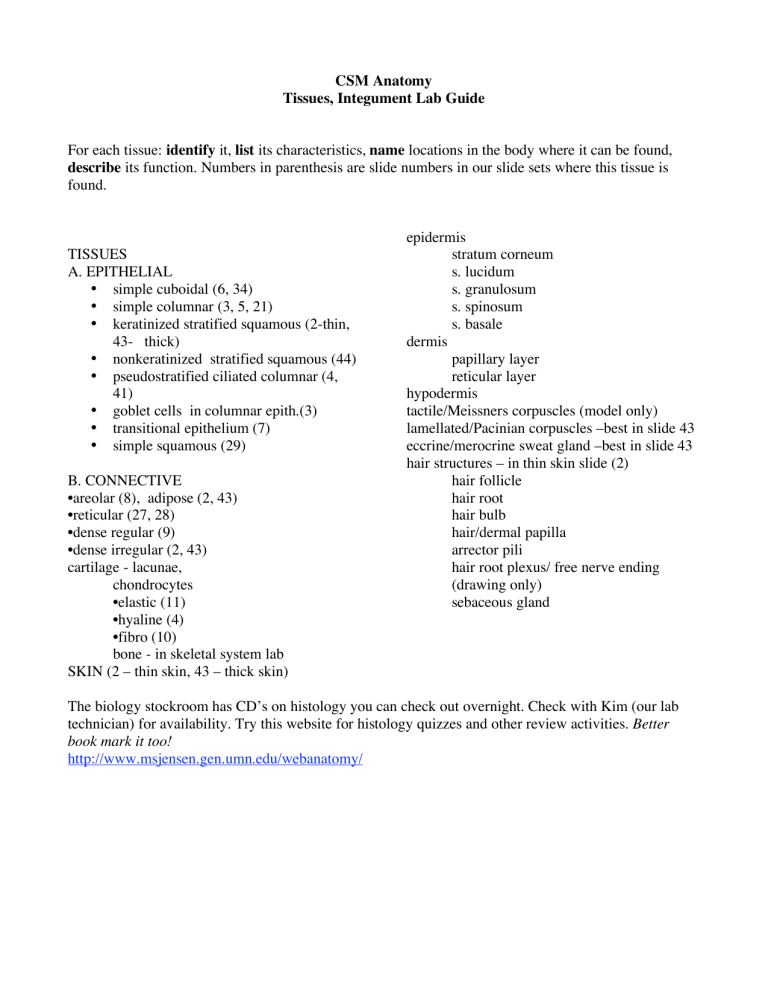
CSM Anatomy
Tissues, Integument Lab Guide
For each tissue: identify it, list its characteristics, name locations in the body where it can be found, describe its function. Numbers in parenthesis are slide numbers in our slide sets where this tissue is found. epidermis stratum corneum TISSUES
A. EPITHELIAL
• simple cuboidal (6, 34)
• simple columnar (3, 5, 21)
• keratinized stratified squamous (2-thin,
43- thick)
• nonkeratinized stratified squamous (44)
• pseudostratified ciliated columnar (4,
41)
• goblet cells in columnar epith.(3)
• transitional epithelium (7)
• simple squamous (29) dermis s. lucidum s. granulosum s. spinosum s. basale papillary layer reticular layer hypodermis tactile/Meissners corpuscles (model only) lamellated/Pacinian corpuscles –best in slide 43 eccrine/merocrine sweat gland –best in slide 43 hair structures – in thin skin slide (2)
B. CONNECTIVE
•areolar (8), adipose (2, 43)
•reticular (27, 28)
•dense regular (9)
•dense irregular (2, 43) cartilage - lacunae, hair follicle hair root hair bulb hair/dermal papilla arrector pili hair root plexus/ free nerve ending chondrocytes
•elastic (11)
•hyaline (4)
(drawing only) sebaceous gland
•fibro (10) bone - in skeletal system lab
SKIN (2 – thin skin, 43 – thick skin)
The biology stockroom has CD’s on histology you can check out overnight. Check with Kim (our lab technician) for availability. Try this website for histology quizzes and other review activities . Better book mark it too! http://www.msjensen.gen.umn.edu/webanatomy/


