Chapter 16: Infrared Spectroscopy
advertisement
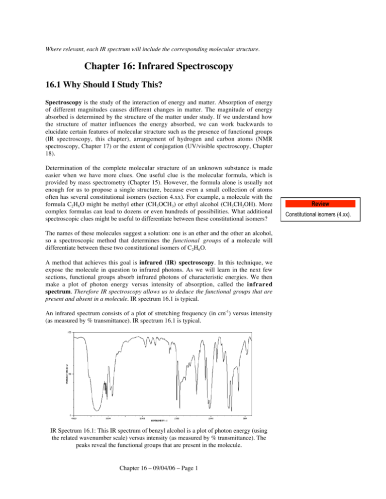
Where relevant, each IR spectrum will include the corresponding molecular structure. Chapter 16: Infrared Spectroscopy 16.1 Why Should I Study This? Spectroscopy is the study of the interaction of energy and matter. Absorption of energy of different magnitudes causes different changes in matter. The magnitude of energy absorbed is determined by the structure of the matter under study. If we understand how the structure of matter influences the energy absorbed, we can work backwards to elucidate certain features of molecular structure such as the presence of functional groups (IR spectroscopy, this chapter), arrangement of hydrogen and carbon atoms (NMR spectroscopy, Chapter 17) or the extent of conjugation (UV/visible spectroscopy, Chapter 18). Determination of the complete molecular structure of an unknown substance is made easier when we have more clues. One useful clue is the molecular formula, which is provided by mass spectrometry (Chapter 15). However, the formula alone is usually not enough for us to propose a single structure, because even a small collection of atoms often has several constitutional isomers (section 4.xx). For example, a molecule with the formula C2H6O might be methyl ether (CH3OCH3) or ethyl alcohol (CH3CH2OH). More complex formulas can lead to dozens or even hundreds of possibilities. What additional spectroscopic clues might be useful to differentiate between these constitutional isomers? The names of these molecules suggest a solution: one is an ether and the other an alcohol, so a spectroscopic method that determines the functional groups of a molecule will differentiate between these two constitutional isomers of C2H6O. A method that achieves this goal is infrared (IR) spectroscopy. In this technique, we expose the molecule in question to infrared photons. As we will learn in the next few sections, functional groups absorb infrared photons of characteristic energies. We then make a plot of photon energy versus intensity of absorption, called the infrared spectrum. Therefore IR spectroscopy allows us to deduce the functional groups that are present and absent in a molecule. IR spectrum 16.1 is typical. An infrared spectrum consists of a plot of stretching frequency (in cm-1) versus intensity (as measured by % transmittance). IR spectrum 16.1 is typical. IR Spectrum 16.1: This IR spectrum of benzyl alcohol is a plot of photon energy (using the related wavenumber scale) versus intensity (as measured by % transmittance). The peaks reveal the functional groups that are present in the molecule. Chapter 16 – 09/04/06 – Page 1 Review Constitutional isomers (4.xx). IR spectroscopy is a powerful tool for the determination of molecular structure, and thus it is of fundamental importance for the modern organic chemist (research scientist and student alike). It has been applied to a wide range of structural problems, such as penicillin in the 1940’s to more modern examples such as residue found in clay pots believed to be from fermentation of wine thousands of years ago, or the composition of extraterrestrial planetary atmospheres (as we will see in section 16.8). Global warming has been linked to the ability of small molecules in the atmosphere (such as CH4 and CO2) to absorb infrared energy from the sun. In your organic chemistry laboratory course, you will probably use IR spectroscopy to verify the identity and purity of organic substances. Let us start our exploration of infrared spectroscopy by understanding electromagnetic radiation, the basis for all spectroscopic methods. 16.2 Spectroscopy and The Electromagnetic Spectrum Energy exists as particles or packets called photons. Each photon has both electric field and magnetic field components, so we call these photons electromagnetic radiation. The complete range of photon energies is called the electromagnetic spectrum (Figure 16.1). Increasing wavelength Review Wavelength (nm): Electromagnetic spectrum (general chemistry) Radiation type: Frequency (Hz): 10 cosmic rays -3 10 -1 102 gamma rays X-rays 1019 1017 400 ultraviolet (UV) 105 800 visible 109 infrared microwave (IR) 1015 1013 1010 radio 105 Increasing frequency; increasing energy Figure 16.1: The electromagnetic spectrum Our daily lives are influenced by the existence of this electromagnetic radiation: Photo: Rainbow. Caption: Visible light, a type of electromagnetic radiation, at work. Cosmic rays consist of high-energy nuclei and subatomic particles that bombard the Earth from space. Gamma rays (γ-rays) are produced by radioactive decay. X-rays are used in medical imaging. Scattering of x-rays by electron clouds is the basis of x-ray crystallography, which reveals position of atoms in space within a crystal. Ultraviolet (UV) radiation causes suntan and sunburn. UV/visible spectroscopy determines conjugation in a molecule (Chapter 18). Visible light is the basis for vision, and consists of the familiar rainbow of colors. UV/visible spectroscopy determines conjugation in a molecule (Chapter 18). Infrared (IR) photons are perceived as heat. IR spectroscopy reveals the functional groups within a molecule (Chapter 16). Microwaves are used in radar, communications and cell phones. Microwave spectroscopy can reveal dipole moments and bond lengths. Chapter 16 – 09/04/06 – Page 2 Radio waves are basis for AM and FM radio and are also used in cell phones. Nuclear magnetic resonance spectroscopy (NMR) uses radio waves to determine the carbon and hydrogen skeleton of a molecule (Chapter 17). Photons have both wave and particle properties, but for our study of spectroscopy it is more useful to think of the wave properties. Waves have wavelength and frequency. Wavelength (λ) is the distance between one point on a wave and the same point on the next wave, and is measured in nanometers (abbreviated nm; 1 nm = 10-9 m = 10 Å) or in micrometers (abbreviated µm; 1 µm = 10-6 m; also called microns). Frequency (ν) is the number of wave crests that pass a given point in a given period of time, and is measured in Hertz (abbreviated Hz; 1 Hz = 1 s-1). These concepts are illustrated in Figure 16.2. wave crest wavelength λ frequency ν = number of waves that pass this point per unit time Figure 16.2: Waves are characterized by their frequency ν and wavelength λ. Picture yourself standing on the beach in the surf. The distance between the tops of the waves is their wavelength, and the number of waves that pass your feet each second is their frequency. Photons can also be characterized by their energy, which is related to wavelength and frequency: E = hν = hc/λ In these equations, h (Planck’s constant) is 6.62607 x 10⁻³⁴ m2 kg s-1 and c (the speed of light) is 2.9979 x 10⁸ m s-1. These equations remind us that high-energy photons (such as x-rays) have high frequency and short wavelength. Low energy photons (such as radio waves) have low frequency and long wavelength. Guided by this inverse relationship between energy and frequency, spectroscopists have found it useful to characterize frequency by its wavenumber (symbol = “nu bar”), the inverse of its wavelength because wavenumber is directly proportional to energy: “nu bar” (cm-1) = 104/λ (cm). The wavenumber unit is reciprocal centimeters (cm-1). Thus photons whose frequency is 4000 cm-1 have higher energy than photons whose frequency is 1000 cm-1. Infrared spectroscopy involves absorption of infrared photons, which changes the bond vibrational state of the molecule. How does this fact give information about molecular structure? To answer this question we must first learn about the nature of bond vibrations. Chapter 16 – 09/04/06 – Page 3 Review Wave-particle duality (general chemistry) 16.3 Physical Basis of IR Spectroscopy: Bond Vibrations Two atoms A and B joined by a covalent bond may move back and forth along the bond axis, producing a bond stretch. In Figure 16.3 the arrows indicate the directions that the atoms move through space. Bond stretching Bond stretching (symmetric) Bond stretching (asymmetric) Bending A A B A B B A C B A A B C C B A C B C B A C Figure 16.3: Bond vibrations. The arrows show the relative motions of the atoms. A three-atom group A–B–C can vibrate along the A–B and B–C bonds, just like the twoatom case previously discussed. Simultaneous elongation or shortening of the A–B and B–C bonds is termed a symmetric stretch. Elongation of one bond with simultaneous shortening of the other bond is called an asymmetric stretch. A, B and C can also move along paths that do not lie on the bond axis. This type of vibration is called bending. In a bending mode, the A–B–C bond angle changes: linear A–B–C becomes bent A–B–C, and bent A–B–C becomes linear or more severely bent, depending upon how the atoms move. Molecular vibration is a complex interaction of these and other atomic motions, but stretching modes are the only ones that are important for our introduction to IR spectroscopy. Concept Focus Question 16.1 Many toxic substances have been found in tobacco smoke, such as carbon monoxide, carbon dioxide, sulfur dioxide and arsenic. In order to determine by IR spectroscopy how much of a substance is present, we need to know the spectrum of each substance. How many absorptions are expected in the IR spectra of each of these substances? Assume that each vibrational mode leads to absorption. (We will discover later that this assumption is not always valid.) Draw each of these vibrational modes. Energy is said to be quantized when it can only be in certain discreet amounts or levels. Like many other types of molecular energy, bond vibration energy is quantized: the frequency and energy of a bond vibration is limited to discreet levels. Molecules composed of a few atoms typically have dozens of vibrational energy states. More complex molecules may have hundreds or even thousands. Excitation from a lower vibrational quantum level (energy state) to a higher vibrational quantum level can occur only by absorption of a photon whose energy exactly matches the energy difference (ΔE) between the energy levels. In any discussion of spectroscopy, the lowest energy level is called the ground state. Any levels with higher energy are Chapter 16 – 09/04/06 – Page 4 called excited states. (You should review these concepts from your introductory chemistry text if they have slipped your mind.) Review Quantized energy, ground state, excited state, relationship between photon energy and frequency (general chemistry). Excited vibrational states ΔE Ground vibrational state Figure 16.4: Absorption of energy results in excitation from the lowest energy vibrational state (the ground state) to higher energy vibrational states (excited states). For excitation to occur the photon absorbed must have energy = ΔE. For organic molecules, the energy gap between the ground and low vibrational excited states is about 1-10 kcal mol-1. Photons that have this energy, and thus can cause changes in vibrational quantum states, lie in the infrared portion of the electromagnetic spectrum. A vibrational quantum state can be described as have a longer average bond length than the next lowest vibrational state. (This is a simplification but adequate for our purposes.) ΔE is not the same for all molecules, but instead varies with molecular structure. If we can figure out the relationship between structure and ΔE, we might be able to use ΔE to deduce information about the structure of a substance. In the next section we explore this relationship. 16.4 Hooke’s Law, Stretching Frequencies and Functional Groups A simple but useful model for the vibration of bond A–B is two masses attached by a spring. The behavior of a spring or bond is described by Hooke’s Law: Stretching frequency = 1 2πc f mA + mB 1/2 mAmB In this equation, c is the speed of light. For a spring, f is the force constant (the stiffness of the spring) and mA and mB are the masses at the ends of the spring. The expression (mA + mB)/mAmB is called the reduced mass. For a bond, f is proportional to the bond order (bonds with higher bond order are harder to stretch) and mA and mB are the masses of the atoms (in grams). As we learned previously, the stretching frequency is related to energy by E = hν. (If you have studied Hooke’s Law in a physics course, this would be a good time to review your notes.) Art: Hooke picture Robert Hooke (1635-1703): English physicist, astronomer, inventor and natural philosopher. Sometimes called “England’s Leonardo da Vinci,” he frequently claimed Sir Isaac Newton’s ideas as his own. What does Hooke’s Law tell us about the relationship between the nature of the bond and the stretching frequency? To explore this point, let’s consider the case of allyl chloride, H2C=CHCH2Cl. The carbon-carbon bonds of allyl chloride differ in their strength and bond order. The Hooke’s Law model of bond vibration shows that the stretching frequency is related to bond order. As bond order increases so does the stretching frequency. In other words double bonds are harder to stretch than single bonds. You can convince yourself of this with a simple exercise. Note how much force is necessary to stretch a rubber band between your thumb and forefinger. Now try the same thing with two rubber bands. So for allyl chloride we predict the carbon-carbon single bond stretch occurs at lower energy (lower frequency) than the carbon-carbon double bond stretch. The actual carboncarbon single bond stretching frequency for allyl chloride is ~1120 cm-1 and the alkene Chapter 16 – 09/04/06 – Page 5 Photo: Stretching a single rubber band requires less energy than two rubber bands. stretching frequency is 1649 cm-1, so the prediction is correct. Thinking about this same idea for other bonds leads to a general conclusion: when comparing bonds between the same two atoms, bond stretching frequency increases with increasing bond order. For example, the stretching frequency of a typical C-C is ~1200-800 cm-1, ~1650 cm-1 for C=C and ~2200 cm-1 for C≡C. A–B A=B A≡B Increasing bond order Increasing stretching frequency Figure 16.5: Higher bond order causes higher stretching frequency. Concept Focus Question 16.2 Which of the two indicated bonds has a higher stretching frequency? (a) (c) O OH (b) Concept Focus Question 16.3 The carbon-oxygen stretching frequencies of formaldehyde and carbon monoxide are 1746 cm-1 and ~2150 cm-1, respectively. Explain this difference based on bond order. We have discovered from Hooke’s Law that bond order influences stretching frequency. Hooke’s Law tells us that stretching frequency is also influenced by atomic masses. The C–H and C–Cl bonds differ in the mass of the atom attached to carbon. Hooke’s Law tells us that as the mass of one atom increases, the reduced mass decreases. (Try this yourself by calculating the reduced mass when mA = carbon and mB = hydrogen versus mB = chlorine.) The smaller reduced mass causes a decrease in the stretching frequency. For example, an sp3 C–H stretch occurs 3000-2800 cm-1 whereas an sp3 C–Cl stretch occurs 830-600 cm-1. (Compare these numbers with the typical C–C stretching frequency given above.) In summary, Hooke’s Law tells us that stronger bonds and lighter atoms give rise to higher stretching frequencies (higher cm-1). In other words, stretching frequency is related to functional groups. This is the fundamental fact that we exploit in using IR to determine the functional groups in a molecule. Caution!!! The Hooke’s Law model for bond stretching frequencies is a good working approximation. However it does not account for all factors that influence a bond such as conjugation. These additional molecular features can have small or large effects on the stretching frequency, so use Hooke’s Law with caution. Chapter 16 – 09/04/06 – Page 6 Concept Focus Question 16.4 Sort each set of bonds in order of increasing stretching frequency. (a) C–N, C=N, C≡N (b) sp3 C–H, sp3 C–N and sp3 C–O (c) The C–H, C–D and C–Cl bonds of CHCl3 and CDCl3 Concept Focus Question 16.5 Assign these stretching frequencies to the corresponding bonds in methanol: 3340 cm-1, 2945 cm-1 and 1030 cm-1. 16.5 Bond Polarity and Absorption Intensity Like any other type of spectrum, an IR spectrum is a plot of energy (expressed as frequency or wavelength of photons) versus intensity of absorption or transmittance. We discovered in section 16.4 that presence of peaks in the IR spectrum at particular stretching frequencies reveals what functional groups are present in a molecule. What can we learn about the molecule from the intensity of the absorption? What then is the relationship between the nature of the bond and the intensity of the corresponding peak in the IR spectrum? Think Ahead Question 16.1 Acetone has six C–H bonds (stretching frequencies ~3000-2900 cm-1) and one C=O bond (stretching frequency 1715 cm-1). In IR spectrum 16.2 the absorptions corresponding to the C–H stretches are less intense than those for the C=O stretch even though there are more C–H bonds than C=O bonds. What difference(s) in C=O and C–H bonds might account for this? IR Spectrum 16.2: Acetone Answer: We are comparing two bonds, so we focus on their differences. Hooke’s Law (section 16.4) tells us that differences in bond order (single versus double bond) or differences in the masses of the bonded atoms (C–H versus C–O) influence the stretching frequency, but it says nothing about the absorption intensity. So what other differences between the C–H and C=O bonds account for the intensity difference? One difference worthy of exploration is the relative polarity of the two bonds. (You may want to review bond polarity from section 1.xx.) The electronegativity difference between carbon and hydrogen is small (ΔEN=0.4), whereas the electronegativity Chapter 16 – 09/04/06 – Page 7 Review Bond polarity (1.xx) difference between carbon and oxygen is much larger (ΔEN=1.0). Therefore the C=O bond is more polar than the C–H bond. In the IR spectrum, the less polar C–H bond has smaller absorption intensity than the more polar C=O bond. Studies of many IR spectra show this correlation to be broadly true. In general, less polar bonds cause weaker absorptions (smaller peaks) than more polar bonds. For example, the C-H bonds of 4-octyne (IR spectrum 16.3) are slightly polar whereas the C≡C bond is nonpolar (because it is symmetrical). In the IR spectrum, the C–H stretches appear at 2963-2669 cm-1 whereas the C≡C stretch which usually occurs ~2200 cm-1 so weak that it cannot be seen. IR spectrum 16.3: IR spectrum of 4-octyne, illustrating the relative intensities of the sp C–H and C≡C stretch peaks. This same bond polarity effect causes the C=O stretch absorption to be more intense than most other peaks in the IR spectrum. Carbonyl stretches occur 1850-1650 cm-1, a region of the IR spectrum that usually does not contain any other intense peaks. Therefore it is easy to decide if an unknown molecule has a carbonyl group. For example, in IR spectrum 16.4 the carbonyl stretch peak (1710 cm-1) is strong. IR spectrum 16.3: 4-Hydroxy-4-methyl-2-pentanone showing strong C=O absorption. Chapter 16 – 09/04/06 – Page 8 Concept Focus Question 16.6 For each pair of functional groups, select the one with the more intense absorption. (a) Alcohol O–H versus amine N–H (b) Alkyne C≡C and nitrile C≡N (c) Terminal alkyne (R–C≡C–H) versus internal alkyne (R–C≡C–R) Concept Focus Question 16.7 The 1718 cm-1 and 1642 cm-1 peaks in IR spectrum 16.5 correspond to the alkene and ketone stretches. Which peak is caused by the ketone? IR Spectrum 16.4: 5-Hexen-2-one Concept Focus Question 16.8 Arrange these alkenes according to the relative intensity of their C=C stretching peaks: ethylene, 1,1-difluoroethylene, cis-1,2-difluoroethylene and tetrafluoroethylene. 16.6 Interpretation of IR Spectra: A Guided Tour of Functional Groups In this section we explore the IR spectra of some simple, compounds of known structure containing common functional groups, with the goal of learning how to spot these functional groups in the IR spectra of more complex or unknown compounds. In section 16.7 we develop an effective method to quickly analyze these unknowns. We learned previously that an IR peak is characterized by its stretching frequency in cm-1; section 16.4) and its intensity (strong or weak; section 16.5). In this section we will learn that shape of the peak (broad or narrow) is also important. Therefore pay careful attention to these three characteristics as we explore the IR spectra of common functional groups. During our tour we will focus on the 4000-1450 cm-1 portion of the IR spectrum, sometimes called the functional group region. The segment of the IR spectrum below 1450 cm-1 is called the fingerprint region. The fingerprint region contains many absorptions, but these are not important for our introduction to IR spectroscopy. The complex pattern of peaks in this region are unique for each compound, so they can be used (like human fingerprints) to confirm that an unknown is, in fact, the suspect compound. We begin our functional group with alkanes, the simplest of organic molecules. Chapter 16 – 09/04/06 – Page 9 A. Alkanes Think Ahead Question 16.2 Examine IR spectrum 16.6. What are the significant absorption(s) in the range of 40001500 cm-1 ? IR Spectrum 16.6: Hexane Answer: The spectrum has a complex set of peaks 3000-2800 cm-1 which correspond to sp3 C–H bond stretches. This complex pattern actually consists of numerous overlapping peaks, because hexane has several different types of s p3 C–H bonds. Additional complexity occurs because there are symmetric as well as asymmetric stretches of similar energy. (Review symmetric and asymmetric stretches in section 16.3). In the IR spectra of other molecules, the intensity of this sp3 C–H stretching absorption varies widely depending upon how many sp3 C–H bonds are present. The hexane spectrum also has a prominent peak around 1450 cm-1, due to C–H bending vibrations. The number and intensity of C–H bending peaks can lend a clue about the presence of methyl, isopropyl, tert-butyl and other alkyl groups in the molecule. When IR was the principle method of organic spectroscopy (before the advent of NMR, chapter 17), these peaks were important. Now we ignore them (for the most part) because it is so much easier to get this same structural information from the NMR spectrum. Like C–H bonds, C–C single bonds are also common features of organic molecules. Their stretching vibrations occur 1200-800 cm-1, and are generally weak. Consequently, they are of little value in routine IR work. Verify all of these absorption ranges by examining the IR spectrum of cyclohexane (16.7). IR Spectrum 16.7: IR spectrum of cyclohexane, showing the characteristic features in the IR spectra of alkanes. Chapter 16 – 09/04/06 – Page 10 Are these C–H stretch and C–H bending absorptions common? The vast majority organic compounds we will encounter have C–H bonds, many of which include sp3 carbons. So these absorptions are ubiquitous, appearing in almost every infrared spectrum. (Flip through this chapter and examine the IR spectra to verify this prediction.) Because these absorptions are so common they are not useful clues to determine the structure of an unknown compound. (If your friend is describing his pet mammal, knowing that it has fur isn’t much of a clue as to what kind of mammal it is because all mammals have fur to some extent.) However, we need to know what these absorptions correspond to so they don’t mislead us when we analyze more complex spectra. In the following sections we will look at more IR spectra to determine characteristic absorptions for common functional groups. You may find it useful to prepare a summary table of characteristic IR absorptions as we go along. So far your table might look like this: Bond sp3 C–H C–H Frequency (cm-1) 3000-2800 ~1450 Intensity variable variable A complete table can be found [insert location: end of chapter/inside cover/tear-out card]. (Don’t peak until you have completed your own table.) Caution!!! The characteristic stretching frequency ranges cited for various functional groups are approximate. These ranges cover most, but not all, cases. Factors such as strain or strong electron density changes (due to powerful electron-withdrawing or electron-donating groups) can cause stretching frequencies to lie outside of these characteristic ranges. For example, most sp3 C–H stretches occur 3000-2800 cm-1 but for cyclopropane the C–H stretches occur 3100-3020 cm-1. This frequency range might mislead you into thinking that these are sp2 C–H stretches instead of sp3 C–H stretches. Concept Focus Question 16.9 Nujol is a liquid mixture of high molecular-weight alkanes, used in some cases for the IR analysis of solids. Which absorbances 4000-1500 cm-1 of IR spectrum 16.8 correspond to nujol, and which to vanillin? IR spectrum 16.8: Vanillin in nujol Chapter 16 – 09/04/06 – Page 11 B. Alkynes and Nitriles Next we will determine the IR absorptions that are characteristic of the triple bond functional groups: alkynes and nitriles. Think Ahead Question 16.3 In the spectrum of 1-decyne (16.9), what significant peaks 4000-1500 cm -1 are characteristic for an alkyne? Ignore absorptions that have been assigned previously. IR Spectrum 16.9: 1-Decyne Answer: Ignoring the absorptions that correspond to sp3 C–H stretching or C–H bending, we see the alkyne spectrum has two new, sharp absorptions: a strong absorption around 3300 cm-1 and a moderate absorption around 2200 cm-1. These absorptions are absent in the IR spectra of alkanes (Think Ahead Question 16.2), so they must be due to the alkyne functional group. The 3300 cm-1 is due to the sp C–H stretch (the hydrogen at the end of the chain) and the 2200 cm-1 absorption is due to a C≡C stretch. Examining the IR spectra of many terminal alkynes reveals that a typical sp C–H stretch peak is usually strong and occurs 3320-3280 cm-1. Absorptions due to C≡C stretches are of variable (but usually moderate) intensity and in the 2260-2100 cm-1 range. (Add this information to your IR data table.) How can we use IR spectroscopy to differentiate between terminal alkynes (R–C≡C–H) and internal alkynes (R–C≡C–R)? Think Ahead Question 16.4 How does the IR spectrum of an internal alkyne such as CH3CH2C≡C(CH2)4CH3 (3nonyne) differ from the spectrum of a terminal alkyne such as HC≡C(CH2)5CH3 (1octyne)? Answer: An internal alkyne does not have an sp C–H bond, so the IR spectrum will not have the corresponding absorption at 3300 cm-1. Like a terminal alkyne, an internal alkyne has a triple bond. However, in 3-nonyne the alkyl groups attached to the triple bond are similar (ethyl and pentyl) so as we learned in section 16.5 the absorption is weak or completely absent. In fact the IR spectrum of an internal alkyne looks much like the IR spectrum of an alkane. Verify these conclusions by comparing the IR spectra of Figure 16.6. Chapter 16 – 09/04/06 – Page 12 IR spectrum of 1-octyne, a terminal alkyne (R–C≡C–H). IR spectrum of 3-nonyne, an internal alkyne (R–C≡C–R). Figure 16.6: IR spectra of 1-octyne and 3-nonyne, illustrating the characteristic features in the IR spectra of terminal and internal alkynes. Like alkynes, nitriles also have triple bonds, so we might expect similarities in the IR spectra of these functional groups. The C≡N stretching absorption for nitriles typically appears ~2280-2220 cm-1. It is sharp and usually of moderate intensity. Verify this by exploring the IR spectrum of butyronitrile (16.10). IR spectrum 16.10: IR spectrum of butyronitrile, showing characteristic absorption of nitriles. Concept Focus Question 16.10 Assign the important peaks in the 4000-1500 cm-1 range of IR spectra 16.11-16.13 to the appropriate bonds. Chapter 16 – 09/04/06 – Page 13 IR Spectrum 16.11: 1-Pentyne IR Spectrum 16.12: 4-Methylpentanenitrile IR Spectrum 16.13: Unknown compound with exactly seven carbon atoms. Chapter 16 – 09/04/06 – Page 14 Concept Focus Question 16.11 Does IR spectrum 16.14 correspond to an alkyne? Explain your reasoning. IR Spectrum 16.14 16.6C Alkenes Alkenes can have from one to four substituents, so the spectral possibilities are more complex than for alkynes. There are two fundamental alkene types: terminal alkenes (the pi bond connects carbons 1 and 2 of the carbon skeleton) and internal alkenes (the pi bond is elsewhere in the carbon skeleton). The IR spectra of both alkenes types are similar, but there are also some useful differences. Review Types of alkenes (4.xx). Think Ahead Question 16.5 Examine the IR spectrum of 1-pentene (16.15). What significant peaks 4000-1500 cm-1 are characteristic for an alkene? IR Spectrum 16.15: 1-Pentene Answer: The IR spectrum of 1-pentene shows three important absorptions that are absent in the spectrum of an alkanes: two strong absorptions around 3080 and 1645 cm-1, as well as a weak absorption at 1782 cm-1. The 3080 cm-1 peaks correspond to stretching of the vinylic C–H bond. (Note its proximity to the similar sp3 C–H stretch.) In general, the vinylic C–H stretch peaks for all alkenes appear 3100-3000 cm-1 and are usually strong. Chapter 16 – 09/04/06 – Page 15 A vinylic hydrogen is bonded to the sp2 carbon of an alkene. H The 1645 cm-1 peaks are caused by the C=C stretch. In general, a C=C stretch peak of an alkene (terminal or internal) appears at 1680-1610 cm-1 and is of variable intensity. It is usually much stronger in terminal alkenes than in internal alkenes (due to the bond polarity effects we learned about in section 16.5). The C=C stretching absorption for internal alkenes may be weak (or not even visible) when the C=C group symmetrical, or close to symmetrical (section 16.4). The IR spectra of terminal alkenes (but not other alkenes) sometimes also include a weak absorption ~1780 cm-1. IR Spectrum 16.16: IR spectrum of 2-ethyl-1-butene showing the characteristic absorptions of alkenes. The C=C stretching frequency is lower if the alkene is strained or conjugated. Verify this by comparing the C=C stretching frequencies for cyclopentene, cyclobutene and cyclopent-2-en-1-one. O 1614 cm-1 Cyclopentene No strain or conjugation 1588 cm-1 Cyclopent-2-en-1-one Conjugation 1565 cm-1 Cyclobutene Ring strain Figure 16.7: Stretching frequency is decreased when an alkene or other functional group is conjugated with another functional group, or is contained within a strained ring. Concept Focus Question 16.12 Label each important peak 4000-1500 cm-1 with the corresponding bond stretch. IR Spectrum 16.17 Chapter 16 – 09/04/06 – Page 16 Concept Focus Question 16.13 Determine if IR spectra 16.18-16.21 correspond to an alkane, alkyne (terminal or internal) or alkene (terminal or internal). IR Spectrum 16.18 IR Spectrum 16.19 IR Spectrum 16.20 Chapter 16 – 09/04/06 – Page 17 IR Spectrum 16.21 16.6D Aromatics Like alkenes, aromatic rings might also be considered to include sp2 C–H and C=C bonds. Does this mean their IR spectra are similar to alkenes? Think Ahead Question 16.6 In the IR spectrum of toluene (16.22), what significant peaks 4000-1450 cm-1 reveal the presence of a benzene ring? IR Spectrum 16.22: Toluene Answer: A benzene ring consists of six sp2 carbons connected with alternating single and double bonds. Therefore in analogy to alkenes we expect to see C=C stretch peaks around 1600 cm-1. However, benzene has significant resonance delocalization of its pi electrons. How does this influence the nature of the C=C bonds? The resonance hybrid suggests all six C–C bonds of benzene are more than single bonds but not complete double bonds (e.g., they have partial pi character, as discussed in section 3.xx). Partial pi bonds Resonance contributors Resonance hybrid Chapter 16 – 09/04/06 – Page 18 A partial double bond is not as strong as a full double bond, so the stretching frequency of aromatic C–C bonds is predicted to be lower than for alkene double bonds (section 16.4). An alkene C=C stretch typically occurs as a single peak, 1680-1610 cm-1, whereas spectrum 16.22 shows more complex patterns at slightly lower frequencies. Looking at the IR spectra of many benzene ring compounds reveals that the benzene ring C=C stretch patterns occur as a pair of peaks, one at 1625-1575 cm-1 and the other at 15251475 cm-1. (Up to four C=C stretch peaks can occur in this region, depending on the nature and number of benzene ring substituents.) The complexity comes from the many different skeletal vibration modes available to a benzene ring. The peaks vary from weak to strong intensity. The each benzene ring carbon may also have an sp2 C–H bond. (Molecules which none of the benzene ring carbons has a hydrogen are uncommon.) Because of their similarity to alkene sp2 C–H stretches, we expect benzene ring C–H stretches to occur ~3100 cm-1. Examining many IR spectra reveal that benzene ring sp2 C–H stretches are of medium to weak intensity, and occur between 3080-3030 cm-1. IR spectrum 16.23 illustrates the typical peaks expected in the IR spectrum of a molecule with a benzene ring. IR Spectrum 16.23: IR spectrum of 1,2-dimethylbenzene, showing the characteristic features in the IR spectra of molecules with a benzene ring. In this section we discovered the IR peaks that are diagnostic for a benzene ring. However, these peaks do not appear in the IR spectra of all aromatic molecules. The IR of heteroaromatic molecules (such as pyridine or furan) and polycyclic aromatic molecules (such as naphthalene or anthracene) are slightly to significantly different from the IR spectra of molecules with simple benzene rings. N O Pyridine Furan Naphthalene Chapter 16 – 09/04/06 – Page 19 Anthracene Aromatic peaks are sometimes confused with amide N–H bending peaks. See section 16.6I. Concept Focus Question 16.14 Assign all the important peaks 4000-1450 cm-1 in IR spectrum 16.24. IR Spectrum 16.24: 3-Phenylpropene Concept Focus Question 16.15 Polystyrene, a component of many common plastics, is formed by polymerization of styrene, C6H5CH=CH2 (section 34.xx). Examine the IR spectrum of polystyrene (16.25) and decide if the polymer retains any of the alkene or benzene rings of the styrene from which it was formed. (Because the peak at 1601 cm-1 is strong and distinct, polystyrene was once used as a reference to calibrate IR spectra.) IR Spectrum 16.25: Polystyrene Concept Focus Question 16.16 “Bicarbuet of hydrogen” (C6H6) was first isolated in 1825 by Michael Faraday from residues in London street lamp illumination gas (section 23.xx). Many structures were proposed for this substance, some of which are shown below. If IR spectroscopy had been available at the time, it would have clearly differentiated between these candidate structures. Describe what you would expect to see in the IR spectra of each of these C6H6 isomers. Chapter 16 – 09/04/06 – Page 20 H H3C C C C C CH3 H Kekulé benzene Dewar benzene Prismane Fulvene 2,4-Hexadiyne Now we turn our attention to functional groups containing carbon-oxygen and carbonnitrogen single bonds. 16.6E Alcohols Think Ahead Question 16.7 What significant peaks 4000-1450 cm-1 in the IR spectrum of 3-hexanol (16.26) reveal the presence of an alcohol? IR Spectrum 16.26: 3-Hexanol Answer: In addition to peaks due to sp3 C–H stretching peaks 3000-2800 cm-1, spectrum 16.26 has a strong, broad peak, centered ~3350 cm-1. This peak is caused by the O–H stretch. Examining many IR spectra tells us that O–H stretch peaks typically occur 36503200 cm-1. (Phenol O–H stretches typically occur about 50-100 cm-1 lower than this.) The peak is intense due to the highly polar nature of the O–H bond, and broad due to hydrogen bonding. The O–H stretch of a carboxylic acid is also broadened by hydrogen bonding (section 16.6H). (In the unusual cases where hydrogen bonding is suppressed, the O–H peak can be quite sharp.) Chapter 16 – 09/04/06 – Page 21 Review Hydrogen bonding (7.xx) Hydrogen bonding often causes broadening of OH and NH signals in NMR spectra as well (section 17.xx). Broad peaks ~3300 cm-1 also may be due to water impurities. Assign these peaks with caution. IR Spectrum 16.27: IR spectrum of cyclohexanol, showing the characteristic features in the IR spectra of alcohols. Concept Focus Question 16.17 Assign the characteristic peaks 4000-1450 cm-1 to the appropriate bonds. IR Spectrum 16.28: 2-Methyl-2-propanol IR Spectrum 16.29: 5-Hexyn-3-ol Chapter 16 – 09/04/06 – Page 22 IR Spectrum 16.30: 4-Hydroxybenzonitrile 16.6F Amines Like alcohols, primary amines (RNH2) and secondary amines (R2NH) contain a polar bond to hydrogen that is capable of hydrogen bonding. In what ways are the IR spectra amines similar to, and different from, the IR spectra of alcohols? What IR peaks are characteristic for amines? Think Ahead Question 16.8 What absorptions in the 4000-1450 cm-1 range of IR spectrum 16.31 reveal the presence of an amine functional group? IR spectrum 16.31: Diphenylamine Answer: Primary (RNH2) and secondary (R2NH) amines have N–H bonds. (Tertiary amines, R3N, do not have an N–H bond.) An N–H stretch is much like an O–H stretch, so we expect to see it ~3500 cm-1. An N–H bond is less polar than an O–H bond because nitrogen is less electronegative than oxygen. From this we predict the N–H stretch to be less intense, and the peak narrower (hydrogen bonding is not as strong). Spectrum 16.31 agrees with our predictions, showing moderately intense, sharp N–H peaks ~3400 cm-1. (In some cases the N–H stretch is so weak it cannot be seen at all.) Examination of many spectra reveal that the N–H stretch for primary amines typically occurs 3500-3000 cm-1 and for secondary amines 3450-3300 cm-1. Chapter 16 – 09/04/06 – Page 23 The number of N–H stretches can be useful to categorize the amine as primary or secondary. A secondary amine gives a single peak whereas a primary amine gives two peaks. This is because the two N–H bonds of an NH2 group can stretch in a symmetric or asymmetric fashion (section 16.3). However, the N–H stretch patterns may be misleadingly complex or obscured by other peaks (especially O–H stretches) so it may not always be possible to decide from the IR spectrum if the amine is primary or secondary. IR Spectrum 16.32: IR spectrum of 2-methylaniline (a primary amine), showing the characteristic features in the IR spectra of amines. Concept Focus Question 16.18 Spectra 16.33-16.35 correspond to amines. Assign all of the important peaks in the 40001450 cm-1 range. Decide if each amine is primary, secondary or tertiary. IR Spectrum 16.33 Chapter 16 – 09/04/06 – Page 24 IR Spectrum 16.34 IR Spectrum 16.35 16.6G Ketones, Esters and Aldehydes We learned in section 16.5 that the intensity of an IR absorption depends on the polarity of the bond being stretched. A carbonyl group (C=O) is highly polar due to the large electronegativity difference between oxygen and carbon, so the IR peaks corresponding to a C=O stretch are usually strong. Among molecules that have C=O, the carbonyl stretch peak is usually the most intense peak in the spectrum. What features of an IR spectrum reveal the presence of a carbonyl group? How can we determine which specific carbonyl functional group(s) are present? Think Ahead Question 16.9 Based on the IR spectra of 3-pentanone (16.36), propyl ethanoate (16.37) and benzaldehyde (16.38), what absorption(s) reveal the presence of a carbonyl group? Chapter 16 – 09/04/06 – Page 25 IR spectrum 16.36: 3-Pentanone IR spectrum 16.37: Propyl ethanoate IR spectrum 16.38: Benzaldehyde Answer: The spectra of 3-pentanone and ethyl propionate have strong absorptions around 1750 cm-1, due to the C=O stretch. Note the intensity of these peaks, due to the high polarity of the C=O bond. Carbonyl stretching peaks are often the most intense peaks in the IR spectrum. This makes them easy to identify. (The weak absorptions ~3400-3300 cm-1 are also due to the carbonyl, but are generally too weak to be of any value.) In general, C=O stretches occur 1840-1630 cm-1. The exact C=O stretching frequency depends the particular functional group, as we shall discover in the following sections. Ketone stretch peaks occur 1750-1705 cm-1 and esters 1750-1735 cm-1. These stretching Chapter 16 – 09/04/06 – Page 26 frequency ranges overlap, and so it may not always be possible to differentiate between a ketone and an ester based just on the IR spectrum. Other information may be necessary to make this distinction, such as the number of oxygen atoms in the molecular formula, or the 13C-NMR chemical shift of the carbonyl carbon (section 17.xx). Ring strain and conjugation with an adjacent pi bond also influence the C=O stretching frequency (Figure 16.7). For example, the C=O stretch for a ketone conjugated with a benzene ring typically occurs at 1700-1680 cm-1. Like ketones and esters, aldehydes also have carbonyl groups, which appear in their IR spectra at 1740-1720 cm-1, or lower if conjugated. The benzaldehyde IR spectrum also shows a pair of peaks at ~2850 cm-1 and ~2750 cm-1, which are absent in the IR spectra of ketones and esters. (Examining many spectra reveal these peaks appear 2850-2800 cm-1 and 2750-2700 cm-1.) This pattern is caused by the aldehyde C–H bond stretch and is usually of moderate intensity. The peak around 2850 cm-1 may not always be distinct because of its proximity to the sp3 C–H stretches, but the 2750 cm-1 peak is usually quite distinct and therefore characteristic for aldehydes. IR Spectrum 16.39: IR spectrum of decanal, showing the characteristic features in the IR spectra of ketones, esters and aldehydes. Concept Focus Question 16.19 Assign the peaks 4000-1500 cm-1 to the appropriate bonds. IR Spectrum 16.40: Cyclopentanone Chapter 16 – 09/04/06 – Page 27 The two aldehyde C-H stretching peaks are sometimes called the Fermi doublet. IR Spectrum 16.41: Ethyl acetate IR Spectrum 16.42: 5-Hexene-2-one IR Spectrum 16.43: Cinnamaldehyde Concept Focus Question 16.20 Based on IR spectra 16.44-16.46, identify each unknown molecule as a ketone, ester, aldehyde or none of these. Chapter 16 – 09/04/06 – Page 28 IR Spectrum 16.44: Unknown molecule, C7H14O IR Spectrum 16.45: Unknown molecule, C5H10O IR Spectrum 16.46: Unknown molecule, C10H12O2 16.6H Carboxylic Acids A carboxylic acid has C=O and O–H bonds, so does its IR spectrum look like a ketone with an alcohol attached, or something different? Chapter 16 – 09/04/06 – Page 29 Think Ahead Question 16.10 In the IR spectrum of decanoic acid (16.47) what significant peaks 4000-1450 cm-1 reveal the presence of a carboxylic acid? IR spectrum 16.47: Decanoic acid Answer: From IR spectrum 16.47 we can conclude that carboxylic acids are different than a just ketone bonded to an alcohol. The carboxylic acid functional group includes a carbonyl so we expect a strong peak ~1750-1700 cm-1 just like we saw for other carbonyl-containing functional groups. In agreement with this prediction, spectrum 16.47 has a strong peak at 1711 cm-1. (Examination of many carboxylic acid IR spectra reveals that this C=O stretch occurs in the 1725-1700 cm-1 range, or a bit lower if conjugated.) Spectrum 16.47 also includes a broad peak centered at ~3000 cm-1. We learned in sections 16.6E and 16.6F that O–H and N–H stretching peaks are broadened by hydrogen bonding, so by analogy we conclude that the broad 3000 cm-1 peak is due to the O–H portion of the COOH functional group. This broad peak typically appears in the 32002500 cm-1 range. IR Spectrum 16.48: IR spectrum of benzoic acid, showing the characteristic features in the IR spectra of carboxylic acids. Concept Focus Question 16.21 Assign the peaks 4000-1500 cm-1 of spectra 16.49-16.51 to the appropriate bonds. Chapter 16 – 09/04/06 – Page 30 IR Spectrum 16.49: Unknown compound, C5H10O2 IR Spectrum 16.50: Unknown compound, C7H6O3 IR Spectrum 16.51: Unknown compound, C6H12O2 16.6I Amides Amides are like esters in that they both have a carbonyl group bonded to an atom with a lone pair (O=C-X:). In primary and secondary amides this nitrogen bears a hydrogen atom, like a carboxylic acid. Do the IR spectra of amides resemble carboxylic acids or do they look more like a combination of a ketone and an amine? Chapter 16 – 09/04/06 – Page 31 Think Ahead Question 16.11 Based on IR spectrum 16.52 what absorptions reveal the presence of an amide? IR spectrum 16.52: N,2,2-trimethylpropionamide Answer: We know that carbonyls give strong IR peaks (section 16.6G), so by analogy with previous observations on carbonyl-containing functional groups, we assign the strong peak at ~1630 cm-1 to the C=O stretch. N–H peaks are often sharper than O– H peaks because N – H hydrogen bonds are generally weaker than O–H hydrogen bonds. In older writings the amide C=O stretch might be called the amide I band, and the N–H bend the amide II band. The primary and secondary amides have N–H bonds, whose stretching frequencies are similar to amine N–H bonds (section 16.6F). Just like a primary amine, a primary amide has two N–H bonds, with symmetric and asymmetric stretching modes, and two corresponding peaks ~3350 cm-1 and ~3180 cm-1. A secondary amide has just one N–H bond, whose stretch appears ~3300 cm-1. In analogy to amines, amide N–H stretches are usually of moderate intensity. This may be useful to differentiate them from alcohol O–H stretches, which are often quite strong. Primary and secondary amides also show an N–H bend 1640-1550 cm-1. (A tertiary amide has no amide N–H bonds so its IR spectrum does not have amide N–H stretching or bending peaks.) The N–H bend peak may not always be obvious. For example, if the C=O stretch and N–H bend have similar stretching frequencies, they may overlap and appear as a single peak. In addition, benzene ring stretching peaks (section 16.6D) can be misinterpreted as N–H bending peaks. IR Spectrum 16.53: IR spectrum of valeramide (a primary amide) showing the characteristic features in the IR spectra of amides. Chapter 16 – 09/04/06 – Page 32 Concept Focus Question 16.22 Prepare a table that summarizes the characteristic IR absorptions for primary, secondary and tertiary amides. Concept Focus Question 16.23 For amide IR spectra 16.54-56 (a) assign the peaks 4000-1500 cm-1 to the corresponding bond motions and (b) determine if each amide is primary, secondary or tertiary. IR Spectrum 16.54 IR Spectrum 16.55 IR Spectrum 16.56 Chapter 16 – 09/04/06 – Page 33 16.6J What Factors Influence Carbonyl Stretching Frequencies? In sections 16.6G-16.6I we learned the characteristic stretching frequencies for the most common carbonyl-containing functional groups. These stretching frequencies all fall with the 1850-1600 cm-1 range, but why are they not all the same? What structural features account for the different characteristic stretching frequency ranges for different functional groups, as well as variations in stretching frequency for the carbonyl bond of the same functional group in different molecules? Think Ahead Question 16.12 Based on Hooke’s Law (section 16.4), suggest a reason why the carbonyl stretching frequencies for acetone (1715 cm-1) and N,N-dimethylacetamide (1646 cm-1) are not the same. O O 1715 cm-1 1646 cm-1 (CH3)2N Acetone N,N-dimethylacetamide Answer: Hooke’s Law tells us that stretching frequency is dependent on the masses of the bonded atoms (which is the same for all C=O bonds) as well as the bond order. Does C=O bond order differ among the various carbonyl-containing functional groups? Review Pi bond-lone pair resonance (3.xx) What is different between a ketone and an amide? One feature is the amide’s nitrogen lone pair adjacent to the carbonyl, which results in significant resonance. (Review resonance patterns in section 3.xx if this has slipped your mind.) Compare this with the ketone, which has no adjacent lone pair and consequently no resonance delocalization of the pi bond. Resonance contributors O Resonance hybrid O δ- O Amide: CH3 N(CH3)2 CH3 N(CH3)2 O O X Ketone: CH3 δ+ N(CH3)2 CH3 No additional contributors CH3 CH3 CH3 The resonance hybrid structures show that the amide has a partial carbonyl pi bond, whereas the ketone has a full pi bond. Recall Hooke’s Law (section 16.4), which tells us that lower bond order causes lower stretching frequency. This agrees with the observed stretching frequencies: an amide has a partial pi bond (due to resonance delocalization) and a lower stretching frequency (1646 cm-1) than a ketone (1715 cm-1) that has a full pi bond (no resonance delocalization). Does this resonance explanation work in all cases? Do all molecules in which the carbonyl pi bond is delocalized by resonance have lower stretching frequencies? Esters and carboxylic acids have resonance much like amides, but their typical stretching frequencies do not reflect this. For example, the carbonyl stretching frequency of acetic acid (CH3COOH, a typical carboxylic acid) is 1714 cm-1, not significantly different from the carbonyl stretching frequency our sample ketone (1715 cm-1). The carbonyl stretching frequency of a methyl acetate (CH3COOCH3, a typical ester) is 1746 cm-1, higher than our sample ketone. Many other features that influence stretching frequency (ring strain, Chapter 16 – 09/04/06 – Page 34 conjugation, hydrogen bonding, electron-withdrawing or electron-donating effects, etc.) must be considered. Detailed exploration of all these factors must be made in order to explain all the carbonyl stretching frequency trends. For most routine IR interpretations, however, we only need to know the typical stretching frequency ranges, without having to concern ourselves too much how all of these factors might be operating. Concept Focus Question 16.24 Explain how resonance causes the difference in each set of carbonyl stretching frequencies. O O (a) O Na OH -1 Acetic acid (1714 cm ) Sodium acetate (1553 and 1413 cm-1) O O O (b) CH3 OCH3 OCH3 O Methyl cyclohexanoate (1739 cm-1) Methyl benzoate (1724 cm-1) Phenyl acetate (1765 cm-1) This completes our tour of the most common functional groups. Did you remember to construct your characteristic stretching frequencies table? If not, now is the time to do it. If your table is complete, compare it with Table 16.2 for completeness and accuracy. You have probably already discovered the value of this table for IR spectral work while working through the Concept Focus Questions in this chapter. 16.7 Analyzing Spectra: The Five Zones Now that we have completed out discussion of the physical basis of IR spectroscopy and toured the characteristic features of the IR spectra of the more common functional groups, we can figure out how to analyze and interpret the spectra of molecules with unknown structures. There many ways this can be achieved. A method commonly employed by experienced chemists is to divide the IR spectrum into certain regions or zones. The presence and/or absence of peaks in these zones reveals to us the functional groups that are present or absent in the substance being analyzed. Dividing the IR spectrum into five zones works well, and gives us a fast method for IR spectrum analysis that is simple to learn and easy remember. Think Ahead Question 16.13 List the functional group bond stretches that appear in each of these five zones of the IR spectrum: 3700-3200 cm-1, 3200-2800 cm-1, 2400-2100 cm-1, 1850-1650 cm-1 and 16501450 cm-1. Answer: Zone 1 (3700-3200 cm-1): O–H (alcohol) N–H (amine or amide) sp C–H (terminal alkyne) Chapter 16 – 09/04/06 – Page 35 Zone 2 (3200-2800 cm-1): sp2 C–H (aryl or vinyl) sp3 C–H (alkyl) sp2 C–H (aldehyde) O–H (carboxylic acid) Zone 3 (2400-2100 cm-1): C≡C (alkyne) C≡N (nitrile) Zone 4 (1850-1650 cm-1): C=O (various functional groups) Zone 5 (1650-1450 cm-1): C=C (alkene) C=C (benzene ring) Concept Focus Question 16.25 Some functional groups have bands in two of our five zones. List these functional groups, along with their characteristic stretching frequencies and the zones that contain them. Now that we have established the five zones for IR analysis, how do we use them? Think Ahead Question 16.14 Using the five zone list developed in the previous Think Ahead Question, determine which of the common functional groups are present and absent in a molecule whose IR spectrum is shown below. IR spectrum 16.57: Unknown substance, C6H12O2. Answer: In your past studies of chemistry and other science courses, you’ve probably discovered the value of having a logical approach to problems. Spectroscopy problems, especially the analysis of unknown spectra, are no exception. So how can our five zone approach be used as a logical and efficient way to analyze unknown IR spectra? Start by listing the five zones and the functional groups that might be seen in each. Then carefully examine the corresponding portions of the IR spectrum, and note the functional groups that are or are not present. Chapter 16 – 09/04/06 – Page 36 Caution!!! When analyzing IR spectra, you may be tempted to write down only the functional group(s) that are present. However, the absence or presence of any given functional group is of equal importance in determining the structure of an unknown substance. Just like any other problem, you should also take advantage of all the clues provided. Your answer must be consistent with this extra information, whether or not you actually need it to solve the problem. The chemical formula of the unknown is often available (or can be determined from the mass spectrum) and can provide some useful information. For example, the absence of nitrogen in the formula tells us the molecule cannot have an amine, amide, nitrile or any other nitrogen-containing functional groups. The DBE count (section xx.xx) is also useful. For example, a formula with two DBE means the molecule cannot contain a benzene ring (which requires four DBE). Other information such as the mass spectrum or NMR spectrum may also be employed to solve the problem. With a little bit of practice you will soon learn to integrate the various clues into your procedure for the analysis of IR spectra. Frequent reference to Table 16.2, the list of characteristic IR stretching frequencies that we assembled during our tour of functional groups earlier in the chapter, is also useful. Now let’s analyze the unknown (IR spectrum 16.57). Zone 1 (3700-3200 cm-1) O–H (alcohol): The peak at ~3550 cm-1 could be an alcohol. N–H (amine or amide): The absence of nitrogen in the formula prevents the molecule from having an amine or amide. sp C–H (terminal alkyne): ~3350 cm-1 is appropriate for the C–H stretch of a terminal alkyne. However, the absence of a C≡C stretch around 2200 cm-1 eliminates this possibility. (The formula has just one DBE. This rules out an alkyne, which requires two DBE.) Zone 2 (3200-2800 cm-1) sp2 C–H (aryl or vinyl): The absence of Zone 2 peaks above 3000 cm-1 suggests the absence of aryl or vinyl C–H bonds. sp3 C–H (alkyl): The presence of Zone 2 peaks below 3000 cm-1 suggests the presence of sp3 C–H bonds. (We expect to see these, as many organic molecules have one or more bonds of this type.) sp2 C–H (aldehyde): An aldehyde requires two C–H stretches in Zone 2, at ~2900 cm-1 and ~2700 cm-1. The 2900 cm-1 peak is often obscured by sp3 C–H stretches, but the clear absence of the 2700 cm-1 peak unambiguously tells us the molecule does not have an aldehyde. O–H (carboxylic acid): The telltale broad O–H stretch ~3000 cm-1 is missing, so the molecule does not have a carboxylic acid. Zone 3 (2400-2100 cm-1) C≡C (alkyne) and C≡N (nitrile): The absence of a peak ~2200 cm-1 tells us the molecule does not have an alkyne or nitrile. (These functional groups are also eliminated because they require two DBE and the formula only has one.) The absence of nitrogen in the formula gives further evidence the molecule does not have a nitrile group. Chapter 16 – 09/04/06 – Page 37 Zone 4 (1850-1650 cm-1) C=O (various functional groups): The strong peak at 1710 cm-1 corresponds to a carbonyl stretch. (Recall from section 16.6G that carbonyl stretches are often the strongest peak in the IR spectrum, when present.) To determine which of the possible carbonyl functional groups might be present, we turn to Table 16.2. 1710 cm-1 could be a carboxylic acid or a ketone. A carboxylic acid is excluded by the absence of a broad O–H stretch in Zone 2, so the unknown carbonyl is probably a ketone. (Recall from Figure 16.7 that conjugation reduces a typical carbonyl stretching frequency by 20-40 cm-1. If conjugation is possible in the unknown molecule then it must be considered when choosing the carbonyl functional group possibilities.) Zone 5 (1650-1450 cm-1) C=C (alkene) and C=C (benzene ring): The absence of a peak ~1600 cm-1 eliminates both of these possibilities. An alkene demands one DBE and a benzene ring requires four DBE, so the DBE count can be another useful clue to ascertaining the presence or absence of these functional groups. From this analysis we can conclude the molecule probably has an alcohol, sp3 C–H and a carbonyl (probably a ketone). Most other common functional groups are probably absent. An IR spectrum gives no direct evidence of ethers or tertiary amines because they have no absorptions in the functional group region. In this case an ether is not possible because both oxygens available in the formula are assigned to the alcohol and carbonyl groups. The absence of nitrogen in the formula means the structure cannot contain a tertiary amine. Can we determine the molecular structure at this point? Sometimes we can, sometimes we cannot. Sometimes there is only one structure that is consistent with the formula and IR analysis, or there may be several structure candidates. Without a formula it is almost impossible to determine the exact structure at all. For our sample problem, there are several possible isomers that fit the available data. (How many can you draw?) The actual compound is 4-hydroxy-4-methyl-2-pentanone, but at our basic level of IR analysis, we cannot differentiate between any of the possible C6H12O2 ketone alcohol isomers. O C=O stretch 1710 cm-1 OH O-H stretch 3535 cm-1 Structure for IR Spectrum 16.57: 4-Hydroxy-4-methyl-2-pentanone Now it’s your turn. Concept Focus Question 16.26 Perform five zone analyses on IR spectra 16.58-16.61. Propose at least one reasonable molecular structure in each case. Chapter 16 – 09/04/06 – Page 38 IR Spectrum 16.58: C12H26O IR Spectrum 16.59: C6H10O2 IR Spectrum 16.60: C10H15N IR Spectrum 16.61: C8H8O3 16.8 In The Real World: The Atmosphere of Titan Based on our studies of infrared spectroscopy so far, you might conclude that this spectroscopic method is only useful for the laboratory work on simple, well-defined organic compounds. However, infrared spectroscopy has been applied to a wide range of practical and theoretical problems, such as: Photo: Old IR instrument used for penicillin analysis, maybe with people. • Penicillin: IR spectroscopy was critical to the elucidation of the correct structure for penicillin just after WWII. This was one of the first major applications of IR. • Archaeology: In the past decade, spectroscopy has become a significant tool in archaeology. It continues to be applied to such diverse problems as determining the geographic locality of ancient amber specimens, or analysis residues in Neolithic Chinese pottery jars (revealing that fermented beverages were being produced 9,000 years ago). Photo: old pottery jars, perhaps from archaeological museum at U. Pennsylvania. • Pollution control: The level of pollutants in vehicle exhaust of vehicles can be measured using infrared spectroscopy. Levels of carbon monoxide, unburnt hydrocarbons, nitrogen oxides (NOx) and other substances can be determined. Photo: Nasty auto exhaust, maybe with remote sensing facility. • Forensic science: Infrared spectroscopy can be applied to trace evidence left at a crime scene. For example, the IR spectrum of a paint sample can be compared Photo: Maybe a still from the CSI TV show, if not passé by publication date. Chapter 16 – 09/04/06 – Page 39 against a database of known paint samples to potentially determine the make, year and model of the vehicle that left it. Photo: Police officer administering intoxilizer test. Photo: Cassini-Huygens spacecraft from JPL web site. • Law enforcement: The modern law enforcement officer uses an intoxilizer (essentially an infrared spectrophotometer) to determine the blood alcohol level of a suspect drunk driver. • Space exploration: Ground-based and (more importantly) spacecraft-based infrared spectrophotometers have been employed to probe the atmospheric and surface composition of other planets and their moons, as well as interstellar space. One of the more spectacular applications of IR in this field has been the exploration of Saturn and its largest moon Titan by the Cassini-Huygens spacecraft, which entered orbit around Saturn on June 30, 2004. The Huygens probe separated and landed on Titan about five months later. Let’s explore this last Real World (or should we say Real Extra-Worldly?) application in more depth. Photo: Titan, showing orange haze. Caption: Titan, largest moon of Saturn, as seen by... The orange haze is the heaviest photochemical smog in the solar system (even worse than Los Angeles). Huygens biographical info if not included in chap 6. Huygens also discovered plane-polarized light, as we learned during our studies of stereochemistry in section 6.xx. The Cassini-Huygens spacecraft bears his name. Why devote time and funding to the study of extraterrestrial atmospheres? Many of these atmospheres started with simple molecules such as nitrogen and methane. Over millions of years, interaction with ultraviolet light, plasmas (energetic, charged particles in magnetic fields) and other agents caused reactions that resulted in a plethora of more complex organic molecules such as higher molecular weight alkanes, alkenes, alkynes and nitriles. It is possible that these complex organic molecules were the basic building blocks necessary for the emergence of life. The processes that are occurring now in the atmospheres of Jupiter, Titan and other extraterrestrial bodies probably provide excellent models of the conditions on prebiotic Earth one billion years ago. Therefore these studies of extraterrestrial atmospheres probe some of the most basic questions of our own origins, and perhaps of the existence of life on other planets as well. Titan is Saturn’s largest moon, and the second largest in the solar system, with a radius of 2575 km. (Earth’s moon has a radius of 1738 km.) It was discovered by Dutch mathematician Christiaan Huygens in 1655, about 45 years after Galileo discovered the largest moons of Jupiter. Gerard Kuiper confirmed the existence of Titan’s atmosphere in 1944 by spectroscopic analysis of reflected starlight. These spectroscopic studies also revealed the presence of methane. Using infrared spectroscopy and other methods, with instruments both on the ground and on spacecraft, the molecules listed in Table 16.1 have been found in Titan’s atmosphere. Table 16.1: Composition of Titan’s Atmosphere Molecule Nitrogen Methane Ethane Acetylene Propane Hydrogen cyanide Ethylene Cyanoacetylene Formula N2 CH4 CH3CH3 HC≡CH CH3CH2CH3 HC≡N H2C=CH2 N≡C-C≡CH Abundance 90-97% 2-10% 1.3 x 10⁻⁵% 2.2 x 10⁻⁶% 7 x 10⁻⁷% 2 x 10⁻⁷% 1 x 10⁻⁷% 1.5 x 10⁻⁹% Ammonia and water have been detected on the surface. Hydrogen, carbon monoxide, methane-d (CH3D), acetonitrile (CH3CN), dicyanoacetylene (N≡C–C≡C–C≡N), amines, Chapter 16 – 09/04/06 – Page 40 diimides, polycyclic aromatics and other molecules containing up to seven carbons have also been detected on the surface and/or atmosphere. Laboratory studies have shown that when some of these molecules are exposed to conditions similar to those in the atmosphere of Titan, complex mixtures of amino acids, pyrimidines and purines are formed. Could these products be the origin of life as we know it here on Earth? It is clear that life does not exist on Titan, because it has no liquid water and the temperature is much too low: a chilly 94K. Concept Focus Question 16.27 Which molecules listed in Table 16.1 cannot be detected by infrared spectroscopy? Concept Focus Question 16.28 (a) How do the infrared spectra of methane and methane-d (CH3D) differ? (b) Infrared spectroscopy might be used to detect isotopes of other elements as well. For example, how do the infrared spectra of 12CH4 and 13CH4 differ? Concept Focus Question 16.29 What peaks in the infrared spectrum of Titan’s atmosphere signal the presence of glycine (H2NCH2COOH), the simplest amino acid? Concept Focus Question 16.30 The atmosphere of Titan was found to contain traces of a molecule with formula C3HN. Its IR spectrum contains two distinct bands between 2250 and 2200 cm-1. Suggest a structure for this molecule. Chapter 16 – 09/04/06 – Page 41 Chapter Summary 16.1 Why Should I Study This? Infrared spectroscopy (IR) is an instrumental technique that reveals the functional groups present and absent in a substance. An infrared spectrum is a plot of stretching frequency versus absorption intensity. 16.2 Spectroscopy and the Electromagnetic Spectrum Electromagnetic energy exists in the form of discreet packets called photons. These photons have a wide range of energies that comprise the electromagnetic spectrum. Infrared photons are less energetic than visible light but more energetic than microwaves. 16.3 The Physical Basis of Infrared Spectroscopy: Bond Vibrations Atoms move about in space causing various bond vibrations. The most important bond vibrations in infrared spectroscopy are motion of atoms along the bond axis (bond stretching). 16.4 Hooke’s Law, Stretching Frequencies and Functional Groups Hooke’s Law is an equation that relates stretching frequency to bond order and masses of the bonded atoms. Multiple bonds vibrate at lower frequency (lower cm-1) than single bonds. Bonds between heavier atoms vibrate at lower frequencies than bonds between lighter atoms. Functional groups always have the same atoms and same bonds, so their stretching frequencies are usually similar, regardless of what molecule they are in. 16.5 Bond Polarity and Absorption Intensity The probability of photon absorption during bond stretch is proportional to the change in dipole moment during stretching. Highly polar bonds such as carbonyl groups give more intense absorptions. Less polar bonds such as symmetrical bonds give little or no absorption. 16.6 Interpretation of IR Spectra: A Guided Tour of Functional Groups A-I. Various Functional Groups Characteristic stretching frequencies for various functional groups are summarized in Table 16.2 (page xx). J. What Factors Influence Carbonyl Stretching Frequencies? Carbonyl group stretching frequencies occur 1850-1650 cm-1, but vary characteristically with the functional group. The specific stretching frequency range is controlled by resonance, bond order, atomic mass and other effects. 16.7 Analyzing Spectra: The Five Zones IR spectra are conveniently and efficiently analyzed by dividing the spectrum into five zones. The zones are summarized in Table 16.2 (page xx). Each zone contains a small set of functional groups that may be absent or present in the molecule. 16.8 In the Real World: The Atmosphere of Titan IR spectroscopy has been applied to many problems outside of the organic chemistry lab, such as the composition of extraterrestrial atmospheres. IR has revealed a complex mixture of molecules on Titan, which could be a model for the atmosphere of prebiotic Earth. Chapter 16 – 09/04/06 – Page 42 New Terms Spectroscopy (page xx) Infrared (IR) spectroscopy (page xx) Infrared spectrum (page xx) Wavenumber (unit = reciprocal centimeter, cm-1) (page xx) Bond stretch (page xx) Symmetric stretching mode (page xx) Asymmetric stretching mode (page xx) Bending mode (page xx) Hooke’s Law (page xx) Stretching frequency (page xx) Functional group region (page xx) Fingerprint region (page xx) Chapter 16 – 09/04/06 – Page 43 Concept Review Questions 1. Briefly explain the molecular events that result in an infrared spectrum. 2. What molecular structure features control the intensity of an infrared absorption? 3. Explain why similar functional groups have similar stretching frequencies. 4. Briefly explain the Five Zone approach for analysis of an infrared spectrum. 5. What is the fingerprint region of the infrared spectrum? Why is it usually ignored? 6. The infrared absorption bands of most common functional groups have characteristic features (absorptions in two zones, a broad peak, etc.) in addition to their stretching energies. Briefly describe the cases relevant to the Five Zone analysis. 7. What functional groups are absent or present in a molecule of formula C5H10O2 with IR spectrum 16.62? IR Spectrum 16.62 Chapter 16 – 09/04/06 – Page 44 Practice Problems To be added later Chapter 16 – 09/04/06 – Page 45 Table 16.2: Characteristic Bond Stretching Frequencies Will probably appear inside front or back cover of text, or as separate card (a tear-out?) Bond Zone 1: 3700-3200 cm-1 O–H alcohol, phenol N–H (1o amine) N–H (2o amine) N–H (1o amide) N–H (2o amide) sp C–H Zone 2: 3200-2800 cm-1 O–H carboxylic acid vinyl C–H aryl C–H sp3 C–H C–H aldehyde Zone 3: 2400-2100 cm-1 C≡C C≡N Zone 4: 1850-1650 cm-1 C=O ester C=O ketone C=O aldehyde C=O carboxylic acid C=O amide Zone 5: 1650-1450 cm-1 alkene C=C N–H bend C=C benzene Frequency (cm-1) Intensity1 Section 3600-3200 3500-3300 (2 peaks) 3450-3300 ~3350 and ~3180 ~3300 3320-3280 s, b v v m m s 16.6E 16.6F 16.6F 16.6I 16.6I 16.6B 3200-2500 3100-3000 3080-3030 3000-2800 2850-2800 and 2750-2700 s-m, b s m-w v m 16.6H 16.6C 16.6D 16.6A 16.6G 2260-2100 2280-2220 v m 16.6B 16.6B 1750-17352,3 1750-17052,3 1740-17202 1725-17002 1690-16302,3 s s s s s 16.6G 16.6G 16.6G 16.6H 16.6I 1680-16102,3 1640-1550 1625-1575 plus 1525-1475 v s-m v 16.6C 16.6F, I 16.6D 1 s = strong; m = moderate; w = weak; v = variable; b = broad. Lower by 20-40 cm-1 if conjugated to an adjacent pi bond. 3 Lower if contained in strained ring. 2 Chapter 16 – 09/04/06 – Page 46

