Variant formation of external jugular vein and branching pattern of
advertisement
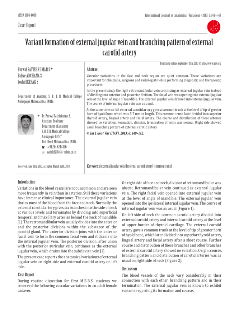
eISSN 1308-4038 International Journal of Anatomical Variations (2013) 6: 140–142 Case Report Variant formation of external jugular vein and branching pattern of external carotid artery Published online September 11th, 2013 © http://www.ijav.org Porwal SATISHKUMAR S Bidwe ARCHANA S Joshi DEEPAK S Department of Anatomy, S. R. T. R. Medical College, Ambajogai, Maharashtra, INDIA. Dr. Porwal Satishkumar S. Assistant Professor Department of Anatomy S. R. T. R. Medical College Ambajogai 431517 Dist. Beed, Maharashtra, INDIA. +91 244 6244326 satish27181@ yahoo.co.in Received June 21th, 2012; accepted March 27th, 2013 Abstract Vascular variations in the face and neck region are quiet common. These variations are important for clinicians, surgeons and radiologists while performing diagnostic and therapeutic procedures. In the present study the right retromandibular vein continuing as external jugular vein instead of dividing into anterior and posterior division. The facial vein was opening into external jugular vein at the level of angle of mandible. The external jugular vein drained into internal jugular vein. The course of internal jugular vein was as usual. At the same time on left external carotid artery gave a common trunk at the level of tip of greater horn of hyoid bone which was 5-7 mm in length. This common trunk later divided into superior thyroid artery, lingual artery and facial artery. The course and distribution of these arteries showed no variation. Formation, division, termination of veins was normal. Right side showed usual branching pattern of external carotid artery. © Int J Anat Var (IJAV). 2013; 6: 140–142. Key words [external jugular vein] [external carotid artery] [common trunk] Introduction Variations in the blood vessel are not uncommon and are seen more frequently in vein than in arteries. Still these variations have immense clinical importance. The external jugular vein drains most of the blood from the face and neck. Normally the external carotid artery gives six branches into the side of neck at various levels and terminates by dividing into superficial temporal and maxillary arteries behind the neck of mandible [1]. The retromandibular vein usually divides into the anterior and the posterior divisions within the substance of the parotid gland. The anterior division joins with the anterior facial vein to form the common facial vein and it drains into the internal jugular vein. The posterior division, after union with the posterior auricular vein, continues as the external jugular vein, which drains into the subclavian vein [1]. The present case reports the anatomical variations of external jugular vein on right side and external carotid artery on left side. Case Report During routine dissection for first M.B.B.S. students we observed the following vascular variations in an adult female cadaver. On right side of face and neck, division of retromandibular was absent. Retromandibular vein continued as external jugular vein. The right facial vein opened into external jugular vein at the level of angle of mandible. The external jugular vein opened into the ipsilateral internal jugular vein. The course of internal jugular vein was as usual (Figure 1). On left side of neck the common carotid artery divided into external carotid artery and internal carotid artery at the level of upper border of thyroid cartilage. The external carotid artery gave a common trunk at the level of tip of greater horn of hyoid bone, which later divided into superior thyroid artery, lingual artery and facial artery after a short course. Further course and distribution of these branches and other branches of external carotid artery showed no variation. Origin, course, branching pattern and distribution of carotid arteries was as usual on right side of neck (Figure 2). Discussion The blood vessels of the neck vary considerably in their connection with each other, branching pattern and in their termination. The external jugular vein is known to exhibit variants regarding its formation and course. Variant vessels of the neck 141 1 2 7 3 8 3 2 4 6 1 4 5 5 6 Figure 2. Common trunk for superior thyroid, lingual and facial arteries. (1: common carotid artery; 2: internal carotid artery; 3: external carotid artery; 4: common trunk; 5: superior thyroid artery; 6: lingual artery; 7: facial artery; 8: submandibular gland) 7 Figure 1. Unusual formation and termination of external jugular vein. (1: superficial temporal vein; 2: maxillary vein; 3: retromandibular vein; 4: external jugular vein; 5: facial vein; 6: internal jugular vein; 7: subclavian vein) Bertha and Suganthy studied 35 specimens and reported various types of variation as follows [2]: • Bilateral drainage of common facial vein into subclavian and bilateral absence of external jugular vein. • Absence of division of retromandibular vein. The retromandibular vein joined with anterior facial vein to form common facial which drained into internal jugular vein. • Opening of common facial vein into external jugular vein. Rajanigandha et al. reported a case in which retromandibular vein joined with facial vein to form common facial vein. After a short course common facial vein divided into anterior and posterior divisions. The posterior division opened into internal jugular vein through a connecting vein. The anterior division continued as a variant external jugular vein up to the jugular notch, where it crossed the midline to drain into opposite subclavian vein [3]. Sanjeev et al. studied 37 formalin preserved head and neck specimens. They found following variations [4]: • Higher division of common carotid artery (16.22% cases) • Lower division of common carotid artery (27.02% cases) • Common trunk of superior thyroid artery and lingual artery (2.7%) • Common trunk of lingual artery and facial artery (18.92%) • Common trunk of occipital artery and ascending pharyngeal artery (24.32%) • Common trunk of occipital artery and posterior auricular artery (2.7%). Troupis et al. performed dissection on 15 human cadavers. They observed a common trunk of lingual and facial artery from the right external carotid artery in 6% cases [5]. Chitra reported a case of trifurcation of right common carotid artery [6]. Delic et al. studied MRI carotid angiograms of 91 patients. They found that superior thyroid and lingual artery originate as a common trunk from external carotid artery in one case (1.09%); lingual and facial artery originate as a common trunk from external carotid artery (3.09%) [7]. The present study showed variations different as compared to available literature. Such vascular variations do occur due to appearance and disappearance of various vascular channels during development. External jugular vein is used for measurement of jugular venous pressure, which reflects pressure in the right atrium. Cannulation of external jugular vein is done for Satishkumar et al. 142 performing various procedures such as transvenous liver biopsy, transvenous pacemaker insertion, caval filter placement. External jugular vein is used as a patch for carotid endarterectomy [2, 8]. Knowledge of course and branching pattern of carotid arteries is essential to prevent vascular trauma during surgeries like thyroidectomy, glossectomy, laryngectomy and ligation of external carotid artery in severe epistaxis. Knowledge of these variations is essential for physicians, radiologists and surgeons while performing various diagnostic and therapeutic procedures in face and neck regions so as to prevent misdiagnosis and misadventures. References [1] Standring S, ed. Gray’s Anatomy. The Anatomical Basis of Clinical Practice. 39th Ed., New York, Elsevier Churchill Livingstone. 2006; 543–547, 551–552. [2] Bertha A, Suganthy R. Anatomical variations in termination of common facial vein. J Clin Diagn Res. 2011; 5: 24–27. [3] [4] Rajanigandha V, Rajalakshmi R, Ranade AV, Pai MM, Prabhu LV, Ashwin K, Jiji PJ. An anomalous left external jugular vein draining into right subclavian vein: A case report. Int J Morphol. 2008; 26(4): 893-895. Sanjeev IK, Anita H, Ashwini M, Mahesh U, Rairam GB. Branching pattern of external carotid artery in human cadavers. J Clin Diagn Res. 2010; 4: 3128–3133. [5] Troupis TG, Dimitroulis D, Paraschos A, Michalinos A, Protogerou V, Vlasis K, Troupis G, Skandalakis P. Lingual and facial arteries arising from the external carotid artery in common trunk. Am Surg. 2011; 77: 151–154. [6] Chitra R. Trifurcation of right common carotid artery. Indian J Plast Surg. 2008; 41: 85–88. [7] Delic J, Savkovic A, Bajtarevic A, Isavkovic E. Variations of ramification of external carotid artery- common trunks of collateral branches. Period Biol. 2010; 112: 117–119. [8] Greenfield LJ, Zocco J, Wilk J, Schroeder TM, Elkins RC. Clinical experience with the KimRay Greenfield vena caval filter. Ann Surg. 1977; 185: 692–698.

