Introduction to Microscopy - University of San Diego Home Pages
advertisement
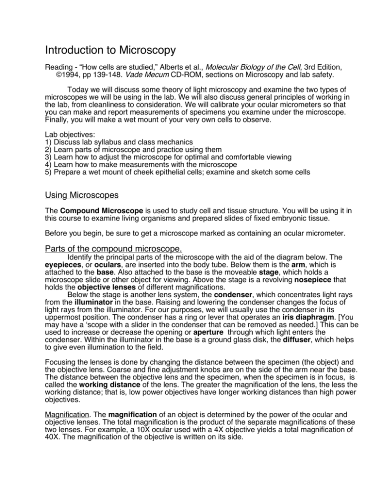
Introduction to Microscopy Reading - “How cells are studied,” Alberts et al., Molecular Biology of the Cell, 3rd Edition, ©1994, pp 139-148. Vade Mecum CD-ROM, sections on Microscopy and lab safety. Today we will discuss some theory of light microscopy and examine the two types of microscopes we will be using in the lab. We will also discuss general principles of working in the lab, from cleanliness to consideration. We will calibrate your ocular micrometers so that you can make and report measurements of specimens you examine under the microscope. Finally, you will make a wet mount of your very own cells to observe. Lab objectives: 1) Discuss lab syllabus and class mechanics 2) Learn parts of microscope and practice using them 3) Learn how to adjust the microscope for optimal and comfortable viewing 4) Learn how to make measurements with the microscope 5) Prepare a wet mount of cheek epithelial cells; examine and sketch some cells Using Microscopes The Compound Microscope is used to study cell and tissue structure. You will be using it in this course to examine living organisms and prepared slides of fixed embryonic tissue. Before you begin, be sure to get a microscope marked as containing an ocular micrometer. Parts of the compound microscope. Identify the principal parts of the microscope with the aid of the diagram below. The eyepieces, or oculars, are inserted into the body tube. Below them is the arm, which is attached to the base. Also attached to the base is the moveable stage, which holds a microscope slide or other object for viewing. Above the stage is a revolving nosepiece that holds the objective lenses of different magnifications. Below the stage is another lens system, the condenser, which concentrates light rays from the illuminator in the base. Raising and lowering the condenser changes the focus of light rays from the illuminator. For our purposes, we will usually use the condenser in its uppermost position. The condenser has a ring or lever that operates an iris diaphragm. [You may have a ‘scope with a slider in the condenser that can be removed as needed.] This can be used to increase or decrease the opening or aperture through which light enters the condenser. Within the illuminator in the base is a ground glass disk, the diffuser, which helps to give even illumination to the field. Focusing the lenses is done by changing the distance between the specimen (the object) and the objective lens. Coarse and fine adjustment knobs are on the side of the arm near the base. The distance between the objective lens and the specimen, when the specimen is in focus, is called the working distance of the lens. The greater the magnification of the lens, the less the working distance; that is, low power objectives have longer working distances than high power objectives. Magnification. The magnification of an object is determined by the power of the ocular and objective lenses. The total magnification is the product of the separate magnifications of these two lenses. For example, a 10X ocular used with a 4X objective yields a total magnification of 40X. The magnification of the objective is written on its side. ANIMAL DEVELOPMENT LAB MANUAL Oculars or Eyepieces Body tube Revolving nosepiece Arm Objective lens Coarse focus knob Stage Fine focus knob Condenser Condenser focus knob Illuminator 2 INTRODUCTION TO MICROSCOPY Working with your Compound Microscope Please follow all of the steps outlined here each time you use your microscope. You will be able to get the best image quality possible and minimize the chance of damaging your slides or your microscope. 1. Plug in and turn on the in-base illuminator. 2. If necessary, clean your objective, ocular and condenser lenses by wiping them gently with a clean sheet of lens paper. NEVER use anything but lens paper for cleaning lenses (no Kleenex, shirt sleeves, etc.). Anything else may scratch the lens. If there is something that doesn't seem to come off easily, contact your instructor or a TA to see if there is something more that we can do. NEVER attempt to clean the lenses with water or other liquid unless specifically directed by your instructor or a TA. The glue holding in lens components dissolve if you use other than approved solvents, destroying the lens. 3. Using the coarse focus, move the stage down as far as it will go. 4. Place the lowest power objective in operating position by rotating the nosepiece. You should feel a definite click when it is in the correct position. Make sure that the condenser is in its uppermost position and that the iris diaphragm is fully open. 5. Place a prepared slide (such as the starfish embryos slide) on the stage, securing it with the slide holder. Gently close the spring finger of the slide holder on the slide. Letting it fly may chip or crack your precious slide. 6. While looking at the microscope from the side, carefully move the stage up toward the objective with the coarse adjustment until it is close to, but not touching the slide. DON'T LOOK THROUGH THE OCULARS WHILE PERFORMING THIS STEP. Watch the slide and the objective. 7. Now look through the oculars and slowly back off, increasing the distance between the objective lens and the slide, until the specimen comes into focus. NEVER focus in the opposite direction, by decreasing this distance while looking through the oculars because you risk smashing the slide into the objective. This may break the slide and scratch the objective. This is more likely to be a problem with higher power objectives. Still, practice this approach even with low power objectives and you'll always be safe. 8. To achieve binocular vision (which increases your visual acuity and decreases the chance of headaches), hold your head so that you can see clearly through one ocular with one eye (if you have a dominant eye, use your weaker eye to make these adjustments). Now adjust the distance between the eyepieces until you can see through both simultaneously. Write down the number on the scale between the oculars. You can use this distance in the future to adjust your microscope. One ocular lens can be focused individually. While not looking through the microscope, turn this ocular all the way down, then all the way up, then return both to about midpoint in the focusing range. Look through both oculars at your specimen and cover (don't close) the eye looking through the focussing ocular with a 3 x 5 card and get your best focus with the other using the fine focus knob. Try to be relaxed and not squint with either eye. Move the card to cover the 'focused' eye and focus on the specimen using the focus ring on the ocular. Record the adjustment you use on the ocular focusing ring. (If you have a ‘dominant eye,’ you may want to focus your weaker eye first.) 9. Once focused, you may want to decrease the aperture of the iris diaphragm, found as a part of the condenser. Doing so will increase the depth of focus and the contrast of the specimen. [If your microscope has a phase/brightfield slider in the condenser, it must be removed to fully close the iris diaphragm. Gently remove the slider and place it within its protective container 3 ANIMAL DEVELOPMENT LAB MANUAL and put it away in the drawer until you are done with the microscope, or need to use phase contrast.] Close the iris diaphragm slowly until details of the specimen are sharply defined. Closing the diaphragm too much will decrease the resolution and introduce greater diffraction of the light. This will also increase the contrast in the specimen. (In the case of poorly stained or clear specimens, this may actually be an advantage. You may need to increase contrast and sacrifice resolution to see what you want in some specimens.) Change the aperture opening until you are satisfied that you have the best image possible. You may want to change the setting for different specimens. Also, you will probably need a smaller aperture for higher power objectives. NEVER turn the microscope on its side or turn it upside down; the ocular lenses can fall out and break. Köhler Illumination The optimal configuration of your microscope is when the condenser is in proper focus. This can be achieved with our microscopes despite the lack of a field diaphragm in the base of the illuminator (many 'scopes have an adjustable aperture just above the illuminator). The state of "Köhler illumination" is usually achieved by focusing the edges of a closed-down field diaphragm using the condenser. We can mimic the effect of a field diaphragm using a card with a small hole punched in it. 1. Place a slide with a specimen on the stage and focus on the specimen with the 4X or 10X objective. After the specimen is in focus, remove the slide. 2. Place a card with a hole in it on the illuminator in the base, centering the hole over the illuminator opening. [If you have a field diaphragm, close it down as far as it will go.] 3. Look through the microscope and focus the image of the edges of the hole using the condenser focus knob. Do not touch the regular focus knob. 4. After the image of the hole is focused, remove the card. [With a field diaphragm, open the aperture until the field is just filled with light - the edges of the in-focus diaphragm are just beyond the edge of the field of view. Obviously this will be different for each objective.] A card with a hole of the appropriate size (to just fill the field) could be created for each objective to complete the "Köhler illumination." Resolution The resolution of a microscope and lens system is proportional to the width of the cone of illumination collected by the objective, and depends on both the objective and the condenser, which focuses the light from below through the specimen. The maximal resolution possible is given by the formula Resolution = 0.61 λ / n sinθ where λ is the wavelength of light θ is 1/2 angular width (2θ) of the cone of light collected by the objective lens. n is the refractive index of the medium separating the specimen from the objective and condenser lenses. The bottom expression, n sinθ , is called the numerical aperture (NA) of the lens. 4 INTRODUCTION TO MICROSCOPY Since the theoretical maximum for the angle 2θ is 180°, the maximum for sinθ is 1. Since the refractive index of air is 1, for dry lenses, NA ≤ 1; but for oil immersion objectives, NA can be as high as 1.4. (Maximal resolution is obtained with oil on both the objective and condenser.) High resolution objectives (which are also high magnification objectives) have very short working distances, and very small (shallow) depths of field. The closer you are to the specimen the wider the possible cone of light, therefore yielding higher resolution. If you plug in the minimal wavelength of visible light (~0.4 µm), and an NA of 1.4 into the equation, you will find the theoretical limit of resolution for light microscopy. Care of the Microscope After Use After you have finished using your microscope: 1. Place the low power objective in the operating position. 2. Lower the stage as far as it will go using the coarse focus knob. 3. Remove any slides from the stage and put them away. If you have a wet mount, remove it and make sure the stage is clean and dry. Especially be sure to clean off any sea water as the salts are corrosive. Dispose of any glass properly - in a box marked for that purpose, not in the general trash. 3. If the slider has been removed from the condenser, return it carefully to its slot. 4. If necessary, clean the lenses with lens paper. 5. Turn off the illuminator, unplug the microscope, tie up the power cord, cover and return the microscope to the cabinet. The activities that follow, a. The Dissecting Microscope, b. Measurements... and c. Preparation of a Wet Mount, can be done in any order. Because we have a limited number of stage micrometers, needed for the "Measurements..." section, please continue with one of the other activities if a stage micrometer is not currently available. The Dissecting Microscope (or Stereomicroscope) The same rules apply to these microscopes with regard to care and cleaning. Although this microscope looks similar to the compound microscope, its uses and the principals of its optics are quite different. Whereas the image seen through a compound microscope is inverted, the image in a dissecting microscope is not. This allows you to manipulate (or dissect) a specimen while looking through the microscope. (This is much harder if the image is "upside-down and backwards.") The effective magnification of this kind of system is limited, usually in the range of 5x to 50x, but the working distance is great, allowing room to get your hands between the lenses and the specimen. The lenses and prisms in a dissecting microscope produce two light paths, one to each eye. This produces a true stereoscopic (or three-dimensional) image, which also aids in manipulation or dissection. The distance between the oculars can be varied as with the compound microscope to achieve binocularity. One of the oculars can be focused, so when adjusting the oculars for your eyes, start with the right eye and follow the same directions as for the compound microscope (see above). You should be able to identify the parts of this microscope as well; a diagram similar to that for the compound microscope above. The dissecting microscopes we will use change magnification by twisting the nosepiece. There is a single focus knob on the sides of this 5 ANIMAL DEVELOPMENT LAB MANUAL 6 INTRODUCTION TO MICROSCOPY microscope. Based on the power of your ocular lenses and the magnification in place indicated on nosepiece, you can calculate the total magnifications possible with your dissecting microscope. We now have a number of stereomicroscopes with a raised base and a separate source of fiber optic illumination. This type of illumination has significant advantages over stereomicroscopes with fixed, built-in illumination. (With built-in illumination, about the only advantage is simplicity - everything is in one place. The main disadvantage is that the angle of the light is fixed or only minimally variable. This limits how well you will be able to see some specimens.) You will find throughout the course that optimal lighting is often the key to seeing a given feature of a specimen. Using the stereomicroscope, one may use light from below (transillumination) or from above (incident lighting). Transillumination is typically used for specimens that are small, thin and fairly clear. Larger and opaque specimens require incident light. In transillumination, a fiber optic light guide is pointed into the back of the base and light is reflected upward from an adjustable mirror in the base through a glass plate. The angle and nature of this light can be adjusted a little by moving the mirror. Incident lighting is provided by bending the fiber optic light guide(s) to light the specimen from above or the side. Try adjusting the illuminator to give maximum light; you may or may not need this much light, although often you will need as much light as you can get. Note that another advantage of fiber optics vs. built-in light source is that it is much cooler to the specimen. Whereas a builtin light source can often be downright deadly to a live organism because the entire base is heated, the only heat a fiber optic-illuminated specimen gets is from the light itself. Even using fiber optic illumination, it is still possible to “cook” your specimens, especially if the light is turned up to a very high intensity. Therefore it is a good idea to remove specimens and turn off the light when you are not viewing them to avoid such problems. The stereomicroscope offers the opportunity to get a close-up view of virtually any common or uncommon object you would like to see. Choose a specimen: a slide, your hair, a piece of paper with writing, your fingernails (everyone’s cuticles look awful here); whatever you're curious about. You'll be amazed at what you can see. Measurements under the Microscope Your compound microscope is equipped with a device in one ocular called an ocular micrometer (or eyepiece graticule) which can be used to measure items within the field of view. First, however, you must calibrate your ocular micrometer with a stage micrometer. This special glass microscope slide is fragile and expensive. Ask your instructor to provide you with one of the lab's stage micrometers. 1. The stage micrometer has fine lines etched in it that cover 2.0 mm total. The larger divisions are 0.1 mm or 100 microns (µm) apart; at the left are fine divisions that are 0.01 mm or 10 µm apart. Place it on the stage and focus on the lines with your lowest power objective. Determine how much distance separates the larger demarcations of the micrometer. 2. Line up the divisions of the ocular micrometer with those on the stage micrometer below. Then use the stage micrometer to measure the ocular micrometer by comparing the two sets of lines. (If your ocular micrometer seems out-of-focus, check with your instructor or TA.) 7 ANIMAL DEVELOPMENT LAB MANUAL Ocular Micrometer Scale Stage Micrometer Scale In this example, the longest lines are 100 µm apart and the shorter ones are separated by 10 µm. The lines on the top represent the stage micrometer; those below the ocular micrometer. Since 15 divisions of the ocular micrometer equal 21 small divisions of the stage micrometer, each division of the ocular micrometer is equal to (21 x 10 µm)/15, or 14.0 µm. To determine the calibration for higher power objectives (10x and 40x), simply divide by the appropriate factor. The 10x objective magnifies 2.5 times (10/4) more than the 4x objective. Therefore divide the calibration by 2.5 to yield 14 µm / 2.5 = 5.6 µm per ocular micrometer division. For use with the 40x objective, divide by 10 -- 14 µm / 10 = 1.4 µm per ocular micrometer division. Or you could repeat the procedure above, measuring the ocular micrometer with the stage micrometer for your 10x and 40x objectives. (Note: The 100x is an oil immersion lens and we will not be using it.) It is useful to redo the calibration with at least one of these objectives to convince yourself that the conversion factor method works - you should get approximated the same numbers for the calibration. If you don't, you've done something wrong. Record the results of your calibration to use today and in the future for measurements. You will find slight variations from microscope to microscope. If you use the same microscope each time (they are numbered) you can count on your calibration next time. Example of using your calibration: You have determined that the calibration for your ocular micrometer with the 4x objective is 14 µm/division. You measure the diameter of a cell using the 10x objective as 24 divisions. What is the actual diameter of the cell? 24 divisions x 14.0 µm/division(at 4x) x (4/10) = 134.4 µm Always report measurements in microns, never in "divisions." Preparation of a Wet- mount Slide At times during the course, you will take a sample of organisms or living tissue to examine under the compound microscope. You will then sketch what you observe. The following exercise will demonstrate one way to mount tissue on a microscope slide. You will now examine cells sloughed from your own cheek epithelium in a way similar to that you may use for other tissues. 1. Clean a microscope slide by rubbing any dust off with a Kimwipe. (Lens paper is unnecessary for slides. Note, however, that Kimwipes are a low-lint cleaning paper). 2. Using a wood applicator, gently scrape back and forth on the soft lining inside your cheek. You don't have to do it hard. Let's not have any bleeding, OK? 3. Smear the scraped tissue in the middle of a clean microscope slide and add a small drop of water. The water is a mounting medium, which improves viewing of the preparation. Note that you need very little water. In this case, you can simply dip the other end of the wood applicator into some water and dab it on the specimen. A common mistake in making wet mounts is to add too much of the mounting medium. 4. Lower a coverslip slowly onto the drop of water, starting with one side of the coverslip contacting the drop. Try to avoid getting bubbles under the coverslip. Doing this well can take some practice. Also prepare at least one slide in which you add a stain to make the cells 8 INTRODUCTION TO MICROSCOPY easier to see. Try a bit of 0.1% neutral red stain, dabbing a small drop on a new smear of cheek cells. 5. Place the slide on the stage of your compound microscope. Observe first at low power then at higher powers. These cells are constantly sloughed off from the skin. Because these cells remain moist in the mouth, they retain their original shape somewhat. They are, however, definitely dead. 6. Sketch at least five cells from your best slide. Make your drawing of each cell at least 1" across, please. Label the nucleus, nuclear membrane, cytoplasm and plasma membrane. If other structures are apparent, sketch them in as well. We'll see whether we can figure out what they are. Measure the diameter of each cell and its nucleus with your ocular micrometer and label your drawings with these sizes. What is the average diameter of the nucleus? Always convert your ocular micrometer measurements into microns (µm) and be sure to check whether your results makes sense. Do you calculate that a nucleus averages 0.013 µm in diameter? That result would be nonsense, since the theoretical limit of resolution of the light microscope is only about 0.2 µm! (The wavelength of visible light determines this ultimate limit. Microscope makers achieved this limit by the end of the 1800's. Check your reading for more information about the theory and history of microscopy.) The Compound Fluorescence Scope (Research Grade) and Digital Camera In this class you will have the privilege of using a very high quality and expensive microscope for some of your studies. It should be used only after appropriate training and with the utmost care to preserve its function for all users. The Olympus BX-61 and accompanying parts cost more than $40,000. Some individual components (such as some objectives) cost $1000 or more. A fine digital camera linked to a computer will allow us to take high-quality color photomicrographs. The computer is for use with the microscope camera, and should not be altered in any way by addition or alteration of any software or settings that might interfere with this primary function. 9


