THE MICROSCOPE
advertisement
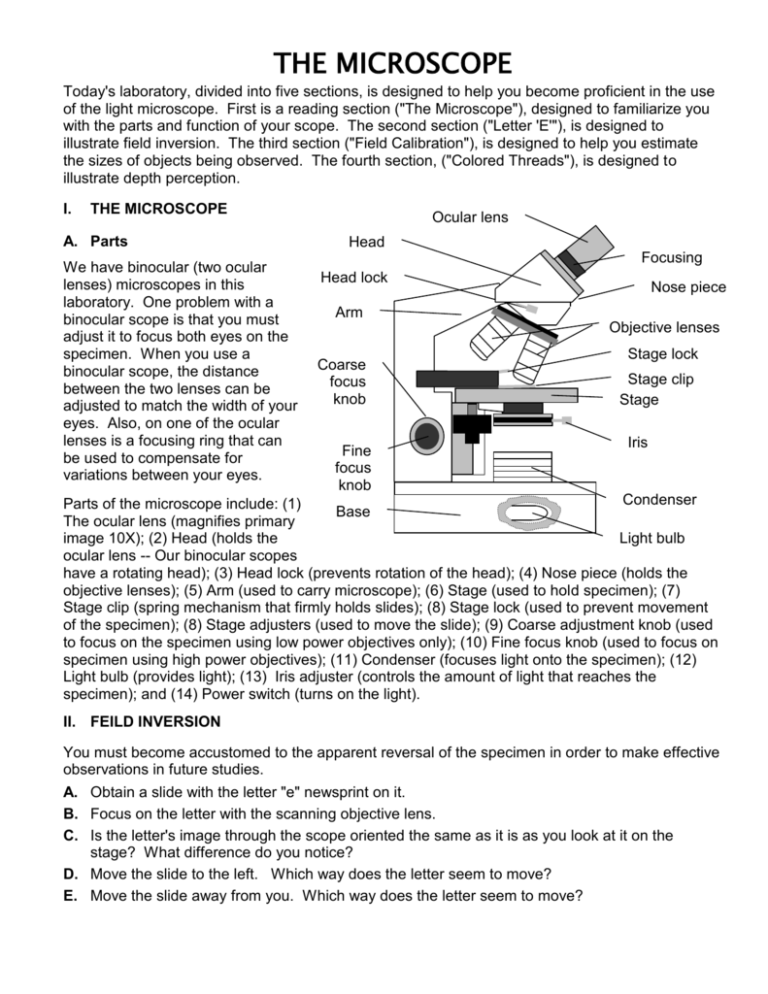
THE MICROSCOPE Today's laboratory, divided into five sections, is designed to help you become proficient in the use of the light microscope. First is a reading section ("The Microscope"), designed to familiarize you with the parts and function of your scope. The second section ("Letter 'E'"), is designed to illustrate field inversion. The third section ("Field Calibration"), is designed to help you estimate the sizes of objects being observed. The fourth section, ("Colored Threads"), is designed to illustrate depth perception. I. THE MICROSCOPE A. Parts We have binocular (two ocular lenses) microscopes in this laboratory. One problem with a binocular scope is that you must adjust it to focus both eyes on the specimen. When you use a binocular scope, the distance between the two lenses can be adjusted to match the width of your eyes. Also, on one of the ocular lenses is a focusing ring that can be used to compensate for variations between your eyes. Ocular lens Head Head lock Focusing ring Nose piece Arm Objective lenses Coarse focus knob Fine focus knob Stage lock Stage clip Stage Iris a d j Condenser Parts of the microscope include: (1) Base u The ocular lens (magnifies primary s Light bulb image 10X); (2) Head (holds the t ocular lens -- Our binocular scopes e have a rotating head); (3) Head lock (prevents rotation of the head); (4) Nose piece (holds the r objective lenses); (5) Arm (used to carry microscope); (6) Stage (used to hold specimen); (7) Stage clip (spring mechanism that firmly holds slides); (8) Stage lock (used to prevent movement of the specimen); (8) Stage adjusters (used to move the slide); (9) Coarse adjustment knob (used to focus on the specimen using low power objectives only); (10) Fine focus knob (used to focus on specimen using high power objectives); (11) Condenser (focuses light onto the specimen); (12) Light bulb (provides light); (13) Iris adjuster (controls the amount of light that reaches the specimen); and (14) Power switch (turns on the light). II. FEILD INVERSION You must become accustomed to the apparent reversal of the specimen in order to make effective observations in future studies. A. Obtain a slide with the letter "e" newsprint on it. B. Focus on the letter with the scanning objective lens. C. Is the letter's image through the scope oriented the same as it is as you look at it on the stage? What difference do you notice? D. Move the slide to the left. Which way does the letter seem to move? E. Move the slide away from you. Which way does the letter seem to move? III. FIELD CALIBRATION The field of view is the area that you can see through the ocular and objective. Knowing the size of the field of view is important because you can use it to determine the approximate size of an object you are examining. A. B. C. D. Obtain a metric ruler. Focus on the ruler with the scanning objective lens. Measure the diameter of the middle of the field of view. The diameter of the field of view of the higher objectives can be calculated using the formula: Fsca X Msca = Fhi X Mhi Flow = diameter of the Field of View of the lower-power objective Msca = magnification of the low-power objective Fhi = diameter of the Field of View of the higher-power objective Mhi = magnification of the higher-power objective E. Calculate the diameters of fields of view for each higher objective on your microscope. F. Make a habit of estimating sizes of cells and structures observed this block. IV. DEPTH OF FIELD Depth of field is the thickness of the object that is in sharp focus. It varies with magnification. A. Obtain a slide of three overlapping colored strings, then, using the scanning objective lens, focus up and down and try to determine the order of the threads from top to bottom. B. Repeat using the 10X and 40X objectives. V. HELPFUL HINTS ON MICROSCOPY A. Start at low power than move on to higher powers 1. Low power lets you see more area of the slide and is easier to focus B. The lenses are parfocal, which means only fine focus is needed when switching between them C. Use less light on lower objectives 1. Too much light will "bleach out" an organism (especially if not stained) 2. Higher objectives need more light to work D. Use your picture book to get an idea of what you are looking for E. Constantly adjust your fine focus knob and occasional adjust light levels 1. Most objects we are observing are three-dimensional, while your field of view becomes more two-dimensional the more your magnify, therefore, to see the "whole organism" fine focusing is needed 2. Low light levels give you better contrast, but higher levels can bleach out artifacts and structures you are finished observing F. Use the stage clip and knobs to move your specimen around when needed 1. Some areas of the slide may be better than others G. Keep in mind your objective in studying the object 1. If the field of view is highly repetitive (e.g., a bacterial smear), just concentrate on one good area of the slide 2. If your goal is to study the anatomy or morphology of an object, you may have to move the slide around to see all areas of the specimen 3. If you are examining a sample of diverse organisms, move the slide around to make sure you see all organisms on the slide


