Cerebellum - Mosaiced.org
advertisement
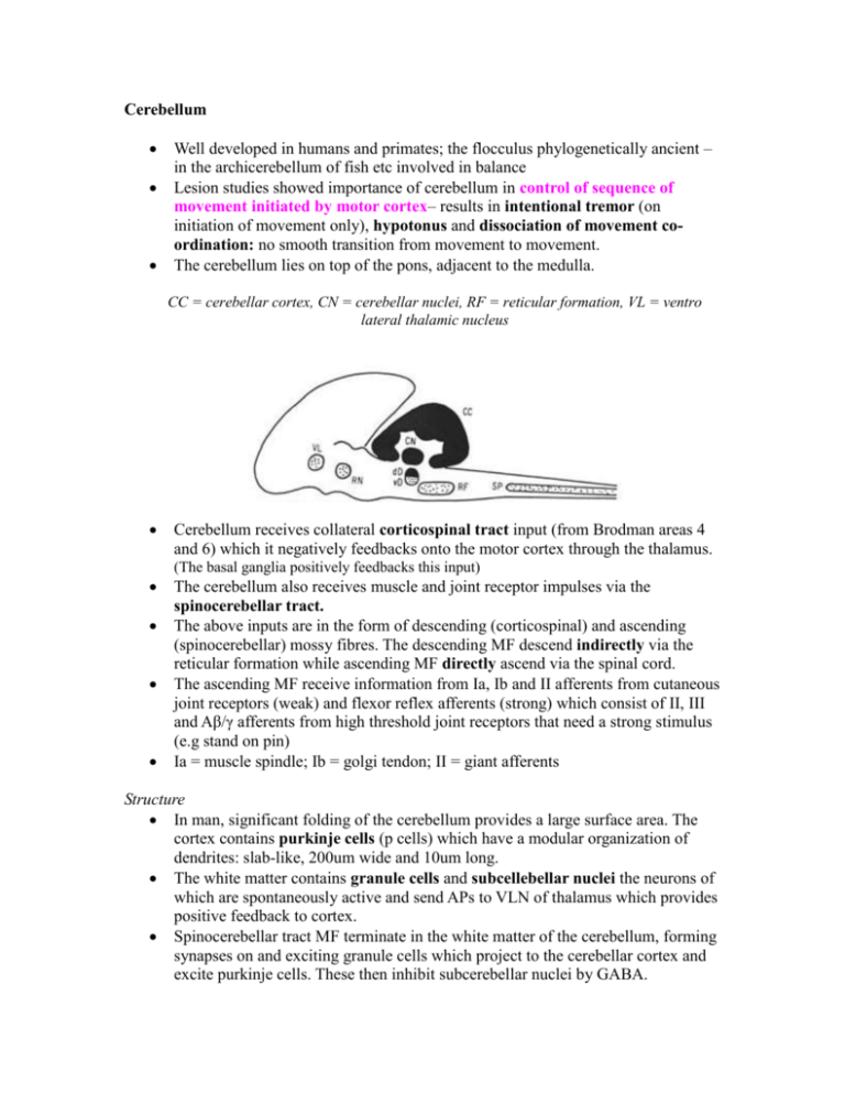
Cerebellum Well developed in humans and primates; the flocculus phylogenetically ancient – in the archicerebellum of fish etc involved in balance Lesion studies showed importance of cerebellum in control of sequence of movement initiated by motor cortex– results in intentional tremor (on initiation of movement only), hypotonus and dissociation of movement coordination: no smooth transition from movement to movement. The cerebellum lies on top of the pons, adjacent to the medulla. CC = cerebellar cortex, CN = cerebellar nuclei, RF = reticular formation, VL = ventro lateral thalamic nucleus Cerebellum receives collateral corticospinal tract input (from Brodman areas 4 and 6) which it negatively feedbacks onto the motor cortex through the thalamus. (The basal ganglia positively feedbacks this input) The cerebellum also receives muscle and joint receptor impulses via the spinocerebellar tract. The above inputs are in the form of descending (corticospinal) and ascending (spinocerebellar) mossy fibres. The descending MF descend indirectly via the reticular formation while ascending MF directly ascend via the spinal cord. The ascending MF receive information from Ia, Ib and II afferents from cutaneous joint receptors (weak) and flexor reflex afferents (strong) which consist of II, III and Aβ/γ afferents from high threshold joint receptors that need a strong stimulus (e.g stand on pin) Ia = muscle spindle; Ib = golgi tendon; II = giant afferents Structure In man, significant folding of the cerebellum provides a large surface area. The cortex contains purkinje cells (p cells) which have a modular organization of dendrites: slab-like, 200um wide and 10um long. The white matter contains granule cells and subcellebellar nuclei the neurons of which are spontaneously active and send APs to VLN of thalamus which provides positive feedback to cortex. Spinocerebellar tract MF terminate in the white matter of the cerebellum, forming synapses on and exciting granule cells which project to the cerebellar cortex and excite purkinje cells. These then inhibit subcerebellar nuclei by GABA. Ia/Ib/II ----SC----+ Granule cells ----+ Purkinje cells ---- − Subcerebellar nuclei MF stimulation produces small EPSPs onto purkinje cells even though 1 MF diverges onto 600 granules which converge onto a purkinje cell. This means that to reach threshold and generate an AP at a purkinje cell many MF have to be stimulated – temporal/spatial summation. There is somatotopic representation in the cerebellar cortex as in the cerebral cortex (precentral/postcentral gyri). PV = paravermis; V = vermis; L = lateral lobes; F = floculus; N = nodulus; F = fastigial nucleus; I = interpositus nucleus; Dn = dentate nucleus Purkinje cells in the vermis receive input from trunk and proximal limb proprioceptors (ascending MF pathway) and inhibit the fastigial nucleus which results in reduced activity in the VLN of thalamus and reduced excitation of the motor cortex. Purkinje cells in the paravermis receive input from distal limb digit receptors (ascending MF pathway) and inhibit the interpositus nucleus which reduces firing to the VLN and motor cortex. Purkinje cells of lateral lobes receive excitatory input from the motor cortex via the pons (descending MF pathway) and inhibit the dendate nucleus, reducing firing to VLN and motor cortex. Climbing fibres CF inputs come from neurons in the olive of the medulla oblongata. The olive receives input from the spino-olivary tract which receives weak input from Ia and Ib afferents and strong FRA. Olive neurons wrap around dendrites of purkinje cells and one neurone synapses with one purkinje cell (1:1). CF pathway involved in learning (how to walk, play piano, drive…) and not in smooth integrated control like the MF pathway Unlike MF stimulation, CF stimulation of the purkinje cells is strong and produces high frequency bursts of action potentials (-70mV to 40mV), followed by a powerful IPSP. This results in powerful inhibition followed by powerful excitation of the subcerebellar nuclei by purkinje cells
