TIF1a is a suppressor of RARa oncogenicity in the liver
advertisement
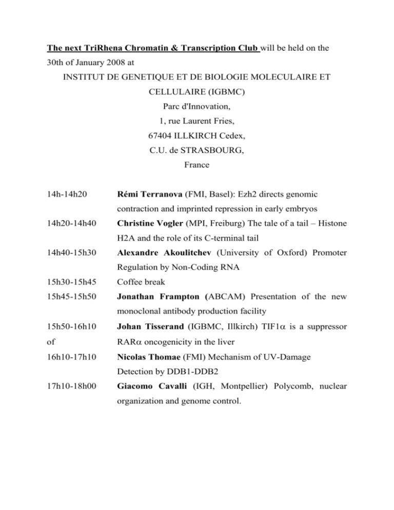
The next TriRhena Chromatin & Transcription Club will be held on the 30th of January 2008 at INSTITUT DE GENETIQUE ET DE BIOLOGIE MOLECULAIRE ET CELLULAIRE (IGBMC) Parc d'Innovation, 1, rue Laurent Fries, 67404 ILLKIRCH Cedex, C.U. de STRASBOURG, France 14h-14h20 Rémi Terranova (FMI, Basel): Ezh2 directs genomic contraction and imprinted repression in early embryos 14h20-14h40 Christine Vogler (MPI, Freiburg) The tale of a tail – Histone H2A and the role of its C-terminal tail 14h40-15h30 Alexandre Akoulitchev (University of Oxford) Promoter Regulation by Non-Coding RNA 15h30-15h45 Coffee break 15h45-15h50 Jonathan Frampton (ABCAM) Presentation of the new monoclonal antibody production facility 15h50-16h10 Johan Tisserand (IGBMC, Illkirch) TIF1 is a suppressor of RAR oncogenicity in the liver 16h10-17h10 Nicolas Thomae (FMI) Mechanism of UV-Damage Detection by DDB1-DDB2 17h10-18h00 Giacomo Cavalli (IGH, Montpellier) Polycomb, nuclear organization and genome control. Local Speakers TIF1 is a suppressor of RAR oncogenicity in the liver Johan Tisserand1, Konstantin Khechumian1, Marius Teletin2, Manuel Mark1,2, Benjamin Herquel1, Florence Cammas1, Daniel Metzger1, Pierre Chambon1,2 and Régine Losson1 1 2 Institut de Génétique et de Biologie Moléculaire et Cellulaire (IGBMC) Institut Clinique de la Souris (ICS) TIF1 has been identified in a yeast genetic screen for proteins modulating the transactivation potential of retinoic acid receptor RXR and was subsequently found to interact via a single LxxLL motif with the AF-2 activation domain of numerous nuclear receptors, including RARs, ERs, TRs, and VDR. TIF1 belongs to a family of chromatin transcriptional corepressors that has emerged as key regulators of developmental and physiological processes. To adress the physiological functions of TIF1, we have generated TIF1-null mice by gene disruption. We have discovered that TIF1-null mice spontaneously develop hepatocellular adenomas and hepatocellular carcinomas. Importantly, we found that a deletion of a single RAR allele in the TIF1-/- genetic background is capable of preventing the entire TIF1 KO-driven oncogenic process. Taken together, these results define TIF1 as a suppressor of tumorigenesis in the liver acting by inhibiting the oncogenic activity of RAR Ezh2 directs genomic contraction and imprinted repression in early embryos Rémi Terranova 1, Shihori Yokobayashi 1, Michael B. Stadler 1, Arie P. Otte 2, Stuart H. Orkin 3 and Antoine H.F.M Peters 1 1. Friedrich Miescher Institute for BioMedical Research, Maulbeerstrasse 66, CH-4058 Basel, Switzerland 2. Swammerdam Institute for Life Sciences, University of Amsterdam, Kruislaan 406, 1098 SM Amsterdam, The Netherlands; 3. Department of Pediatrics, Division of Hematology/Oncology, Children's Hospital, the Dana Farber, Cancer Institute, Harvard Medical School, Boston, MA 02115, USA In mammals, genomic imprinting regulates parental-specific expression required for normal development. Here we characterize novel higher-order chromatin features associated with establishing imprinted silencing at the paternal Kcnq1 cluster in early mouse embryos. In this cluster, silencing depends on expression of the Kcnq1ot1 non-coding RNA in-cis. By immuno-FISH, we show that proteins of Polycomb Repressive Complexes (PRC) 1 and 2, and repressive histone modifications associate with Kcnq1ot1, and form a distinct repressive nuclear compartment devoid of active marks and RNA polymerase II. In vivo, both PRCs associate to Kcnq1ot1 from the zygote stage onwards, but PRC1 recruitment to Kcnq1ot1 is PRC2-independent. Nevertheless, in the trophoblast lineage of E6.5 embryos, the PRC2 protein Ezh2 is essential for repression of paternal alleles of Kcnq1 cis-genes. Furthermore, we observe Ezh2-dependent genomic contraction of the paternal Kcnq1 cluster, assigning a novel role to Ezh2 in organizing large genomic clusters for gene repression. Mechanism of UV-Damage Detection by DDB1-DDB2 Nicolas Thomae Friedrich Miescher Institute for BioMedical Research, Maulbeerstrasse 66, CH-4058 Basel, Switzerland The UV-damaged DNA binding complex (UV-DDB) plays an important role in detection and recruitment of the nucleotide excision repair (NER) machinery to sites of UV-damage in vivo. Mutations in the DDB2 subunit result in xeroderma pigmentosum (complementation group E) a severe UV-sensitivity syndrome with a high incidence of skin neoplasias. The DDB1 subunit directly interacts with the E3-type cullin type ligase Cul4A and Cul4B. A family of about 40 DCAF proteins has been identified that binds to DDB1 in the presence of Cul4 and serves as ubiquitin ligase substrate adapters. Here we present the 3.2Å crystal structure of DDB1 bound to the damage specific adapter DDB2. DDB2 uses a helix-turn-helix motif Nterminal to a 7 bladed WD40-fold as main DDB1 binding epitope providing insight into the architecture of a DDB1-DCAF complex. DNA binding by the complex is achieved via the WD40 propeller of DDB2 that by itself is sufficient for DNA damage recognition. The finding that a WD40 propeller directly binds nucleic acids has implication for a number of βpropeller proteins found associated with chromatin and nucleic acids. The tale of a tail – Histone H2A and the role of its C-terminal tail Christine Vogler1, Tanja Waldmann1, Pedro de Souza Rocha Simonini1, Mirek Dundr2, Robert Schneider1 1 2 Max Planck Institut für Immunbiologie, Stübeweg 51, 79108 Freiburg Rosalind Franklin University, 3333 Green Bay Rd., North Chicago, IL60064 In the eukaryotic nucleus the DNA is organized in the form of chromatin. The basic unit of chromatin is the nucleosome consisting of 146 bp of DNA wrapped around an octamer of the four core histones H2A, H2B, H3 and H4. The histone proteins are not only important for the compaction of chromatin but also play an important role in the regulation of DNA-dependent processes such as transcription and repair. Posttranslational modifications of the flexible tails of the histones are one way to achieve this regulation. H2A is the only core histone that not only has an N-terminal tail but additionally contains a flexible C-terminal tail. This tail is thought to be located at the entry and exit site of the nucleosomal DNA. Almost nothing is known about the role of this tail in chromatin structure and function nor about proteins interacting with it. We were able to show that this tail is important for nucleosome stability in vitro and in vivo and that its deletion significantly increases nucleosome mobility. Furthermore, we found that expression of C-terminally truncated H2A influences cell proliferation and cell cycle progression in vivo. This is the first demonstration of a biological function for the H2A C-terminus. Invited Speakers Promoter Regulation by Non-Coding RNA Alexandre Akoulitchev Oxford BioDynamics Limited Oxford University Begbroke Science Park Sandy Lane, Yarnton Oxford OX5 1PF Sir William Dunn School of Pathology, University of Oxford Oxford OX1 3RE The complex population of human, regulatory non-coding RNAs includes transcripts implicated in highly specific regulation of gene promoters. One of the examples is the cellcycle dependent DHFR gene. The specificity and efficiency of its regulation depends on a number of epigenetic mechanisms, which include high-order chromatin structures, RNAdependent interactions and promoter-specific interference. Multiple evidence points at a mechanism of DHFR promoter repression in which a noncoding RNA initiates a cascade of epigenetic events similar to transcriptional gene silencing. Polycomb, nuclear organization and genome control. Giacomo Cavalli Institute of Human Genetics, CNRS, 141, rue de la Cardonille, 34396 Montpellier Cedex 5, France. Polycomb and trithorax group (PcG and trxG) proteins maintain the memory of regulatory states of developmental genes via modulation of higher-order chromatin structure. In Drosophila, PcG complexes are recruited to specific DNA regions called PcG response elements (PREs) ). DNA binding proteins bind to these specific sequences, and recruit the PcG complex PRC2, which is capable of trimethylating lysine 27 of histone H3. This in turns recruit the PRC1 complex that can somehow elicit gene silencing. However, the "code" that allows recruitment of PRC2 and PRC1 proteins at PREs is largely unknown, and we are trying to understand it by systematic mapping genomic binding of the putative recruiter factors. PcG proteins bound to chromatin are not homogeneously distributed in the cell nucleus, but they form nuclear compartments called “PcG bodies”. We are trying to understand the role of PcG bodies in gene regulation and, more generally, the role of PcG proteins in the regulation of cell differentiation versus proliferation during development.
