Keywords
advertisement
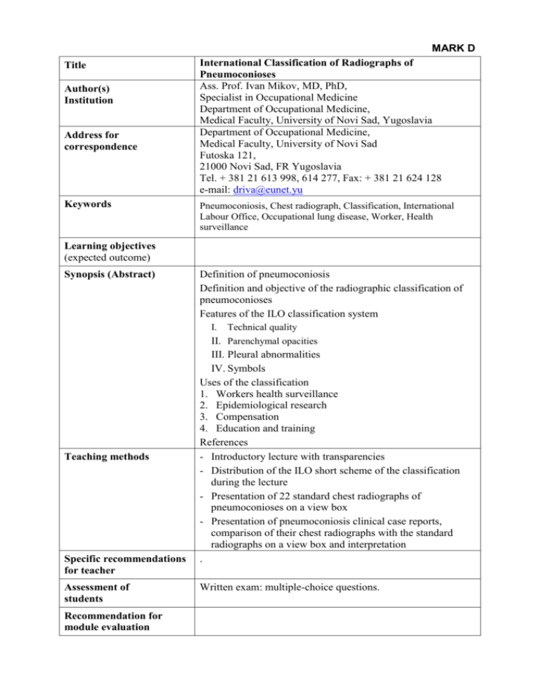
MARK D Title Author(s) Institution Address for correspondence Keywords International Classification of Radiographs of Pneumoconioses Ass. Prof. Ivan Mikov, MD, PhD, Specialist in Occupational Medicine Department of Occupational Medicine, Medical Faculty, University of Novi Sad, Yugoslavia Department of Occupational Medicine, Medical Faculty, University of Novi Sad Futoska 121, 21000 Novi Sad, FR Yugoslavia Tel. + 381 21 613 998, 614 277, Fax: + 381 21 624 128 e-mail: driva@eunet.yu Pneumoconiosis, Chest radiograph, Classification, International Labour Office, Occupational lung disease, Worker, Health surveillance Learning objectives (expected outcome) Synopsis (Abstract) Definition of pneumoconiosis Definition and objective of the radiographic classification of pneumoconioses Features of the ILO classification system I. Teaching methods Specific recommendations for teacher Assessment of students Recommendation for module evaluation Technical quality II. Parenchymal opacities III. Pleural abnormalities IV. Symbols Uses of the classification 1. Workers health surveillance 2. Epidemiological research 3. Compensation 4. Education and training References - Introductory lecture with transparencies - Distribution of the ILO short scheme of the classification during the lecture - Presentation of 22 standard chest radiographs of pneumoconioses on a view box - Presentation of pneumoconiosis clinical case reports, comparison of their chest radiographs with the standard radiographs on a view box and interpretation . Written exam: multiple-choice questions. International Classification of Radiographs of Pneumoconioses Ivan Mikov, MD, PhD, Department of Occupational Medicine, Medical Faculty, University of Novi Sad, Novi Sad, FR Yugoslavia Outlines of the lecture: Definition of pneumoconiosis: The term pneumoconiosis is used to cover group of lung diseases, which result from the inhalation of dust and should be used for all dust damage to the alveolar part of the lung, including the airways that have no mucociliary lining. Definition and objective of the radiographic classification of pneumoconioses: The International Labour Office (ILO) has issued an internationally accepted standard method for the classification of the radiographic changes seen in pneumoconiosis. The ILO has published the last revision in 1980. The ILO classification, which is based on the posteroanterior chest radiograph, is a descriptive system consisting of a glossary of terms and a set of 22 standard radiographs illustrative of the parenchymal and pleural changes of the pneumoconioses. The objective of the classification is to enable the radiographic changes seen in pneumoconioses to be coded in a simple reproducible form. The method of reading the radiographs is to compare each worker's radiograph with a set of standard radiographs supplied by the ILO. Features of the ILO classification system: I. Technical quality: The classification requires films of excellent quality. The accurate classification of poor quality films remains a problem. The reader's opinion about the film quality is recorded as 1,2,3 or 4 i.e. unreadable. II. Parenchymal opacities: The classification grades the parenchymal pattern of the pneumoconioses based on shape, size extent and profusion. a.) Small opacities: - Profusion (numbers: 0, 1, 2 and 3 combined in binary fashion) e.g. 2/2 or 2/3 - Extent (lung fields - RU, RM, RL, LU, LM and LL) - Shape and size (two-letter combinations: round shape - p, q, r and irregular shape - s, t, u) e.g. q/q or q/t b.) Large opacities (capital letters: A, B and C) III. Pleural abnormalities: Pleural thickening and pleural calcification are also categorized and quantified. IV. Symbols: Recording of other abnormalities seen on the chest radiograph. e.g. em (emphysema), tb (tuberculosis), ca (cancer) etc. Uses of the classification: 1. Workers health surveillance The ILO classification is useful for screening and surveillance programmes, which must try to recognize early radiographic abnormalities, to identify sentinel health events and to monitor trends over the time. 2. Epidemiological research It has proved very useful as a method of classifying chest radiographs for epidemiological research, identifying health hazards and establishing exposure-response relationships. 3. Compensation 4. Education and training References: 1. International Labour Office. Guidelines for the use of ILO international classification of radiographs of pneumoconioses. Occupational safety and health series No. 22 (Rev. 80). Geneva: International Labour Office, 1980. 2. Rossiter CE, Jones RN. Radiographic classifications. In: McDonald JC (ed.). Recent advances in occupational health. Edinburgh: Churchill Livingstone, 1981: 129-40. 3. Mikov M, Mikov I. Occupational Medicine. Belgrade: Naucna knjiga, 1995: 12434. (In Serbian) 4. Wagner GR. Screening and surveillance of workers exposed to mineral dusts. Geneva: World Health Organization, 1996: 52-7.
