Here is the 2008 version with answers
advertisement
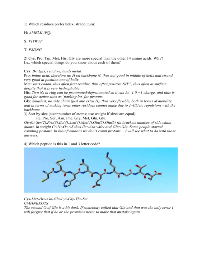
1) Which residues prefer helix, strand, turn: H: AMELK (FQ) S: VITWYF T: PSDNG 2) Cys, Pro, Trp, Met, His, Gly are more special than the other 14 amino acids. Why? I.e., which special things do you know about each of them? Cys: Bridges, reactive, binds metal Pro: imino acid, therefore no H on backbone N, thus not good in middle of helix and strand, very good at position one of helix Met: start codon, thus often first residue, thus often positive NH3+, thus often at surface despite that it is very hydrophobic His: Two Ns in ring can be protonated/deprotonated so it can be -1,0,+1 charge, and thus is good for active sites as ‘parking lot’ for protons. Gly: Smallest, no side chain (just one extra H), thus very flexible, both in terms of mobility and in terms of making turns other residues cannot make due to 1-4/5/etc repulsions with the backbone. 3) Sort by size (size=number of atoms; use weight if sizes are equal): Ile, Pro, Ser, Asn, Phe, Gly, Met, Gln, Glu. Gly(0)-Ser(2),Pro(3),Ile(4),Asn(4),Met(4),Gln(5),Glu(5) (in brackets number of side chain atoms. In weight C<N<O<<S thus Ile<Asn<Met and Gln<Glu. Some people started counting protons. In bioinformatics we don’t count protons… I will see what to do with those answers. 4) Which peptide is this in 1 and 3 letter code? Cys-Met-His-Asn-Glu-Lys-Gly-Thr-Ser CMHNEKGTS The second O of Glu is a bit dark. If somebody called that Gln and that was the only error I will forgive that if he or she promises never to make that mistake again. 5) Below you find all 8 based (ACGT in DNA and ACGU in RNA) each drawn once in the left two columns, and ones in the right two columns. They are not all drawn at the same size, an not all in the same 3' up or 3' down orientation. The left two columns thus make 20 Hbonds with the right two columns if all DNAs would pair up and all RNAs would pair up. Label the 20 columns in the left two columns that make the Watson and Crick base pairs in the DNA (and their equivalents in the RNA) from 1 - 20. That should not be too overly difficult, but the problem is that I want you to label their twenty H-bond partners in the right two columns with the corresponding numbers 1-20. And label each of the 16 pictures with the name of the base, also indicating if it is DNA or RNA. DNA is called DNA because of de-oxi, so those with an oxygen less on the sugar are the DNAs. In the figure I indicated D and R at the position where that ‘decision is being made’, and I labelled the rings with ACGT (T in RNA is actually called U, but that is a minor nomenclature detail). I labelled the H-bonding atoms in the left copy of each. We know CG make 3 H-bonds, AT 2. You need to remember that the one in D-T across from the C that sticks out isn’t bonding. Further, R-T (U) is like D-T, but is missing the extra C that sticks out. More donkey bridges I don’t know. The answer is in the DNA section of the course.
