Cardiopulmonary Physiology
advertisement
Cardiopulmonary Physiology
Millersville University
Dr. Larry Reinking
Chapter 5 - Pumping Action of the Heart
General Structures and Terminology
As you know, the heart has four contractile chambers and a series of valves that permit a
one-way flow of blood. Systole is a term used for the contracting period of a chamber while
diastole refers to the resting, expansion interval. The cardiac cycle refers to all of the events
that occur during one complete systole and one complete diastole. Cardiac output is the volume
of blood pumped in a given time interval.
Atria
The left and right atria are thin walled structures and this wall thickness reflects the low
pressures generated by the chambers (5-6 mm for the right side, 7-8 mm Hg for the left). Most of
ventricular filling depends on 'passive' venous return since the contraction of both atria add only
an additional 20-30% to the ventricular volume. For this reason, the atria are sometimes thought
of as 'primer pumps'. At rest, the heart will work satisfactorily without atrial pumping.
Ventricles
The ventricles are the power pumps of the heart and have thick muscular walls. These
walls are arranged in multiple sheets of myocardium that spiral from the base of the heart to the
apex. The thickness of the left ventricular wall is about three times that of the right. Cardiac
output for each chamber is the same, however the driving pressures differ significantly. Typical
pressures for the left are 120/80 mm Hg (systolic/diastolic) but only 25/1 mm Hg for the right.
Shape of the Ventricles and Pressure
Wall thickness alone does not account for all of the pressure difference between the left
and right ventricles. At least one other factor of important consideration is the shape of these
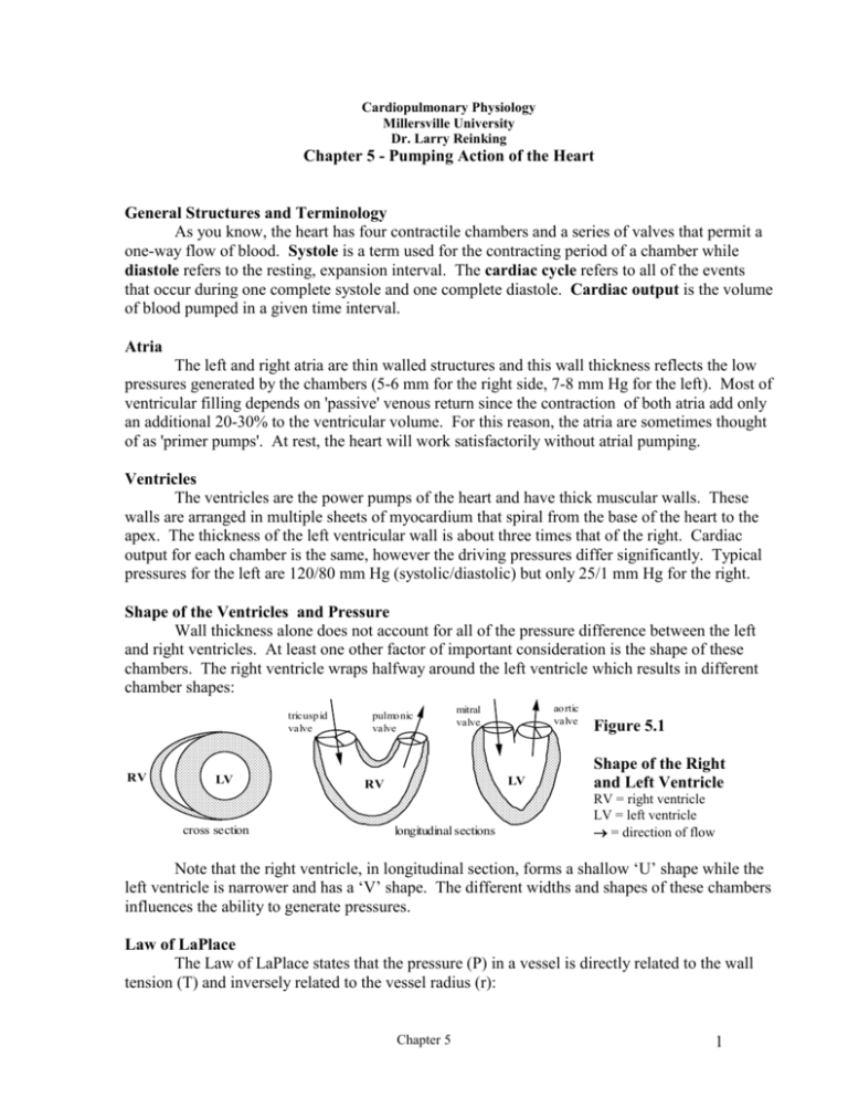
chambers. The right ventricle wraps halfway around the left ventricle which results in different
chamber shapes:
tricusp id
valve
RV
LV
cross section
pulmo nic
valve
ao rtic
valve
mitral
valve
LV
RV
longitudinal sections
Figure 5.1
Shape of the Right
and Left Ventricle
RV = right ventricle
LV = left ventricle
= direction of flow
Note that the right ventricle, in longitudinal section, forms a shallow ‘U’ shape while the
left ventricle is narrower and has a ‘V’ shape. The different widths and shapes of these chambers
influences the ability to generate pressures.
Law of LaPlace
The Law of LaPlace states that the pressure (P) in a vessel is directly related to the wall
tension (T) and inversely related to the vessel radius (r):
Chapter 5
1
P
T
r
Law of LaPlace
Equation 5.1
Thus, small vessels are able to withstand (or generate) more pressure than large vessels
having the same wall thickness. This is why oxygen (a high pressure gas) is compressed in long,
small radius tanks while propane (a low pressure gas) is bottled in squat, wide containers.
Likewise, the left ventricle, having a smaller radius will have a distinct geometrical advantage
over the right ventricle. A smaller radius, combined with a more muscular wall allows this side
of the heart to produce pressures 5-6 times those of the right.
Interesting Nature Note - A giraffe must pump blood to the head against large
hydrostatic columns and left ventricular pressure can exceed 300 mm Hg. In
accordance with LaPlace’s Law, the left ventricle has an almost tubular shape.
We will encounter the Law of LaPlace several times this semester in other applications.
Mechanical Events of the Cardiac Cycle
The heart really is an amazing organ. At a rate of 65 beats/min, a human heart will
complete 3,900 beats/hr... 93,600 beats/day... 34,164,000 beats/year... 2,391,480,000 beats/70
year life span! This is especially impressive when you consider that just a few missed beats
‘spells curtains’. At a cardiac output of 6 L/min, a heart will pump 3.2 million liters/year or 221
million liters in a 70 year life span. For comparison, a regulation Olympic water polo pool holds
1.2 million liters.
At a typical heart rate of 65 beats/min, each cardiac cycle takes 0.92 sec. During this
interval the chambers partition their time between systole and diastole in the following manner:
atrial systole 0.15 sec
atrial diastole 0.77 sec
ventricular systole
ventricular diastole
0.36 sec
0.56 sec
In other words, the atria are at rest for 84% (.77/.92) of the time and the ventricles for
61% (.56/.92) of the time. This is important, as we will see in the next chapter, because
ventricular contraction strength is governed by degree of filling during diastole. As the heart rate
increases, the duration of the cardiac cycle decreases and the diastolic filling times are reduced.
Heart rate is limited, in part, by the refractory period (the plateau, Figure 4.5 in chapter 4)
of the cardiac action potential. The absolute refractory period of the ventricle and atria are about
0.3 and 0.15 seconds, respectively. As a result, the contraction rate of the atria can be much
faster than the ventricles during pathological situations.
Wiggers Diagram and the Cardiac Cycle
A series of events take place during a cardiac cycle that include pressure changes in the
chambers, valve movement, electrical events, heart sounds, flow of blood and pressure changes
in the major vessels. Figure 5.2, below, incorporates these events, over time, into a single figure
(also see your lecture packet). Named in honor of Carl J. Wiggers, this diagram is one of the
cornerstones of cardiology. Become totally familiar with the Wiggers Diagram!
Chapter 5
2
• On the top of this diagram are three lines that deal with pressures: aortic pressure, left
ventricular pressure and left atrial pressure.
• The next line across is left ventricular volume, that is, the volume of blood contained within the
left ventricular chamber at any given point in the cardiac cycle. The difference between the
maximum and minimum value is the volume of blood ejected by the left ventricle.
• Next is the electrocardiogram, a recording made at the skin surface, of the electrical events of
the heart. It is a surface recording that ‘sums’ all of the action potentials of individual muscle
fibers. The P wave corresponds to the electrical depolarization that takes place just before atrial
contraction, the QRS complex is the depolarization that precedes ventricular contraction and the
final wave, the T wave, is caused by ventricular repolarization. We will examine the
electrocardiogram in detail in lab.
• On the bottom is the phonocardiogram, a recording of heart sounds using a surface
microphone on the chest. These sounds are the result of turbulent blood flow caused by valve
closure or rapid chamber filling.
Figure 5.2 The Cardiac Cycle (Wiggers Diagram)
This diagram depicts events for the left ventricle, left atrium, aorta, aortic valve, mitral valve,
electrocardiogram and phonocardiogram. , , and refer to the first, second and third heart
sounds.
Chapter 5
3
Phases of the Cardiac Cycle
The events of the cardiac cycle, on the Wiggers Diagram, are divided into a number of
phases. There is no real end or beginning to a cycle, but we will use atrial systole as a starting
point. Remember that this diagram depicts only the left side of the heart.
1. Atrial Systole - This phase begins as the atrium contracts causing a small rise in atrial
pressure (atrial pressure wave a) as well as ventricular pressure. Prior to this interval, blood had
been flowing through the atrium into the ventricle under the influence of venous pressure.
During this phase the mitral valve is open and the aortic valve is closed. Note that as the atrium
contracts a small additional volume is added to the left ventricle (the primer pump concept).
Also note that aortic pressure gradually drops ('diastolic run-off') as the elastic walls of the artery
recoil from the previous systolic ballooning (p. 5, chapter 1). The P wave of the
electrocardiogram overlaps the beginning of this phase. Recall that this wave represents the
summed depolarization of the atrial myocardium and that there is a slight time lag between
cellular depolarization, cross-bridging and contraction. The QRS complex, in preparation for
ventricular contraction, is seen at the end of this phase. The phonocardiogram is silent during
atrial systole.
Chapter 5
4
At the end of atrial systole the ventricle is at its maximum volume which is called the
end-diastolic volume (diastolic, in this case, refers to ventricular diastole). End-diastolic
volume is an important concept we will use in later lectures.
2. Isovolumic Contraction -This term means that although the ventricle is contracting, the
volume does not change, as can be seen on the ventricular volume curve. As contraction of the
ventricle begins (following the start of the QRS complex), ventricular pressure rapidly rises and
the mitral valve slams shut. The valve closure creates turbulence and vibrations that are detected
as the first heart sound on the phonocardiogram. This phase is isovolumic because both valves
are closed. Aortic pressure continues to drop and reaches its minimum pressure (≈80 mm Hg).
The blip in atrial pressure (atrial pressure wave c) is due to the massive contraction of the
ventricle which distorts the shape of the attached atrium. At the end of this phase, the ventricular
pressure equals and then exceeds aortic pressure, causing the aortic valve to open.
3. Ejection - As the aortic valve opens, blood is rapidly ejected into the aorta (note rapid drop in
ventricle volume) and pressures in the ventricle and aorta rise and are essentially matched. At
this point the aorta will stretch and the elastic componets will store energy. Pressure gradually
rises in the atrium because it is filling, via venous pressure, against a closed mitral valve. About
midway in the cycle, the muscle stops contracting and begins to repolarize. As a result, the aortic
and ventricular pressures drop and the T wave appears on the ECG. Although the pump has
stopped, blood flow continues at a reduced rate due to inertia. The ejection phase can be
subdivided into the period of maximum ejection (steep slope on volume curve) and the period of
reduced ejection (lesser slope on volume curve). At the end of the ejection phase ventricular
pressure falls below aortic pressure and the aortic valve slams shut.
The amount of blood ejected with each heart beat is referred to as the stroke volume and
is typically about 70 ml. The fraction of the end-diastolic volume ejected is called the ejection
fraction and has a value of about 60% (more correctly, 0.6, however the former is the usual
convention).
4. Isovolumic Relaxation - The ventricle is now at rest and because both valves are closed, the
volume is constant and at its lowest value (see ventricular volume curve). The noisy closure of
the aortic valve at the beginning of this phase is detected as the second heart sound. Atrial
pressure continues to rise as blood dams up against the closed mitral valve. Following closure of
the aortic valve, aortic pressure is no longer under ventricular influence, however, a precipitous
pressure drop is prevented by the energy stored in the elastic walls of this artery. The notch
(called the dichrotic notch) in the aortic pressure curve is due to a momentary back-flow of
blood caused by the aortic valve snapping closed. Ventricular pressure rapidly drops during this
phase and when atrial pressure is matched, the mitral valve opens. Repolarization of the
ventricular myocardium is complete (see T wave) by the end of isovolumic relaxation.
5. Rapid Filling - With the mitral valve open, blood rapidly flows into the ventricle (steep slope
on the ventricular volume curve) and exits from the dammed up atrium causing atrial pressure
wave c. Aortic pressure gradually drops during this resting phase. Sometimes, a third heart
sound is heard at the end of this phase, possibly caused by turbulent flow into a nearly filled
chamber. No activity is seen on the electrocardiogram.
Chapter 5
5
6. Diastasis - This period falls in the middle of ventricular diastole. Only a small amount of
blood enters the ventricle and ventricular pressure increases slightly during diastasis. Atrial
pressure also rises slightly because of continued venous return. Since the blood flows from
atrium to ventricle, left atrial pressure will be a little higher than that in the left ventricle. Aortic
pressure continues to fall and the phonocardiogram is silent. Near the end of diastasis the P wave
appears on the electrocardiogram as the atrial myocardium prepares for atrial systole.
The following are characteristic time intervals for the cardiac cycle phases in an adult
human. These values vary with heart rate.
Table 5.1. Durations of Cardiac Phases
Heart Rate = 75 beats/min
Phase
Duration (sec)
Atrial systole
0.11
Isovolumic Contraction
0.05
Ejection
0.26
Isovolumic Relaxation
0.08
Rapid Inflow
0.11
Diastasis
0.19
Heart Valves
Blood flows through the heart from the right atrium right ventricle pulmonary artery
and from the left atrium left ventricle aorta. This one-way flow is the result of the cardiac
valves. There are two types of valves; the atrioventricular valves (A-V valves) and the semilunar
valves. Both types of valves are fibrous tissue, covered with endothelium, that passively open
and close in response to pressure differences between the heart chambers. Around each valve is
a supporting ring of fibrous connective tissue.
Atrioventricular Valves
The tricuspid valve lies between the right atrium and ventricle and is comprised of three
flaps. Positioned between the left atrium and ventricle is the mitral (or bicuspid) valve which
has two flaps. The thin flaps (also called leaflets or cusps) of the A-V valves are extremely
flexible and will close when the pressure in the ventricles only slightly exceeds that in the atria.
On the edges of the valve leaflets are the strong chordae tendineae which connect to the
papillary muscles at the apex of the ventricular walls (Figure 5.3, below). During ventricular
systole, the anchoring action of the chordae tendineae and the forceful contractions of the
papillary muscles prevent the valve leaflets from everting into the atria. In both the A-V valves,
the leaflets have considerable overlap when closed and create a tight seal.
Semilunar Valves
Both of these valves consist of three pocket-like cusps (Figure 5.3). The aortic valve lies
between the left ventricle and aorta while the pulmonic valve is between the right ventricle and
pulmonary artery. A larger pressure difference is needed to make these valves snap shut as
compared to the more responsive A-V valves. At the end of the ejection period, as left
Chapter 5
6
ventricular pressure rapidly drops, some blood actually flows backwards from the aorta into the
ventricle before the cusps close.
Above the semilunar valves are dilated areas on the pulmonary artery and aorta. These
areas, the sinuses of Valsalva, create eddy currents that prevent the cusps from pressing
completely against the walls of the arteries during the ejection period. This action apparently
prevents the valve leaflets from blocking the right and left coronary arteries which originate at
the sinuses of Valsalva of the aorta.
Figure 5.3
A-V and Semilunar
Valves
RV = right ventricle
LV = left ventricle
Heart Sounds
As mentioned in the description of the Wiggers diagram, the closure of the A-V valves
and semilunar valves produce the first and second heart sounds. It was previously assumed that
these sounds were the result of the valve leaflets snapping together, however, current
interpretations suggest that they are caused by vibrations in the chamber walls and blood brought
about by the abrupt valve closure. The third heart sound appears to be caused by blood
decelerating as it enters the distended ventricle at the end of the rapid filling period. Normally,
this sound is heard only in children who have fairly thin chest walls. In adults it is
characteristically heard in individuals with overloaded, distended hearts. When heard in adults,
the third heart sound is usually a very bad omen. A fourth heard sound (or the atrial sound) is
sometimes heard in normal individuals during atrial systole. It appears to be caused by vibrations
of the chamber walls and blood induced by atrial contraction.
Valvular Heart Disease
Although there can be many causes, defective valves are usually the result of rheumatic
fever or congenital abnormalities. These events produce either a valve with a narrowed opening
(valvular stenosis) or a leaky seal (valvular insufficiency). During rheumatic fever, bodily
immune responses to streptococcal infection damage the mitral valve and/or aortic valve.
Rheumatic damage to the tricuspid or pulmonic valve is rare. Congenital defects can occur to
any of the valves or to multiple valves. The following is a brief description of the four most
common types of valvular disease. As you read this section, consult the Wiggers diagram to help
you understand the consequences of these defects. Some of the physiological consequence of
valvular disease will make more sense after you have an understanding of Chapter 6.
Mitral Stenosis
Chapter 5
7
Since the opening between the left atrium and ventricle is narrowed, blood flow between
these chambers will be impaired. During atrial systole, left atrial pressure will be abnormally
high as this chamber pumps against the smaller exit. Cardiac output will drop because the
ventricle cannot completely fill during diastole (see Chapter 6 to understand the mechanism).
As blood flows across the narrowed mitral valve a sound (a murmur) will be caused by
turbulent flow. Thus, a murmur will be heard during atrial systole and during rapid filling. Since
the valve leaflet are likely to be deformed by the narrowing, the first heart sound (caused by
mitral valve closure) can be exaggerated. Also, the valve may open noisily, creating an opening
'snap'. The following diagram, Figure 5.4, illustrates these heart sounds.
E
IR RF
D
AS IC
Figure 5.4
1
2
Heart sounds associated with mitral
Normal
stenosis.
Mitral
Stenos is
os
m
mitral valve
closes
m
mitral valve
opens
1 = first heart sound, 2 = second heart sound
m = murmur, OS = opening snap
AS = atrial systole, IC = isovolumic contraction,
E = ejection, IR = isovolumic relaxation,
RF = rapid filling, D = diastasis
Intensity of sound is indicated by height of shaded area.
Mitral Insufficiency
As a result of a leaky mitral valve, blood will regurgitate from the left ventricle into the
atrium during ventricular systole. This regurgitation causes the left atrial pressure to rise and the
cardiac ejection, into the aorta, to drop. During ventricular systole, the blood in the distended
atrium will return to the ventricle, add to the normal ventricular filling, and overload the
chamber.
As blood regurgitates across the distorted valve turbulent flow will create a murmur.
This murmur is heard for the duration of ventricular systole. During ventricular diastole, excess
blood will rush into a dilated ventricle, resulting in a third heart sound.
Figure 5.5
Heart sounds associated with mitral
insufficiency.
See Figure 5.4 for notations.
Aortic Stenosis
In this situation blood cannot easily exit from the ventricle into the aorta. As a result, left
ventricular pressure will have a high peak value (200 mm Hg) during ventricular systole. Aortic
pressure will rise slowly during the ejection phase and cardiac output will be reduced. This
slowly rising aortic pressure can actually be detected when palpating the radial pulse and is called
the anacrotic pulse. It is a classic sign of aortic stenosis.
Blood rushing across the narrowed valve will create a murmur for the duration of the
ejection period. During ventricular ejection, the pressure gradient between the left ventricle and
aorta will be small just as the valve opens, have a great difference in mid ejection and be small,
again, as the valve closes. This changing pressure gradient results in a murmur that peaks in
intensity during mid ejection.
Chapter 5
8
Figure 5.6
Heart sounds associated with aortic
stenosis.
See Figure 5.4 for notations.
In contrast, the systolic murmur of mitral insufficiency (Figure 5.5) has a constant
intensity. Since the pressure in the atrium is always fairly low, the pressure gradient that forces
blood back into the atrium during ventricular systole will be high for the entire period.
Aortic Insufficiency
Due to a leaky aortic valve, blood regurgitates from the aorta and stretches the ventricle
during diastole. As a consequence, the next contraction is more forceful (see Chapter 6 for
mechanism) and the pressure is abnormally high during the first half of the ejection period. The
regurgitation of blood also causes the diastolic pressure of the aorta to drop rapidly. As a result,
the pulse pressure (systolic - diastolic) becomes large and can be detected by palpating the radial
pulse. A hammering pulse is a classic indicator of aortic insufficiency.
An early diastolic murmur is heard as blood regurgitates into the ventricle. As the
pressure gradient lessens during diastole so does the intensity of the murmur. The high pressure
gradient driving ejection of blood from the ventricle across a misshapen valve can also create a
systolic murmur similar, but of less intensity, to that heard in aortic stenosis.
Figure 5.7
Heart sounds associated with aortic
insufficiency.
See Figure 5.4 for notations.
Chapter 5
9
