Study Cards
advertisement
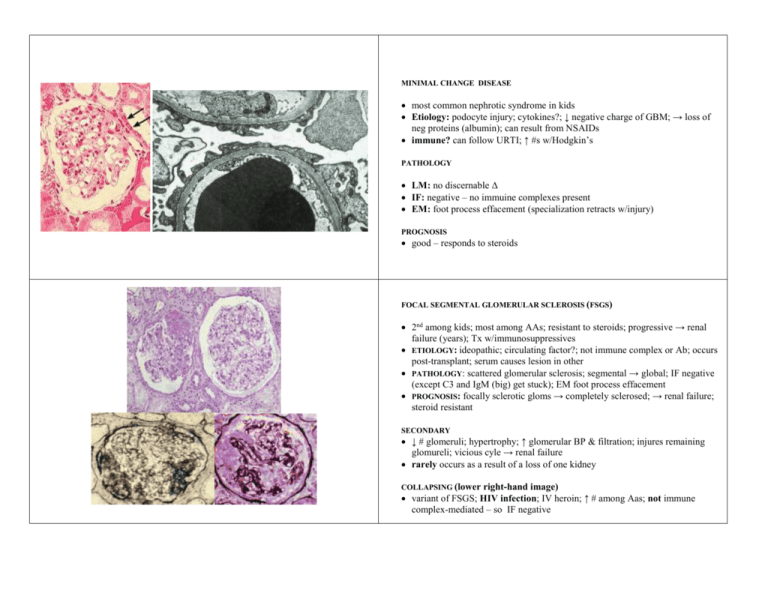
MINIMAL CHANGE DISEASE most common nephrotic syndrome in kids Etiology: podocyte injury; cytokines?; ↓ negative charge of GBM; → loss of neg proteins (albumin); can result from NSAIDs immune? can follow URTI; ↑ #s w/Hodgkin’s PATHOLOGY LM: no discernable Δ IF: negative – no immuine complexes present EM: foot process effacement (specialization retracts w/injury) PROGNOSIS good – responds to steroids FOCAL SEGMENTAL GLOMERULAR SCLEROSIS (FSGS) 2nd among kids; most among AAs; resistant to steroids; progressive → renal failure (years); Tx w/immunosuppressives ETIOLOGY: ideopathic; circulating factor?; not immune complex or Ab; occurs post-transplant; serum causes lesion in other PATHOLOGY: scattered glomerular sclerosis; segmental → global; IF negative (except C3 and IgM (big) get stuck); EM foot process effacement PROGNOSIS: focally sclerotic gloms → completely sclerosed; → renal failure; steroid resistant SECONDARY ↓ # glomeruli; hypertrophy; ↑ glomerular BP & filtration; injures remaining glomureli; vicious cyle → renal failure rarely occurs as a result of a loss of one kidney COLLAPSING (lower right-hand image) variant of FSGS; HIV infection; IV heroin; ↑ # among Aas; not immune complex-mediated – so IF negative MEMBRANOUS GLOMERULONEPHRITIS autoummine disorder:Abs against podocyte cell membrane Ags poor prognostic indicators: ↑ age, ↑ BUN & Cr, ♂, HTN ETIOLOGY 85% idiopathic; infections (malaria, syphilis Hep B); autoimmune (SLE, RA); paraneoplastic (lung, colon, melanoma); meds: gold (Tx RA), penicillamine PATHOLOGY auto-Abs to podocyte FP Ags; activate C5b-9 MAC; → subepithelial space; GBM grows between deposits LM diffuse, unifrom thickening w/spikes & holes IF coarse, regular, granular staining IgG & C3 EM lots of dense deposits over FP injury/effacement COURSE may resolve, stasis, progress to RF; Tx primary cause if possible MEMBRANOPROLIFERATIVE GLOMERULONEPHRITIS (MPGN) may present nephrotic, nepritic, mixed, asymptomatic… suspect in Pts w/nephrotic range proteinuria and ↑ #s RBCs in UA or RF w/↑Cr CLINICAL kids & young adults: ↓ complement, persistent hematuria, HTN, RF (↑Cr) ETIOLOGY immune-complex deposition; endothelial injury & GBM reduplication infectious (Hep B&C, IE); immune (SLE, cryoglobinemia); paraneoplastic PATHOLOGY: IC deposition in MESANGIUM & SUBENDOTHELIAL SPACE LM mesanfial hypercellularity & tram-tracking IF IgG & C3 in capillary & mesangium EM e--dense deposits; GBM reduplication COURSE idiopathic = bad (progressive) 2° – Tx 1° and reversal may follow POST-STREPTOCOCCAL GLOMERULONEPHRITIS acute nephritic syndrome: hematuria, protenuria, HTN, mild renal insufficiency CLINICAL 1-3 wks post throat infection, 3-6 post skin infection edema, HTN, hematuria, azotemia, mild proteinuria & oliguria positive for titres of anti-SLO & anti-DNAse B PATHOGENESIS circulating ICs or Ags plant in glomerulus → subepithelial deposits PATHOLOGY LM: subepithelial humps; inflammatory cells (PMNs & monos) IF: lumpy-bumpy granular peripheral capillary loop deposis (IgG, C3) EM: large, random “humps” or “honkers” PROGNOSIS children: 95% recover spontaneously, 5% progression (fast or slow) adults: 60% recover spontaneously, 40% progress to RPGN or RF LUPUS NEPHRITIS auto-immune, immune-complex mediated disease nephrotic syndrome, acute nephritic syndrome, asymptomatic h/p, or frank ARF PATHOGENESIS IC deposition throughout kidney; complement→ WBCs→ inflammation; PROLIFERATIVE CHANGE mesangial proliferation: hypercellularity & ↑ matrix endocapillary: subendothelial deposits of inflammation; damage to GBMs; margination, necrosis, & rupture pathology IF: “full house effect” – IgG, IgA, IgM, C3… EM: subendothelial IC deposition; “fingerprint” substructure MEMBRANOUS CHANGE: parallel to MPGN; Abs vs. podocyte Ags PROGNOSIS better predicted by chronic changes rather than active lesions TREATMENT steroids & cytotoxic agents (cytoxan) IgA NEPHROPATHY (BERGER’S DISEASE) asymptomatic hematuria/proteinuria CLINICAL FACTORS affects: children & young adults; indigenous populations often anctecedent URTI or GI; Hx of intermittent macroscopic hematuria may present as frank nephritic syndrome; (rarely) RPGN ETIOLOGY unknown trapping of IgA throughout mesangium PATHOLOGY LM: mesangial hypercellularity & ↑ matrix IF: IgA & C3 (alternative path!); may also find IgG & IgM EM: immune-type e--dense mesangial deposits, hypercellularity, ↑ matrix PROGNOSIS bening or progress to CRF (10%-50%) ALPORTS DISEASE hereditary defect in Type IV collage – GBM, cochlea, eye PRESENTATION hematuria, ↑F hearing loss, ocular defects PATHOGENESIS absence of α-5 (XLR), α-3,4 (AR/AD) isoform → disturbance of production PATHOLOGY LM nonspecific IF negative – non-immune EM “basket weave” characteristic lamellation of GBM OUTCOMES ♂ w/XLR, both sexes with AR/AD → ESD at varying rates TREATMENT transplant; rarely Type IV collagen of new kidney recognized as “not self” note: Thin Basement Membrane disease is similar, with Δ error in Type IV collagen CRESCENTIC GLOMERULONEPHRITIS histopathologic hallmark of RPGN TYPE I: ANTI-GBM Abs to GBM; w/pulmonary – Goodpasture; mostly ideopathic may follow hydrocarbon solvent exposure, Influenze A2, malaria, Hodgkins LM: crescents; IM: linear fluorescence of GBMs for IgG; EM: no deposits prognosis good, if caught early TYPE II: IMMUNE-COMPLEX etiology: IC-mediated diseases (SLE, post-strep, MPGN, IgA nephropathy) biopsy to ID and Tx TYPE III: PAUCI-IMMUNE defined by lack of findings of ICs or anti-GBM Abs >90% p or c-ANCA positive idiopathic or systemic vasculitis (Wegener’s or Microscopic polyangiitis) ACUTE TUBULAR NECROSIS most common cause of ARF Ischemic: patchy; due to sepsis, burns, surgery, or hemorrhage Tubulo-toxic: uniform; due to, e.g., aminoglycosides, dyes, rhabdomyolysis 1° MECHANISM injury → ↓synthetic f’ns → ↓ phosphate stores (↑E) → lysosomal destruction PATHOLOGY mild: loss of brush border (1st slide), vacuolization (2nd slide), swelling cells severe: necrotic cells slough off into lumen (3rd slide), leaving BM naked regeneration: flat epithelia (4th), mitotic figures, reactive nuclei w/↑ nucleoli OTHER HARMFUL MECHANISMS IN ATN vasoconstriction necrotic cells in casts obstructing tubules, ↑ pressure, ↓ filtration backleal of filtered urine into kidney vasculature & interstitium PROGNOSIS: often reversible if support is maintained (dialysis) ACUTE PYELONEPHRITIS most common cause: ascending UTI (e. coli); ♂ < 1 – ♀ – 50 < ♂ also fungi (candida albicans) and CMV (transplant) PATHOLOGY gross: pinpoint or largere pustules, may meld; streaks of yellow pus micro: intense streaky neutrophilic interstitial & tubular infiltrate COMPLICATIONS pyonephrosis: kidney → bag of pus perinephric abscess: infection spreds into perinephric soft tissue necrotizing papillitis: requires: infection, obstruction, compromised blood flow; appears as coagulatoin necrosis w/sharp, inflamed edges ACUTE TUBULO-INTERSTITIAL NEPHRITIS (ATIN) inflammation of interstitium → tubules ETIOLOGY/ANTECEDENTS Hx of hypersensitivity, ICs in TBMs, anti-TBM Abs, T-cell mediated damage most commonly, rxn to a drug (β-lactam, sulfonaminde, NSAIDS, diuretic) systemic infections (group A strep, diphtheria, toxoplasmosis, legionnaire’s) MICROSCOPIC PATHOLOGY tubules seperated by edema inflammatory infiltrate (lymphocytes, monocytes, eosinophils, PMNs) CLINICAL PRESENTATION hypersensitivity: fever, hematuria, eosinophilia, pyuria, skin rash, mild proteinuria, eosinophiluria DIABETIS MELLITUS – DIFFUSE AND NODULAR GLOMERULOSCLEROSIS renal disease 2° to Db is ↑ cause of ESRD in US; > ½ dialysis pts clinical features: proteinuria; microalbuminemia; frank nephrotic syndrome; ↓GFR along with other complications of Db (retinopathy) PATHOGENESIS 1° lesion: excess ECM throughout the kidney (many GFs, esp. TGF-β) renal disease w/in 10 yrs of Db; ↓ retinopathy; unexplained hematuria or unusually accelerated renal impairment BIOPSY CONDITIONS: PATHOLOGY GROSS: enlarged, esp. Type 1 Db; evidence of arteriosclerosis/arteriolosclerosis LM: progressive, uniform ↑ in GBM thickness & corresponding ↑ in ECMatrix ½ pts: ↑ mesangial matrix → frank hypocellular nodules (Kimmelstiel-Wilson) IF: linear deposition of IgG (sticky) EM: uniform thickening of GBMs, podocyte foot process efffacement PINK/HYALINE DEPOSITS: fibrin cap, capsular droplet TUBULES & INTERSTITIAL thickening of TBMs & tubular atrophy VASCULATURE: ↑ deposition of ECM in both afferent & efferent arterioles AMYLOIDOSIS deposition of protein → proteinuria, nephrotic syndrome, & renal insufficiency β-pleated sheet: stains w/Congo Red; polarized-light “apple-green” refringence FORMS OF AMYLOID AL: 1° amyloidosis – most common; often from multiple myeloma AA: 2° amyloidosis – serum AA (acute phase reactant) from chronic inflammatory conditions (RA, TB, osteomyelitis) PATHOLOGY glomerulus: mesangial expansion & nodule formation vasculature: lumen diminution of arteries & arterioles LM: amorphous pink or hyaline material EM: masses of thin, nonbranching fibrils IF: reagents of κ, λ light chains, or SAA protein MYELOMA KIDNEY unconrolled secretion of κ, λ light chains accumulate with Tamm-Horsfall proteins PATHOLOGY precipitate as large, occlusive casts in tubules numerous tubules w/in cortex & medulla expanded by large pink casts casts have “cracked” or fractured appearance, some laminated often surrounded by multinucleated giant cells TREATMENT ↓ secretion of light chains by treating MM reducing accumulation by ↑ free water intake, alkalinizing the urine, avoiding additional insults (no radiocontrast dyes) RENAL CELL CARCINOMA clear cell carcinoma most common kidney tumor of adults GROSS PRESENTATION solid, cystic, bright yellow, with some necrosis/hemorrhage MICROSCOPIC PRESENTATION clear cells (lipids & glycogen) in small nests and sinusoidal arrangement TYPICAL CLINICAL PRESENTATION associated w/von Hippel-Lindau; ♂ > ♀ (2:1); smoking; obese ♀ IMPORTANT PREDICTORS OF BEHAVIOR STAGE: local → perinephric fat → renal vein → surrounding structures GRADE: nuclear appearance (differentiation) WILM’S TUMOR most common tumor of childhood HISTOLOGICAL FEATURES: blastemal, epithelial, and mesenchymal TRANSITIONAL CELL CARCINOMA (TCC) (AKA) UROTHELIAL CARCINOMA (MOSTLY PAPILLARY) CLASSIFICATION papilloma; PUNLMP; low-grade papillary ca; high-grade papillary ca; BIOLOGICAL IMPLICATIONS OF CLASSIFICATION ↑ risk of recurrence & progression HISTOLOGICAL DESCRIPTION fronds consisting of fibrovascular cores surrounded by uroepithelium IMPORTANT PREDICTORS OF BEHAVIOR ↑ size; ↑ #; ↑ grade; dysplasia of “uninvolved” mucosa progress: carcinoma in situ → invasive; 20% ADENOCARCINOMA OF THE PROSTATE most common tumor of the prostate in adults HISTOLOGIC PRESENTATION fairly uniform proliferation of small, round glands PRESUMED PRECURSOR LESION prostatic intraepithelial neoplasia (PIN) within ducts IMPORTANT PREDICTORS OF TUMOR BEHAVIOR Gleason score: sum of 2 scores measuring tumor architecture absolute predictor of 5-year survival
