3-4 Extrapyramydal system, cerebellum
advertisement
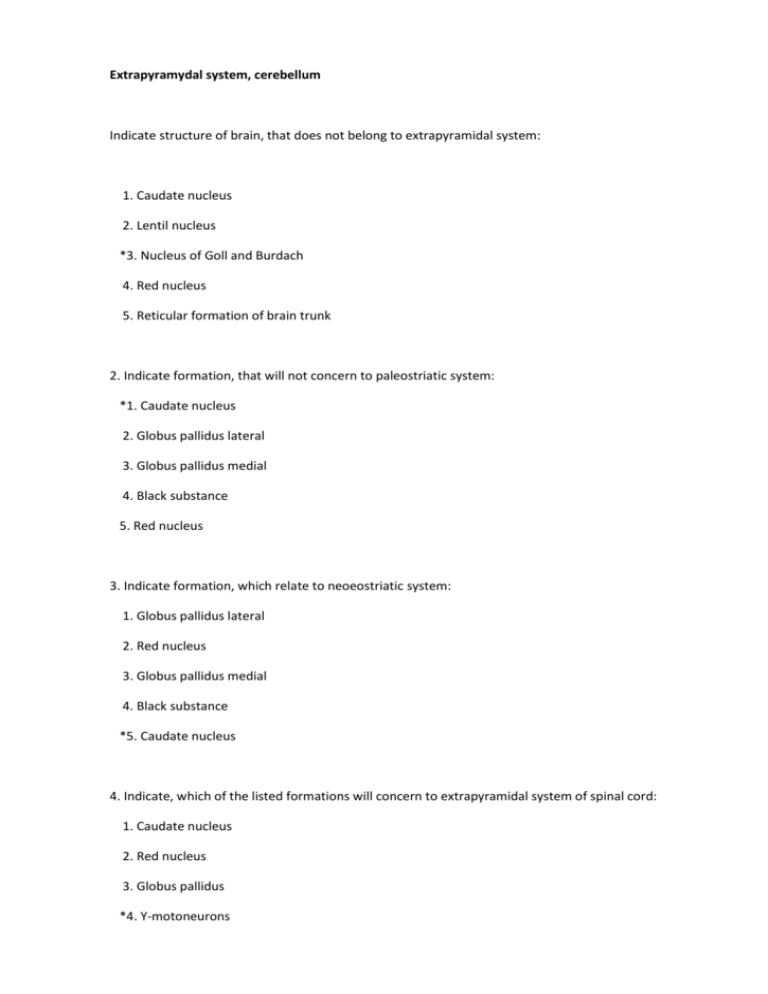
Extrapyramydal system, cerebellum Indicate structure of brain, that does not belong to extrapyramidal system: 1. Caudate nucleus 2. Lentil nucleus *3. Nucleus of Goll and Burdach 4. Red nucleus 5. Reticular formation of brain trunk 2. Indicate formation, that will not concern to paleostriatic system: *1. Caudate nucleus 2. Globus pallidus lateral 3. Globus pallidus medial 4. Black substance 5. Red nucleus 3. Indicate formation, which relate to neoeostriatic system: 1. Globus pallidus lateral 2. Red nucleus 3. Globus pallidus medial 4. Black substance *5. Caudate nucleus 4. Indicate, which of the listed formations will concern to extrapyramidal system of spinal cord: 1. Caudate nucleus 2. Red nucleus 3. Globus pallidus *4. Y-motoneurons 5. Black substance 5. Indicate physiological function, which is not executed by extrapyramidal system: 1. Myostatic regulation 2. Postural control 3. Regulation of muscular tone *4. Coordination of movement 5. Realization of automated movements 6. Indicate symptom, characteristic for lesion in neoeostriatic system: 1. Hypomimium 2. Silent monotonous language 3. Muscular hypertension *4. Hyperkinesis 5. Propulsion 7. Indicate symptom, that is not characteristic for lesion in pallidonigral system: *1. Hypomimium 2. Hypomyotonia 3. Bradykinesia 4. Silent monotonous language 5. Muscular hypertension behind plastic type 8. Indicate symptom, that are not characteristic for parkinsonism: 1. Silent monotonous language 2. Out of flexors *3. Hypomyotonia 4. Static tremor 5. Oligokinesia 9. What infringement below does not concern with hyperkinesisnes: 1. Hemiballism *2. Intention tremor 3. Trochee 4. Athetosis 5. Myoclonia 10. Indicate, what is characteristic of below listed symptoms for lesion of caudate nucleus: 1. Muscular hypertension *2. Hypomyotonia 3. Hypokinesia 4. Bradykinesia 5. Hypomimium 11. Indicate, what infringement of speech arises at parkinsonism: 1. Mutism 2. Aphasia 3. Scanned language 4. Dysarthrium *5. Silent, monotonous language 12. Indicate syndrome, that is characteristic for lesion in extrapyramidal system: 1. Vestibular ataxium 2. Oligophrenia 3. Dynamic ataxium *4. Parkinson 5. Sensitive ataxium 13. Indicate basic function of cerebellum: 1. Realization of voluntary movements *2. Coordination of movement 3. Postural control 4. Automatic movements 5. Maintenance of expressiveness of mimic reactions 14. Indicate formation, that does not relate with the structure of cerebellum: 1. Legs *2. Nucleus of Goll and Burdach 3. Worm 4. Hemisphere 5. Dentate body 15. Indicate cerebellar nucleus: 1. Tail nucleus 2. Red, Swalbe 3. Deiters, Bechterev *4. Fastigial nucleus, gear nucleus 5. Nucleus of Goll and Burdach 16. Indicate, which of the below-mentioned will pass through the middle leg of cerebellum: 1. Dentorubralis *2. Anterior-pons-cerebellar 3. Rubrospinal 4. Govers 5. Vestibulocerebellar 17. Indicate, which of the below mentioned will pass through bottom leg of cerebellum: 1. Spinocerebellar of Govers *2. Spinocerebellar of Flexig 3. Frontal bridge 4. Rubrospinal 5. Occipital bridge 18. Indicate ways, that will pass through top leg of cerebellum: 1. Olivocerebellar 2. Reticulocerebellar 3. Ponscerebellar 4. Vestibulocerebellar *5. Spinocerebellar of Govers 19. Indicate symptom, not characteristic for cerebellar disease: 1. Intention tremor *2. Muscular hypertension 3. Hypomyotonia 4. Uncertain shaky gait 5. Nistagmus 20. Indicate, what dissonance of language arises at cerebellar disease: 1. Aphasia 2. Nasal language *3. Scanned 4. Dysphonia 5. Dysarthrium 21. Which of the listed methods for research of function of cerebellum are not used: 1. Finger-nose test 2. Test on diadochokinesia *3. Test of Barre 4. Calcaneal-knee test 5. Test of Stewart-Holme 22. Indicate, which of the below mentioned are afferent leading ways of communications of cerebellum *1. Spinal way of Flexig 2. Tectospinal 3. Dentorubralis 4. Vestibulospinal 5. Rubrospinal 23. Indicate, which of the below mentioned are efferent leading ways of communications of cerebellum: 1. Spine-cerebellar Flexig 2. Spinocerebellar of Govers 3. Vestibulocerebellar *4. Dentorubralis 5. Front-pons-cerebellar 24. Name method of revealing dynamic cerebellar ataxium: *1. Finger-nose test 2. Station test 3. Test of Barre 4. Test of Budda 5. Liquorodynamic tests 25. Indicate infringement, characterized by lesion of vermis: *1. Static ataxium 2. Sensitive ataxium 3. Muscular hypertension 4. Bradykinesia 5. Vestibular ataxium 26. Indicate detection method of infringement of statics: 1. Test on diadochokinesia 2. Test on hypermetria 3. Finger-nose test *4. Stability in pose Romberg 5. Test of Babiski 27. Name clinical attribute of cortical-frontal ataxium: 1. Deviation at movements aside center 2. Nausea, vomitting 3. Nistagmus *4. Astasium-abasia 5. Scanned language 28. Indicate biochemical mechanism of development of Parkinson syndrome: 1. Infringement of copper metabolism 2. Infringement of exchange of phenylalanine *3. Infringement of exchange of dofaminum 4. Infringement of carbohydrate metabolism 5. Infringement of exchange of lipids 29. Indicate syndromes, that are characteristic for lesion of tail and red nuclei, «оЁб®perл© body: 1. Parkinson *2. Hyperkinetic 3. Non-tactal 4. Central tetraparesis 5. Peripheral tetraparesis 30. On what side of cerebellum infringements at hemilesion and cortex of cerebellar hemispheres are developed: 1. Ataxium of extremities of opposite focus *2. Ataxium of extremities on side of focus 3. Ataxium of trunk from opposite side 4. Ataxium of trunk on same side of foucs 5. Bilateral ataxium of extremities 31. Impossibile to walk a straight line, alternately putting right heel of one leg to toes of other, is connected to: *1. Cerebellar dysfunction 2. Lesion of parietal share of brain 3. Lesion of temporal share of brain 4. Oculomotor dysfunction 5. Loss of sensitivity in cadences 32. Tremor of hands in calmness, especially when awakening the patient, lesion is usually developed where: 1. Visual hump *2. Black substance 3. Caudate nucleus 4. Spinal cord 5. Inside capsule of brain 33. Adiadochokinesia testifies about infringement of: 1. Consecutive movement of fingers 2. Walking from heel to sock 3. Hold back of tremor *4. Synchronisation of movementsa of arms 5. Coordination of colloquial muscles 34. Indicate medication, that is used for treatment of Parkinson syndrome: 1. Nootropic preparations 2. Sedative mean *3. Cholinoblockers (anticholinergic) 4. Anticholine esterase preparations 5. Corticosteroids 35. Sharp static ataxium is characteristic with muscular atony and asynergia for lesion of : 1. Bottom legs of cerebellum 2. Middle legs of cerebellum 3. Top legs of cerebellum *4. Vermis 5. Cerebellar hemispheres 36. Uncertainty and unsteadiness while walking in darkness and good illumination are characteristic for ataxium: *1. Sensitive 2. Vestibular 3. Dynamical cerebellar 4. Static-locomotor cerebellar 5. Cortical (frontal) 37. Functions of cerebellum are all of the below listed, except: 1. Coordination of movement 2. Regulation of muscular tone 3. Synergy of movements 4. Equilibrium of body *5. Postural tone 38. Indicate, with what choreic hyperkinesis is clinically characterized: 1. Slowness *2. Speed, unevenness, precipitancy, absence of stereotype 3. Speed, unevenness, precipitancy, stereotype 4. Twitching in muscular groups and in lonely muscles 5. Involuntary tonic reduction of mimic muscles of person 39. Patient has hypomimicry, flexor, shuffling gait, muscular hypertension behind plastic type. Indicate localization of pathological center: 1. Neoeostriatic system *2. Pallidonigral system 3. Cerebellum 4. Internal capsule 5. Brain trunk 40. Ten year old child has violent movements of extremities face, trunk. Indicate localization of pathological center: 1. Pallidonigral system *2. Neoeostriatic system 3. Cerebellum 4. Fronto-prefrontal site of cerebrum 5. Anterior central convolution 41. Patient with parkinson's disease syndrome. Indicate, how gait is affected: 1. Spastic 2. Spastico-atactic 3. Hemiparesthetical 4. Steppage * 5. Shuffling, fine steps 42. Due to disease unsteadiness at circulation appeared, intensive tremor at finger-nose and calcanealknee tests, More expressed on the right side, Nistagmus at sight to the right. In pose Romberg patient falls to the right. Determine localization of lesion: 1. Vermis 2. Lefthand cerebellar hemisphere *3. Right cerebellar hemisphere 4. Top legs of cerebellum 5. Bottom legs of cerebellum 43. Fast arrhythmic involuntary movements of extremities and trunk are observed in patient. He grimaces, frequently sticks out tongue. Tone of muscles of extremities is lowered. Described syndrome is named: 1. Athetosis 2. Myoclonia *3. Trochee 4. Hemiballism 5. Tic 44. Patient is worried by difficulty while walking. Objectively -- Hypomimium, delay of voluntary movements, increase of muscular tone of extremities behind plastic type, walks in small steps, shuffling. Described syndrome is named: 1. Hyperkinetic *2. Parkinson 3. Non-tactal 4. Alternating 5. Brown-Sechar 45. Patient walks with short steps, body is inclined forward, hands half-bent, face is masklike. Speech monotonous, silent, calmed down. Stereotyped tremor of fingers of hands of "account of coins "type is observed. Syndrome is named: 1. Alternating 2. Non-tactal 3. Hyperkinetic *4. Parkinson 5. Brown-Sechar 46. Patient after having influenza developed violent vermiculations in fingers of bones. These movements are named: *1. Athetosis 2. Hemiballism 3. Myoclonia 4. Tic 5. Trochee 47. Patient has sharp headaches, nausea, vomitting. At objective research horizontal nystagmus is revealed, it is primary at sight to the left, adiadochokinesia at the left, scanned language, unsteadiness of gait, in pose Romberg falls to the left. Determine localization of lesion: 1. Top legs of cerebellum 2. Bottom legs of cerebellum *3. Cerebellar hemisphere at the left 4. Cerebellar hemisphere on the right 5. Vermis 48. Schoolboy began to slovenly, turns head in different directions during lessons, grimace, write letters of different size. Objectively -Lowered tone of muscles in extremities, fast arrhythmic involuntary movements of extremities and trunk are observed. Described syndrome is named: 1. Athetosis 2. Hemiballism 3. Myoclonia 4. Tic *5. Trochee 49. Man-40 years old choreolike hyperkinesis, dementia. Indicated symptoms gradually progress. Name disease: 1. Sharp cerebral circulatory disturbance 2. Paralysis agitan 3. Paraplegia of Strumpell *4. Huntington disease 5. Meningoencephalitis 50. 36 yr old man in residual period of encephalitis, arose attacks of myoclonia of muscles of face, and left hand. At review in interattack period - somewhat lowered muscular tone in left sided extremities, intensive tremor, hypermetria of left sided extremities. Indicate localization of lesion: *1. Red nucleus on the right 2. Black substance at the left 3. Vermis 4. Lefthand cerebellar hemisphere 5. Dentate body on the right 51. Man 42 years, 3 year back survived head trauma. Complains on unsteadiness of gait, infringement of movements of right hand and leg. At review - right-hand pyramidal deficiency behind central type, coordination tests at the left execute with intention, adiadochokinesia through negative chronotropism at the left, hypomyotonia at the left. Indicate striked structures: *1. Anterior central convolution at the left, left side of hemisphere of cerebellum. 2. Anterior central convolution at the left, lefthand frontal -cerebellar way 3. Anterior central convolution on the right, right frontal-cerebellar way 4. Spinocerebellar tracts on the right, rubrospinal at the left 5. Spinocerebellar tracts at the left, rubrospinal on the right 52. Woman 42 years, complains of residual phenomena of trunk encephalitis on complication of gait, unsteadiness, impossible to stand in vertical position and balance. At review - light central tetraparesis, bilateral expressed static-locomotor ataxium. Indicate localization of lesion: *1. Vermis, overlap of pyramidal route 2. Vermis, cerebellar hemisphere 3. Cerebellar hemisphere, red nuclei 4. Internal capsule on the right, vermis 5. Internal capsule at the left, vermis 53. Patient has shaky gait, on the right missdirect, intensive tremor, adiadochokinesia, horizontal nystagmus. Where can lesion be located: 1. Vermis 2. Right cerebral peduncle 3. Lefthand cerebellar hemisphere *4. Right cerebellar hemisphere 5. Lefthand anterior central convolution 54. Patient is moved with effort, cannot stand because of unsteadiness, falls in pose of Romberg, also cannot sit. Volume of active movements in extremities is full, force of muscles is 5 points. Sensitivity (superficial and deep) is kept. What is damaged?: 1. Weigh cerebellum 2. Back cords of spinal cord 3. Cerebellar hemisphere *4. Vermis 5. Precentral convolution of the brainof cortex 55. Patient is worried by shaky gait, rotatory vertigo, nausea, cannot stand in Horizontal position.Nystagmus checks out. Falls In Romberg pose What is damaged? 1. Vermis 2. All parts of cerebellum 3. Back cords of spinal cord *4. Vestibular apparatus 5. Cerebellar hemisphere 56. In woman, 42 years, appeared infringement of movements in left hand, general constraint. Therapist, at which woman was treated from psoriatic polyarthritis, suspected neurological disorder. Neurologic inspection found: Hypomimicry, bradykinesia, flexor pose, extrapyramidal hypertonus. Pathogenetic therapy was ordered Which medication is most used in this instance: 1. Selegelin *2Cyclodol 3. Bromocryptine 4.Haloperidol 5.Amantadin
