Effects of hypocretin–saporin injections into the medial septum on

Effects of hypocretin–saporin injections into the medial septum on sleep and hippocampal theta
Brain Research 913 (2001) 106–115
Dmitry Gerashchenko , Rafael Salin-Pascual , Priyattam J. Shiromani
Neurons containing the peptide hypocretin, also known as orexin, were recently implicated in the human sleep disorder narcolepsy. Hypocretin neurons are located only in the lateral hypothalamus from where they innervate virtually the entire brain and spinal cord. This peptide is believed to be involved in regulating feeding and wakefulness. However, to fully understand what other behaviors are regulated by this peptide it is necessary to investigate each hypocretin target site. In the present study, we focus on one hypocretin target site, the medial septum, where there is a dense collection of hypocretin-2 receptor-containing cells, and degenerating axons are present here in canines with narcolepsy [J. Neurosci. 19 (1999) 248]. We utilize a saporin toxin conjugated to the hypocretin receptor binding ligand, hypocretin-2, and find that when this toxin is injected into the medial septum, it lesions the parvalbumin and cholinergic neurons. We contrast the effects of the hypocretin–saporin with another saporin conjugated toxin, 192
IgG–saporin, that lesions only the cholinergic neurons in the basal forebrain. 192 IgG–saporin reduced theta activity, a finding consistent with previous reports [J. Neurophysiol. 79 (1998) 1633; Neurodegeneration 4 (1995) 61;
Neuroscience 62 (1994) 1033]. However, hypocretin–saporin completely eliminated hippocampal theta activity by day
12, indicating that parvalbumin-containing cells in the medial septum generate theta. The daily amount of sleep and wakefulness were not different between hypocretin–saporin, 192 IgG–saporin, or saline-treated rats. The homeostatic response to 12 h prolonged wakefulness was also not affected in hypocretin–saporin lesioned rats. These findings suggest that hypocretin neurons could facilitate theta generation during episodes of purposeful behavior by activating
GABAergic neurons in the MS/VDB. In this way, hypocretin, which is implicated in feeding, energy metabolism and wakefulness, serves to influence cognitive processes critical for the animal’s survival.
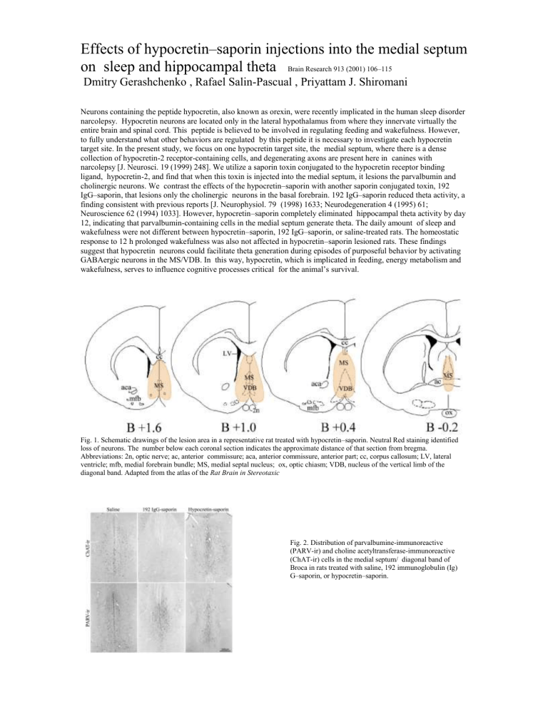
Fig. 1. Schematic drawings of the lesion area in a representative rat treated with hypocretin–saporin. Neutral Red staining identified loss of neurons. The number below each coronal section indicates the approximate distance of that section from bregma.
Abbreviations: 2n, optic nerve; ac, anterior commissure; aca, anterior commissure, anterior part; cc, corpus callosum; LV, lateral ventricle; mfb, medial forebrain bundle; MS, medial septal nucleus; ox, optic chiasm; VDB, nucleus of the vertical limb of the diagonal band. Adapted from the atlas of the Rat Brain in Stereotaxic
Fig. 2. Distribution of parvalbumine-immunoreactive
(PARV-ir) and choline acetyltransferase-immunoreactive
(ChAT-ir) cells in the medial septum/ diagonal band of
Broca in rats treated with saline, 192 immunoglobulin (Ig)
G–saporin, or hypocretin–saporin.


