AZA Koala SSP Veterinary Manual
advertisement
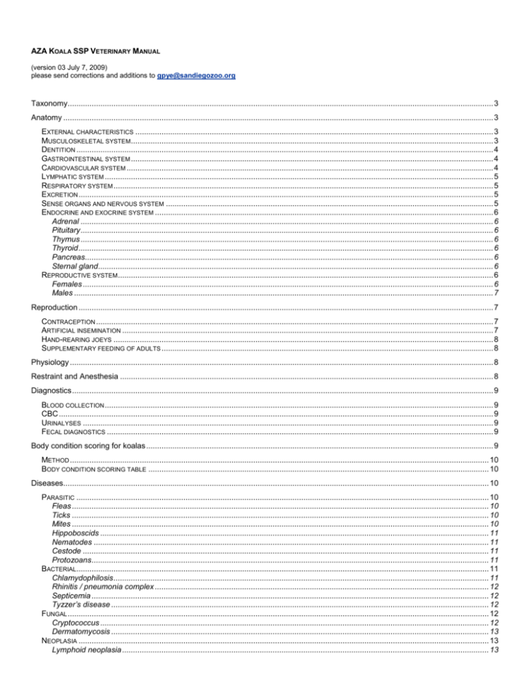
AZA KOALA SSP VETERINARY MANUAL (version 03 July 7, 2009) please send corrections and additions to gpye@sandiegozoo.org Taxonomy................................................................................................................................................................................................... 3 Anatomy ..................................................................................................................................................................................................... 3 EXTERNAL CHARACTERISTICS .................................................................................................................................................................... 3 MUSCULOSKELETAL SYSTEM ...................................................................................................................................................................... 3 DENTITION ............................................................................................................................................................................................... 4 GASTROINTESTINAL SYSTEM ...................................................................................................................................................................... 4 CARDIOVASCULAR SYSTEM ........................................................................................................................................................................ 4 LYMPHATIC SYSTEM .................................................................................................................................................................................. 5 RESPIRATORY SYSTEM .............................................................................................................................................................................. 5 EXCRETION .............................................................................................................................................................................................. 5 SENSE ORGANS AND NERVOUS SYSTEM ...................................................................................................................................................... 5 ENDOCRINE AND EXOCRINE SYSTEM ........................................................................................................................................................... 6 Adrenal ............................................................................................................................................................................................. 6 Pituitary ............................................................................................................................................................................................. 6 Thymus ............................................................................................................................................................................................. 6 Thyroid .............................................................................................................................................................................................. 6 Pancreas........................................................................................................................................................................................... 6 Sternal gland ..................................................................................................................................................................................... 6 REPRODUCTIVE SYSTEM ............................................................................................................................................................................ 6 Females ............................................................................................................................................................................................ 6 Males ................................................................................................................................................................................................ 7 Reproduction .............................................................................................................................................................................................. 7 CONTRACEPTION ...................................................................................................................................................................................... 7 ARTIFICIAL INSEMINATION .......................................................................................................................................................................... 7 HAND-REARING JOEYS .............................................................................................................................................................................. 8 SUPPLEMENTARY FEEDING OF ADULTS ........................................................................................................................................................ 8 Physiology .................................................................................................................................................................................................. 8 Restraint and Anesthesia ........................................................................................................................................................................... 8 Diagnostics ................................................................................................................................................................................................. 9 BLOOD COLLECTION .................................................................................................................................................................................. 9 CBC ....................................................................................................................................................................................................... 9 URINALYSES ............................................................................................................................................................................................ 9 FECAL DIAGNOSTICS ................................................................................................................................................................................. 9 Body condition scoring for koalas ............................................................................................................................................................... 9 METHOD ................................................................................................................................................................................................ 10 BODY CONDITION SCORING TABLE ............................................................................................................................................................ 10 Diseases................................................................................................................................................................................................... 10 PARASITIC ............................................................................................................................................................................................. 10 Fleas ............................................................................................................................................................................................... 10 Ticks ............................................................................................................................................................................................... 10 Mites ............................................................................................................................................................................................... 10 Hippoboscids .................................................................................................................................................................................. 11 Nematodes ..................................................................................................................................................................................... 11 Cestode .......................................................................................................................................................................................... 11 Protozoans ...................................................................................................................................................................................... 11 BACTERIAL............................................................................................................................................................................................. 11 Chlamydophilosis ............................................................................................................................................................................ 11 Rhinitis / pneumonia complex ......................................................................................................................................................... 12 Septicemia ...................................................................................................................................................................................... 12 Tyzzer’s disease ............................................................................................................................................................................. 12 FUNGAL ................................................................................................................................................................................................. 12 Cryptococcus .................................................................................................................................................................................. 12 Dermatomycosis ............................................................................................................................................................................. 13 NEOPLASIA ............................................................................................................................................................................................ 13 Lymphoid neoplasia ........................................................................................................................................................................ 13 Craniofacial tumors ......................................................................................................................................................................... 13 NON-INFECTIOUS .................................................................................................................................................................................... 14 Tubulointerstitial nephrosis ............................................................................................................................................................. 14 Dental disease ................................................................................................................................................................................ 14 Hip and shoulder dysplasia ............................................................................................................................................................. 14 Metabolic bone disease .................................................................................................................................................................. 17 Hypovitaminosis E .......................................................................................................................................................................... 19 DISEASES OF POUCH YOUNG .................................................................................................................................................................... 19 Pouch infection ............................................................................................................................................................................... 19 Mastitis............................................................................................................................................................................................ 19 Enteritis ........................................................................................................................................................................................... 19 Candidiasis ..................................................................................................................................................................................... 19 Neonatal hypoglycemia................................................................................................................................................................... 19 Koala Preventive Medicine Protocols (San Diego Zoo) ............................................................................................................................ 20 ROUTINE EXAMINATIONS .......................................................................................................................................................................... 20 QUARANTINE (SAN DIEGO ZOO) ............................................................................................................................................................... 20 PRESHIPMENT EXAMINATION FOR OUTGOING SHORT-TERM LOAN AND BREEDING LOAN KOALAS (AT SAN DIEGO ZOO) ........................................ 20 PRESHIPMENT EXAMINATION FOR RETURNING SHORT-TERM LOAN KOALAS (AT LOANING INSTITUTION) .............................................................. 20 Koala age determination by tooth wear .................................................................................................................................................... 21 Koala SSP necropsy protocols ................................................................................................................................................................. 23 ANATOMICAL NOTES:............................................................................................................................................................................... 23 COMMON DISEASE SYNDROMES IN CAPTIVE KOALAS ................................................................................................................................... 23 TISSUE CHECKLIST .................................................................................................................................................................................. 24 Koala hip and shoulder reporting.............................................................................................................................................................. 25 Using The Digital Camera To Photograph A Radiograph ............................................................................................................... 26 Using A Scanner To Copy A Radiograph........................................................................................................................................ 26 Koala blood SDZ Reference Ranges ....................................................................................................................................................... 27 Resources ................................................................................................................................................................................................ 28 2 Taxonomy Class: Mammalia o Infraclass: Marsupialia Order: Diprodontia Suborder: Vombatiformes o Family: Phascolarctidae Genus species: Phascolarctos cinereus Although three subspecies have been described, these are arbitrary selections from a cline and are no longer generally accepted as valid. The variation from one form to another is typically continuous, though there can be substantial differences (such as hair color) between individual koalas in any given region. Following Bergmann's Rule, southern individuals from the cooler climates are larger. A typical Victorian or Southern koala (formerly P. cinereus victor) has longer, thicker fur; is a darker, softer grey, often with chocolatebrown highlights on the back and forearms; has a more prominently light-colored ventral side; and fluffy white ear tufts. Victorian and New South Wales koala (formerly P. cinereus cinereus) weights are 12 kg for males [range 9.5-14.9 kg] and 8.5 kg for females [range 7.0-11.0 kg]. In tropical and sub-tropical Queensland, however, the Queensland or Northern koala (formerly P. cinereus adustus) is smaller (at around 6.5 kg for an average male [range 4.2-9.1 kg] and just over 5 kg for an average female [range 4.1-7.3 kg]), a lighter, often rather scruffy grey in color, and has shorter, thinner fur. Free-ranging female koalas commonly live for 13-18 years, whiles males generally live a shorter life due to the hazards encountering during the breeding seasons. Longevity in captivity is similar, though individuals have lived to 20-27 years with either supplementation or special Eucalyptus selection. Koalas are mainly nocturnal, though they will exhibit some activity during the day. Feeding usually is of 1-4 hours per day in several episodes averaging about 20 minutes each time (range 5 minutes to 1-2 hours) and this occurs typically in a 4-5 hour period around dusk. Sleeping, resting, and sitting occupy 19-20 hours per day. Changing trees or branches, grooming, or social behavior account for only a small part of each day. Anatomy External characteristics The koala is a medium to large marsupial that is the only nonhuman occupant of Australia that is a “frontal” mammal (tends to have a face rather than a muzzle). The eyes are forwardly directed and the “rhinarium” is large and apparently vertically oriented. There is a relatively high forehead and the animal is normally observed in an upright stance. These characteristics are considered to generate the empathy in humans for this species. The stout form, thick woolly fur, large thickly furred ears, and rudimentary tail superficially give koalas a resemblance to small bears. Sexual dimorphism is well developed in koalas with adult males up to 50% larger than adult females in the same population. Males have a prominent sternal gland, which is particularly conspicuous in the breeding season. Males have a well-developed pendulous scrotum located cranial to the cloaca. The pouch of the female is not usually outwardly obvious unless it contains a developing joey. The pouch of the female is formed from the invagination of the skin through the cutaneous trunchi muscle layer, whose fibers then form the “sphincter marsupii”. The hair and glandular portion of the pouch varies from the rest of the skin. There is a single pai r of mammary glands with associated teats on the dorsal surface of the pouch. When there is no occupancy by a joey, the opening is positioned centrally and there are well-developed lateral extensions into the groin on either side. As a joey grows, the increasing size distends the pouch wall such that the opening becomes distinctly backwardly directed. A bulge in the pouch is not usually noticeable until the joey is at least 3 months of age. Milk composition varies during lactation similar to other marsupials. There are virtually no subcutaneous fat deposits in koalas, but the cutaneous musculature is well developed. The fur has the highest degree of insulation known for a marsupial in still air and it maintains a high insulative value in wind. The hair density on the back (54.4 3.12 hairs/mm2) is double that of the ventral surface. In contrast, the much paler ventral fur (52.3% reflectance) is better able to reflect solar radiation than the dorsal surface (38.3% reflectance). These properties allow the koala to have considerable flexibility in regulating heat balance by postural adjustments. In koalas in good body condition or “fat”, localized subcutaneous fat may be found in the axilla and inguinal areas and inter nally around the edges of the kidney, in the splenic mesentery, and occasionally as a veneer over the lumbar muscles Musculoskeletal system The skull is very box-like in nature; deep and straight-sided. The palatine vacuities are restricted to the palatines rather than extending beyond their boundaries. The auditory bullae are prominent and alisphenoid in origin. The supratragus of the ear is small. There is virtually no inflection of the angle of the mandible and the two mandibles are firmly fused together. The spine consists of 7 cervical vertebrae, 13 thoracic vertebrae, 11 ribs, 6 lumbar vertebrae and 6-7 caudal vertebrae (vestigial tail). There are perforations in the transverse processes of the seventh cervical vertebrae and the formation of part of the ventral arch of the axis is from cartilage rather than bone. Epipubic bones (ossa marsupilia) are well developed and readily palpable in the abdominal region. The koala has proportionally very long limbs that can propel the animal rapidly over the ground and up trees. The koala walks by moving diagonally opposite limbs alternatively and runs by moving the forelimbs and then the hindlimbs in unison. When climbing, the hands are released 3 and the body is thrust upward by extending the hindlimbs, permitting the hands to clasp at a new level. There is no patella (neither bone nor cartilage). The deltoid muscle is completely undivided, the cleido-occipital muscle is absent, and the omo-atlantic muscle has only a single insertion on the outer part of the spine of the scapula. Despite being an arboreal marsupial, there is no prehensile tail. Instead there are adaptations of the feet: All digits except the broad, shorter hallux are equipped with large, strong and recurved claws. The syndactylous digits (II and III) on the pes are quite large, but the 4th digit is longer and more powerful than rest. The manus is relatively very large and the first two digits are opposable to the other three, giving rise to a hand with two “thumbs”. Except for the apical pads, most of the palmar and plantar surfaces are granulated, rather than having striations and ridges and lack well-differentiated pads. Koalas never descend a tree head first, but they may leap in a more or less upright posture from branch to branch. Dentition The dental formula for koalas is I 3/1, C 1/0, PM 1/1, M4/4. There are 3 pairs of incisors in the upper jaw and 1 pair of incisors in the lower jaw. There are no first or second premolars. All teeth have roots that are not persistently growing. There are cheek pouches. The longitudinal cutting ridge of the premolar and the pyramidal cusps of the molars provide a cutting-shearing action on the Eucalyptus leaves. There is no crushing component in the occusal action, thus as the premolars and molars wear down the efficiency of mastication is reduced leading to larger leaf fragments passing through the gastrointestinal tract requiring these animal to eat significantly more to achieve the same caloric uptake, this in turn accelerates tooth wear. Life span is related to tooth wear. Gastrointestinal system Koalas are monogastric hindgut fermenters. The most notable feature of the alimentary tract is the extraordinary development of the cecum, which is claimed to be the most capacious, in a relative size, among mammals. In spite of its size, it is not particularly specialized. The mucosa is smooth and raised in 11 or 12 folds, which run down its length into the proximal colon. The colon is also capacious and long, although only about half the length of the cecum and it continues into a much narrower distal colon, where fecal pellet formation occurs. It has been suggested that the total daily volatile fatty acid production accounts only for 9% of the digestible energy intake, which suggests that post-gastric fermentation does not contribute greatly to energy production. This suggests that the contents of the cell, rather than cell wall, is the principle source of energy and that the enormous cecum has some other function other than as a site for microbial digestion of plant fiber. Digesta reaching the junction of the small intestine with the cecum and proximal colon divides into two fractions: large particles pass down the proximal colon and are excreted rapidly (average 99 hours) while the solute fraction and small particles are retained in the cecum and proximal colon for a much longer period (average 213 hours). It is unknown why the solute and small particles are retained, but it suggests some dependence upon these fractions and emphasizes the importance of fine communition of leaves by the teeth. Fecal flora has been shown to change in response to seasonal change, differing body condition, and systemic antibiotic therapy. The stomach is small in relation to rest of tract and, except for the “gastric gland”, has no distinctive features. The gastric gland measures about 40 mm in diameter and lies distal to the esophageal opening on the lesser curvature of the stomach. It contains 25 or so crypts which are presumed to secrete gastric juice into the lumen of the stomach. The stomach contents should be fairly dry, densely packed, finely chewed leaf material. Koalas are eucalyptus foliage eaters and they detoxify volatile oils in the liver and then excrete them in the urine following conjugation with glucuronic acid. In young koalas, the liver is a dull red color, similar to other mammals. As koalas age, the liver color will change as a pigment accumulates turning it a very deep purplish- or blue-red color. The linea alba is broad and almost translucent. There is no curtain-like omentum. Cardiovascular system The heart is fairly typical of other mammals and is about 50 mm long and relatively rounded at the apex which is positioned level with the caudal border of the fourth left costal cartilage. As in most marsupials, right auricular extensions encompass the aorta and there is no trace of a fossa ovalis. The right atrioventricular valve appears to have one very large non-septal cusp. There is no obvious phrenicopericardiac ligament so the pericardium appears to be directly adhered to the diaphragm. Resting heart rates are reported between 60-90 beats per minute. The pattern of branching of the conducting arteries of the aortic arch appears to be variable in koalas with various descriptions including a brachiocephalic trunk (from which branches the two carotids and right subclavian artery) followed by the left subclavian artery; a brachiocephalic trunk (from which branches the right carotid and subclavian) followed by the left carotid and left subclavian arteries; and the right subclavian, right common carotid, and left common carotid branching independently from the aorta in close proximity, followed shortly thereafter by the left subclavian. The origins of gonadal and iliac vessels from the abdominal aorta usually differ between marsupials and eutherian mammals. A common coeliacomesenteric arterial trunk is a feature of koalas. The most significant characteristic of the venous system of koalas is the location of the caudal vena cava. It runs to the right of the abdominal aorta and thus resembles the pattern of the eutherians, rather than the pattern typical of marsupials in which the caudal vena cava runs ventral to and obscures the abdominal aorta. It is normal to find small numbers of reticulocytes, cells containing Howell Jolly bodies, and nucleated red blood cells. Anisocytosis and poikilocytosis also appear to be common in blood smears from koalas. Koalas have a very long mean prothrombin time (60.2 11.6 4 sec) and an inability of streptokinase to activate koala plasminogen, therefore clotting in koalas may differ from the pattern described in eutherians. The spleen is variable in shape and of all the Australian marsupials it conforms the least to the forked pattern that is rather distinctive of the group. Lymphatic system The superficial inguinal, superficial axillary, rostral mandibular, mandibular, and facial lymph nodes are palpable in healthy koalas. Respiratory system Koalas have an obvious and striking nose. The external appearance is deceptive because the nasal cavity is actually quite small with the nasal conchae being simple in form. Most of the space is taken up by large maxillary and frontal sinuses, of which there is no readily apparent function. There is an absence of swell bodies (thought to alternate air flow between the right and left nasal passages in most mammals) and sparse development of secretory glandular tissues, which normally modify temperature and humidity of inspired air. This lack of glandular tissue may be a mechanism to help water conservation in this species. Nevertheless, it has been found that evaporative water loss, principally from the respiratory system is the main mechanism for maintenance of stable body temperatures when koalas are exposed to high ambient temperatures. A normal resting respiratory rate of 10-11 breaths per minute can rise to 230 breaths per minutes when panting in response to elevated ambient temperatures; normal body temperatures range from 35.5 – 36.5 C in unrestrained animals. For captive koalas, respiration rates of 15-30 breaths per minutes and a body temperature of 36.7 C as being normal. Pyrexia is not normally noted with illness; more commonly hypothermia is observed with temperatures as low as 29 C. The lack of lateral nasal glands and maxillary sinus glands and therefore the lack of immunoglobulin A that these glands secr ete may be of significance as a predisposing factor in the susceptibility of this species to respiratory infections. The arrangement of the nasopharynx in koalas is unusual and the location of the larynx makes it more difficult to place an endotracheal tube. There is an unusual caudal prolongation of the soft palate which opens by quite a small orifice in close proximity to the esophagus, more or less at right angles to the axes of the esophagus and trachea. This arrangement makes it difficult to envisage how the large epiglottis functions and may be related to the habit of these animals of stretching their necks back, so that the long axis of the head parallels the body, when making the bellowing grunt typical of males in the breeding season. The other common vocalization of koalas is a rather shrill cry most often heard from adult females or juveniles when alarmed or distressed and is usually uttered with the head at its normal perpendicular angle to the body. The thorax capacity is very small in comparison to that of the abdomen. The lungs are composed three right lobes and two left lobes. The lower lobes of each side are about equal in size and half as big again as the upper, or two upper lobes. Some report two right lobes and two left lobes. The mediastinum is intact, so pathologic conditions may be limited to one side of the thorax. Excretion The kidneys are about 35 mm long, 20 mm wide, and 15 mm thick, with the right kidney lying completely craniad to the left. The kidneys are unilobar and have a single shallow papilla. There venous plexuses associated with the ureters and the urinary bladder is typical of mammals. Renal features which characterize koalas are the relatively wide cortex and outer medulla and the highly organized nature of the blood vessels and capillary networks in these regions. Medullary rays are very clearly defined. Interlobular vessels arise perpendicularly from the arcuates and follow straight paths to the capsule. Although this is a typical feature of mammalian kidneys, most marsupials have a more haphazard arrangement of blood vessels. Glomerular and tubular dimensions are similar to other marsupials and proximal and distal tubules intertwine in tight convultions close to the parent glomerulus. Although the renal papilla is shallow, the loops of Henle transverse its full depth and koalas can produce urine more concentrated than humans, but less concentrated than is typical of rodents and carnivores. Sense organs and nervous system The eyes of koalas are unusual. The pupils are vertical slits rather than circular as in other marsupials. The iris is typically brown, but may be blue in some individuals (or unpigmented in albinos). The cornea makes up a large segment of the globe with the diameter being only slightly smaller than the globe diameter. The retina is avascular except for a small area on the optic disc. A tapetum extends across the retina above the optic disk. The mean intraocular pressure is 24.2 ± 6 mmHg. The nictating membrane is positioned in the medioventral portion of the socket and sweeps in a nasal to temporal manner. The lacrimal puncta are slit-like openings that are positioned just inside the eyelids, approximately 2-3 mm from the medial canthus. The opening of the lacrimal duct (0.5-1.0 mm) is found on the ventrolateral aspect of the nasal floor at the mucocutaneous junction. In view of the observation that captive individuals sniff the petiole of many leaves before taking them into their mouths, it is surprising that the olfactory epithelium is not as extensive as that found in a range of other marsupials and no separate septal olfactory organ is 5 present in koalas. A vomeronasal organ with a patent duct is present, but it is only moderately developed and is most likely involved with pheremonal signals associated with reproduction or social organization. All sensory vibrissae are at best poorly developed. The brain is exceptionally small in size and the cerebral hemispheres are extremely smooth. Not only are the cerebral hemispheres reduced in size so that they fail to cover much of the brain stem and are lissencephalic, but the brain is also some 25-30% smaller than the endocranium that houses it. The histologic appearance is as expected. Endocrine and exocrine system Adrenal Grossly proportionally smaller than other mammals: 10 mm long, 8 mm wide, and 5 mm deep, and about 0.25 g in weight. The ratio of adrenal to body weight is about 0.006% which is less than half that typical of mammals (data from males only). The right glan d is usually smaller than left and concealed under lobes of the liver and more or less adherent to the caudal vena cava. Histological appearance is typical of other mammals. The koala adrenal gland responds to the challenge of disease in the usual way: a broadening of the zona fasculata, depletion of the lipid in this zone, and overall enlargement of the gland with frequent formation of cortical nodules. The peripheral blood of minimally stressed koalas has almost no cortisol (less than 0.1 ng/ml), with fairly small amounts of corticosterone (0.7 ng/ml) and 11-deoxycortisol (0.1 ng/ml). In response to trophic stimulation, the major response is a dramatic increase in cortisol (7 ng/ml) with comparatively minor responses in other glucocorticoids. The mineralocorticoid detected in koalas is aldosterone (0.06 ng/ml) which is comparable to other marsupials. It is suggested that koalas have low peripheral blood corticosteroids concentrations and a relatively poor response to acute stress and trophic stimulation. Pituitary Apart from a finding of antidiuretic function, very little is known of the structure / function relationships of either the adenohypophysis or the neurohypophysis. Thymus Both superficial cervical and thoracic lobes may be present in koalas, although thoracic lobes are usually absent or very small. This is opposite to eutherians and other marsupials (only thoracic component present), except for the wombats (no thoracic component) and phalangeroids (both superficial cervical and thoracic lobes are well represented). Thyroid The thyroid consists of two lobes about 25 mm long, 5 mm wide, and 3 mm deep. Typically, it lies between the 4 th and 9th tracheal rings and it lacks an isthmus. The right lobe tends to exhibit more variability in size and location than the left and accessory thyroid tissue may be present. Blood supply is from the common carotid artery and venous return via the internal jugular vein. Pancreas The pancreas is typically of mammals (pale or dull pink), except for the presence of a 20 mm long, thin-walled, saccular dilation of the pancreatic duct prior to its entry into the duodenum in close proximity of the common bile duct. Sternal gland This gland is present in males and can be up to 85 mm long x 50 mm wide and contains both sudoriferous and sebaceous components. It lacks underfur on its surface. The gland undergoes seasonal changes in activity and is presumably under androgenic control. A range of fatty acid derivatives, monoterpenes, and sesquiterpenes has been identified as secretory products (some of these compounds are possibly contaminants from eucalyptus leaf diet, rather than endogenous). Reproductive system Females The female reproductive tract has 2 ovaries, 2 oviducts, and 2 completely separate uteri and cervices. The ovaries tend to be flattened and their enveloping fimbria are located within small peritoneal diverticula that has a small opening, the peritoneal ostium, in the posterior wall. The Fallopian tubes are not very convoluted and there is a relatively slight and gradual change in diameter as they join the duplex uteri. The uteri do not fuse (as they do in eutherians) because the ureters course medial to them. The uterine cervices are well-developed and easily palpable through the wall of the vaginae. Each uterus opens into a vaginal cul-de-sac, separated by a median septum, from which spring 2 separate lateral vaginas about 5 mm caudal to the each cervix. The lateral vaginal empty into the urogenital sinus, which also receives the urethra. This then connects to the cloaca, anterior to the rectum and caudal to the bifid clitoris, a distinctly reddish triangular structure. The lateral vaginas are separated by the urogenital strand – a mass of fibrous connective tissue – that forms a pseudovaginal canal at parturition – connecting the vaginal cul-de-sac to the urogenital sinus. The pseudovaginal canal 6 disappears and then reforms at the next parturition (painful!!). The bilobed clitoris is relatively prominent (about 5 mm long) on the ventral surface of the outlet of the urogenital canal. Koalas are one of the few marsupials to develop a chorioallantoic placenta (usually considered the hallmark of eutherians). The allantois has an extensive area of fusion with the chorion, but no chorionic villi form and there is no fusion with the endometrium in this region. Males The male reproductive tract is of fairly typical form, with a relatively small unlobed cordate prostate (encircles the urethra immediately caudal to the bladder), three pairs of Cowper’s glands (bulbourethral glands) and a distinctly bifid penis with each half having a grooved appearance that in turn gives the penis a four-pronged appearance. Slightly retroverted cornified spines are found on the penile shaft. Reproduction The diploid chromosome number of koalas is 16 with XY / XX sex determination mechanism. Koalas are monovular and seasonally polyestrus with a gestation period that is shorter than the estrus cycle. Ovulation is induced by copulation (either copulation or factors in the semen induce this) and there is no embryonic diapause. The mean duration of non-mated estrus cycles is 332.9 ± 1.1 days. The mean duration of estrus for non-mated cycles is 10.3 ± 0.9 days. The inter-estrus interval for mated, non-parturient estrus cycles (with a luteal phase) is 52.5 ± 0.8 days. Behavioral estrus in captive koalas is characterized by weight loss, vocalization, restlessness, pseudomale mounting of cage mates, and episodes of “hic-coughing” or convulsive jerking, particularly in response to the presence or call of a male. Gestation lasts 34.09 ± 0.53 days (range 32.98-35.64 days). Parturition causes some discomfort, but it is rapid. It takes 30-90 seconds between emergence from the cloaca to entry into the pouch. Blood, fluid, or membranes may be seen in the fur around the cloaca and pouch, but not always. At most, females normally give birth to one young per year and most births occur during the summer. Twin births have been recorded and early pouch emergence may occur due to pouch-size limitations. Males mate with any receptive female without elaborate courtship. Coitus usually takes place in a tree and a plug of coagulated semen may occlude the urogenital tract of the canal of the female following copulation. Typically both sexes appear to reach sexual maturity at two years of age, though the age at which first mating occurs may vary. Following birth, the offspring spend the first six months of life in the pouch feeding on milk. When the pouch young first starts to poke its head out of the pouch at about 5 months of age, it is fed on “pap”, a soft green fluid produced by the female and it is considered to be the evacuated contents of the cecum and this acts as an inoculum for the young’s cecum. The pap coincides with a phase of exponential growth, eruption of the first teeth, and the ingestion of leaves by the young. The young stays in close association with its mother after it vacates the pouch until at least 11 months of age, at which time it is about half grown. It may disperse at this time or continue to associate with its mother for up to another year. Juvenile males usually disperse before females. Free-ranging male koalas are sexually mature at approximately 2 years of age, but competition usually restricts mating until 4-5 years of age. There is a seasonal increase in testosterone concentrations. Free-ranging female koalas commence breeding late in their second year or early in their third year and can produce one offspring each year, up until 14 years of age. Ovulation can be induced in koalas by the administration of 250 IU hCG IM (Johnston et al, 2000). This can be used fir females with prolonged estrus that cannot be mated. Contraception Vasectomy and cauterization of the oviducts via laparoscopy have been performed in koalas. Deslorelin implants: Females remain infertile for one year. Does not appear to affect male fertility. See Herbert CA, Webkey LS, Trigg TE, Francis K, Lunney DH, Cooper DW. 2001. Preliminary trails of the GnRH agonist deslorelin as a safe, long-acting an reversible contraceptive for koalas. Conference on the Status of the Koala in 2001. The National Koala Act. Canberra, November 2001. Artificial insemination Artificial insemination with chilled-extended semen has been successfully performed in koalas. Semen chilled up to 3 days and inseminated up to day 8 of estrus produced pouch young at similar percentages to natural mating (~50%). Semen is collected by electroejaculation with males koalas under anesthesia. Insemination per cloaca is performed with females held with manual restraint. Cryopreservation, estrus synchronization, and ovulation induction are currently being investigated. See: Johnston SD, McGowan MR, Phillips NJ, O'Callaghan P. Optimal physicochemical conditions for the manipulation and short-term preservation of koala (Phascolarctos cinereus) spermatozoa. J Reprod Fertil 2000a, 118:273-81. Johnston SD, McGowan MR, O'Callaghan P, Cox R, Nicolson V. Natural and artificial methods for inducing the luteal phase in the koala (Phascolarctos cinereus). J Reprod Fertil. 2000b, 120:59-64. Allen CD, Burridge M, Mulhall S, Chafer ML, Nicolson VN, Pyne M, Zee YP, Jago SC, Lundie-Jenkins G, Holt WV, Carrick FN, Curlewis JD, Lisle AT, Johnston SD. Successful artificial insemination in the koala (Phascolarctos cinereus) using extended and extended-chilled semen collected by electroejaculation. Biol Reprod 2008, 78:661-6. Zee YP, Holt WV, Gosalvez 7 J, Allen CD, Nicolson V, Pyne M, Burridge M, Carrick FN, Johnston SD. Dimethylacetamide can be used as an alternative to glycerol for the successful cryopreservation of koala (Phascolarctos cinereus) spermatozoa. Reprod Fertil Dev 2008, 20:724-33. Hand-rearing joeys For hand-raising of koala joeys, use low-lactose milk formula and establish an environment emphasizing familiarity and security (one care giver and avoid sudden changes in routines). Milk composition changes throughout lactation and so follow Husbandry Guidelines for hand-raising joeys. Road-kill fresh cecal contents can be used as substitute pap (start when first molars begin to erupt). Supplementary feeding of adults SDZ uses Portagen to supply additional calories to adult koalas. Contact SDZ for more information. Physiology Koalas are arboreal folivorous marsupials that derive food, water, and shelter almost exclusively from Eucalyptus trees. Eucalyptus tress discourage folivory with fibrous leaves containing toxic phenolics and oils, but koalas have adapted with specialized dentition, efficient hepatic detoxification mechanisms, and prolonged retention time in the proximal colon and elongated cecum where bacterial fermentation and tannin-protein complex degradation occur. Energy is conserved with a low basal metabolic rate and much inactivity. Marsupials have a lower basal metabolic rate when compared to placental mammals (eutherians). The basal metabolic rates of marsupials are generally 65-74% of the value expected from an equivalent body mass in eutherians. Koalas and wombats have even lower rates than other marsupials. Basic metabolic rate (kcal/d) = 70.5 x body weight 0.49 This reduction in basal metabolic rate results in their core body temperatures also being lower (2-3C less). Koalas appeared to be able to maintain body temperatures between 35.5-36.5°C when exposed to environmental temperatures of 4.530°C. When air temperature is 40°C, then body temperature may rise to 37.0°C. Panting is the major means of evaporative cooling. Koalas expend minimal energy on thermoregulation when held in ambient temperatures between 10-15° and 25°C. Koalas are estimated to have mean food ingested (wet matter) amounts within the range of 64.2-102 g/kg0.75/d. Therefore northern koalas would be expected to eat approximately 220-415 g of leaf per day. Victorian koalas would consume approximately 320-660 g of leaf per day. Free-ranging koalas rarely drink as moisture in or on leaf provides adequately for their needs. Estimates of water needs for koalas range from 45-70 g/kg/day. Severe tooth wear in old age is the principal factor in limiting longevity of otherwise healthy koalas. Body condition should be assessed by palpation of the contour and bulk of the scapular muscles (See body condition scoring). Koalas have inelastic skin so hydration is better assessed by the ease of the skin sliding over the scapula. Restraint and Anesthesia For captive koalas, many simple procedures (e.g. blood collection from a cephalic vein) can be performed without restraint with the animal sitting in the fork of a tree. If restraint is needed, then the minimum required appears to work best. One of two techniques can be used. With the koala at a height where it can be grabbed by the handler, the forelimbs are grasped at the carpus and removed from the tree. The animal is then pulled from the tree and sat in the crook of one arm. For inhalant anesthesia induction, the koala is sat on the table with one handler holding the forelimbs at the carpus and a second handler holding the rear limbs at the hock. This technique has been used routinely at San Diego Zoo for many years. Alternatively, the forelimbs are grasped at the carpus and the koala is pulled from the tree and lowered to the ground, where the hindlimbs are incorporated into the hand grasp at the hock. The koala is now restrained with a wrist and ankle in each hand, taking care not to allow the koala’s mouth into range of the handler. Individual koalas appear to become accustomed to the particular manner of restraint that they are used to. Captive koalas are induced with isoflurane using a medium sized dog facemask while held in the manner described above. Diazepam at 0.25 mg/kg IV (cephalic vein) or midazolam at 0.1-0.25 mg/kg IV or 0.5 mg/kg IM can be given as premedicants to reduce stress, struggling, and the risk of excessive catecholamine release. Prolonged pre-anesthetic fasting is not necessary as koalas are not prone to regurgitation – short duration fasting is recommended to ensure there is no food material in the mouth at the time of induction and intubation. All koalas should be intubated independent of the anticipated time of the exam. Once lain down under anesthesia, the chance of the palatine tissues obstructing the larynx / glottis is high. While sitting up for the intubation process, obstruction is much less likely and, with practice, intubation is not overly difficult. With a long bladed laryngoscope, the glottis is visualized when the soft palate is elevated. Positioning of the koala and the laryngoscope blade can aid visualization and the endotracheal tube can be used to move the palatine tissues away from the glottis. Visualization of the glottis may be lost when the endotracheal tube (3-5 mm) is passed. The use of a stylet to help keep the endotracheal tube straight can help in passing it. Alternatively, a small diameter polypropylene urinary catheter is visually placed first with the aid of the laryngoscope and the endotracheal tube blindly passed using the catheter as a guide. 8 Auscult the chest frequently with every case to establish that air is moving and you're not just looking at chest excursions. Koalas are held in an upright manner during recovery and after extubation (until able to grasp) to avoid soft palate entrapment of the glottis. Koalas are not given access to trees during recovery, due to their inclination to climb while still ataxic. A small crate or cage with an upright Y branch for the koala to grip and sit in during recovery is ideal. Once fully recovered the koala is returned to its usual enclosure. Emergency intervention for upper airway / pharyngeal obstruction should be available. Use a large bore IV catheter (16 or 14 gauge) ready to stab into the trachea at about the level of the thoracic inlet (palpate this early on during induction so you know where it is in case you have to intervene). If so, stab, pull out the trochar, pull the oxygen hose off the T-piece, attach an adaptor to the hose, connect it to the catheter and give 100% oxygen at 2-4 liters per minute, discontinue anesthesia, and then observe the animal for evidence of reapposition of the nasopharynx and glottis (they stick their tongues out and do an odd chewing motion for a few seconds when they are doing this). Auscultate the chest during this to ensure good air movement. The animal will have excess oxygen "blow by" going out the nose/mouth during this, but their oxygen saturation will remain high. Remove the catheter once the animal is stable. Cases of CNS signs post-recovery from anesthesia have been seen in a few koalas. Animals became increasely agitated during recovery and developed CNS signs (prolonged nystagmus, opisthotonus, tremoring) as well tachypnea and tachycardiac. The cause is unknown and this syndrome can be fatal. Intensive care can result in recovery, though prolonged, in some cases. Remember koalas are armed with sharp claws that can inflict severe wounds and care is taken to securely restrain the feet. In addition, they can also inflict a nasty bite that will easily penetrate through the palm of a human hand. To remove a wild koala from a tree, the animal can be coaxed down the tree by waving a flag on a long stick immediately above its head. When the koala is at a suitable height for grabbing, scruff the koala at the back of the neck with one hand and dorsally over the pelvis with the other hand to remove the animal from the tree. The animal is then forced ventrally to the ground until placed into a suitable crate or hessian sack. This protects the handler from being bitten or scratched. In wild koalas, tiletamine / zolazepam at 5.0 – 7.7 mg/kg IM results in a rapid and smooth induction and anesthesia for a period of 30-45 minutes. Alphaxolone / alphadolone at doses of 3-6 mg/kg IM or 1.2-1.8 mg/kg IV provides good immobilization for periods up to 30 minutes. Diagnostics Blood collection Blood may be collected from the cephalic vein in conscious koalas while they are sitting comfortably in the fork of a tree. The vein may be raised by gently encircling the arm at the elbow with one hand, while drawing the blood with the other hand. Alternatively a rubber band and a hemostat work effectively as a tourniquet. Blood may also be collected from the femoral vein, but this is typically only done with the animal under anesthesia. CBC Lymphocyte percentages commonly exceed neutrophil percentages in healthy animals – an inversed ratio is an indication of an inflammatory response. Nucleated red blood cells are seen commonly in healthy koalas. Urinalyses Koalas can have false positive ketones, glucoses, and proteins on urine dipsticks. Normal koalas sometimes pass red pigment in the urine. Calcium carbonate-like crystals and unidentified small rectangular crystals were seen in urine samples from 12 of 20 normal koalas. Fecal diagnostics Koalas pass fecal pellets that are khaki to olive green that taper at both ends. Normal koalas sometimes pass red pigment in the urine and mauve or grey fecal pellets. 90-200 pellets are typically passed in a 24-hr period. Pap is the soft feces produced by the dam and fed to the newly-emergent pouch young. It is consumed intermittently for 1-5 weeks and the pouch young will eat its first eucalyptus leaf a few days after first pap feeding. The pap inoculates the juvenile’s proximal colon and cecum with tannin-protein complex degrading enterobacteria. Koalas at SDZ frequently have Saccharomyces-like yeast in their fecal samples and it is considered part of the normal flora. Body condition scoring for koalas Body condition in koalas is estimated by palpating the muscle mass over the scapula. Although changes in muscle mass will also be apparent on palpation of other areas (e.g.: limb muscles and muscles of mastication) it is more difficult to accurately measure body condition and changes in body condition using other sites. 9 Koalas infrequently become overweight and often have virtually no subcutaneous fat. Only koalas fed a significant amount of energyrich, non-eucalypt nutrient in their diet run the risk of becoming significantly overweight. A “normal” koala in excellent body condition will still feel “boney” if palpated over the ribs or hips. The body condition score should be assessed by palpating the muscles over the scapula. Ribs will feel prominent in both healthy and emaciated koalas. Method Place a hand across the koala’s shoulders. Locate the spine of the scapula, which runs from the top of the shoulder down towards the upper arm. The spine of the scapula is about 10 cm long in an adult koala and feels like the keel bone of a bird. The scapula itself extends like a plate on either side of the spine. The muscles of the scapula lie on either side of the scapula spine, and generally cover the whole of the scapula, so that the scapula underneath can only be felt in very thin animals. Palpate the muscles to the front and to the back of the scapula spine firmly with your fingers. Palpate both the length of the muscle (along the length of the scapula spine) and across the width of the muscle (from the scapula spine to the edge of the muscle). Judge the roundness of the muscles and also the tone of the muscles. A koala in excellent body condition will have round, firm muscles, which bulge in a convex fashion (outward) on either side of the spine of the scapula. As body condition deteriorates the muscles become smaller, flatter and eventually convex (dishing inwards) on either side of the spine of the scapula (very similar to pectoral muscles in birds). In a koala in poorer condition, the spine of the scapula becomes more prominent and easily palpable, and muscle tone decreases. The scapula itself will be palpable on either side of the scapula spine in very thin animals. Body condition scoring table Strong muscle tone. Obviously convex muscle masses on either side of the scapula. scapula spine palpable on careful palpation. Score 5 Excellent Score 4 Good Score 3 Fair Flat to slightly convex muscles on either side of the scapula. Scapula spine prominent on palpation. Score 2 Poor Slight dishing or concave muscles on either side of scapula. Scapula spine very obvious on palpation. Edges of scapula bone palpable. Score 1 Emaciated Noticeable dishing of muscles on either side of scapula. In very emaciated animals there may be almost no muscles palpable on either side of the scapula spine. In these cases the entire scapula will be palpable through the skin. Good muscle tone. Slightly convex muscles on either side of the scapula. Scapula spine easily palpable. Diseases Parasitic Fleas Ctenocephalides felis is occasionally found on debilitated or orphaned wild koalas being cared for in private dwellings Ticks Ixodes holocyclus, Ixodes cornuatus, Ixodes hirsti, Haemophysalis bancrofti, and Haemophysalis longicornis have been reported in koalas. Moderate numbers unlikely to cause harm, but heavy burdens may cause anemia. Tick paralysis (Ixodes spp.) may occur in naïve koalas translocated from paralysis tick-free areas to endemic areas. Mites Demodex spp. Have been reported in brown antechinus, tiger quoll, and koalas. Skin nodules are seen in dasyurids, but periocular alopecia is reported in the koala. Mites in the hair follicles cause enlargement and ulceration. Ivermectin has been used for treatment. Sarcoptes scabei var. wombati is reported in wombats and koalas. It is not common in koalas, there are only a couple of reports. It can cause mange lesions especially front paws. Ivermectin, moxidectin, and acaricidal baths are recommended treatments. Austrochirus perkinsi is a small fur dwelling mite that is possibly host specific and causes no pathology. Notoedres cati has only been reported once as causing raised scabby lesions on the ventrum of captive koalas. 10 Hippoboscids Hippoboscid spp. are reported in all marsupials. Typically there are no clinical signs and they reside in fur. They are a possible trypanasome vector. Topical insecticides will treat and control if needed. Nematodes Marsupostrongylus longilarvatus is a lungworm that has been reported only once in a koala. It caused a mild interstitial pneumonia with no gross lesions and histology found adults in airways. Durikainemia spp. is an incidental finding in koalas and tightly spiraled worms are seen in the pulmonary arteries. Breinlia mundayi has been only reported once in koala. Two filaroid nematodes were found in free in peritoneal cavity. This same koala also had a filaroid nematode, Johnstonema sp., in the left ventricle. Cestode Bertiella obesa can cause debilitation if large numbers of the cestodes is present. It causes mild enteritis and has an arthropod intermediate host. Protozoans Coccidia has been reported as an incidental finding on one koala. Cryptosporidium sp. has been reported to have caused four mortalities with duodenitis, enteritis and colitis. Toxoplasma gondii can affect all marsupials, but is not a significant disease in koalas; only three mortalities have been reported. Transmission is from cat feces, eating the flesh of another animal that contains the parasite encysted in its muscle, or eating infected earthworms, flies, and cockroaches. Clinical signs in marsupials can include: sudden death, blindness, neurological signs, depression, anorexia, fever, and dyspnea. In koalas, clinical signs seen include: sudden death, acute tachypnea, tachycardiac, pyrexia, and lymphocytosis with disseminated infection. At necropsy, there may be no gross lesions or myocardial hemorrhage, splenomegaly, and gastric ulceration may be seen. Histopathology may show multiple foci of necrosis in the lung, myocardium, skeletal muscle, lymphoid tissue, CNS, adrenal gland, pancreas, and liver. Tissue cysts are most common in brain, muscle, and adrenal gland. Treatment is often unsuccessful, but clindamycin, TMS, and atovaquone have been used in marsupials. Prevention is best, so prevent access to cat feces and vectors (cockroaches and earthworms). Cook or freeze meat for carnivorous marsupials. Toxoplasmosis is zoonotic. Bacterial Chlamydophilosis Chlamydophilosis can be caused by Chlamydophila pneumoniae and Chlamydophila pecorum in koalas. Both can cause ocular and urogenital disease. C. pecorum appears to be more prevalent and more virulent than C. pneumoniae. Combined infections suggest that cross-immunity does not occur. Three main clinical syndromes are seen: Keratoconjunctivitis Epiphora and erythema followed by chemosis Mucopurulent conjunctivitis, keratitis, and blindness Granular hyperplasia of the conjunctiva and nictating membrane and mucopurulent discharge may occlude the entire palpebral fissure Cystitis (dirty-tail disease) Infection of the bladder mucosa causes dysuria and incontinence Orange-brown staining of fur around rump, dysuria, and hematuria Thickened bladder mucosa with scattered petechial hemorrhage or it may be deeply ulcerated Cystitis, pyelonephritis, hydronephrosis, and renal fibrosis Urethritis, cloacitis, ascending bacterial pyelonephritis Infertility Unilateral or bilateral cystic ovarian diverticulus may cause infertility in the presence of normal ovaries Cystic enlargement of the ovarian bursae Salpingitis, hydrosalpinx, metritis, pyometra, vaginitis, and pyovagina o Often with secondary bacterial infection Chlamydophila is transmitted by discharges with the most likely route being venereal based on the extremely low incidence of infection in sexually immature koalas. Other possible other routes of transmission include: fecal-oral from dam to young (pap), arthropods (eyes), 11 parturition, and close contact. Asymptomatic carriers likely occur because the prevalence of clinical disease is less than the prevalence of infection in both free-living and captive populations. The incubation period for conjunctivitis is 7-19 days. The incubation period for urogenital disease is 8-119 days. There are no consistent hematologic or biochemical abnormalities, but anemia and hypoproteinemia may occur. The urine may contain erythrocytes, leukocytes, cellular debris, bacteria, and / or yeasts. Advanced cysts or reproductive tract anomalies may detected by palpation, negative contrast radiography (pneumoperitoneum) or ultrasonography. Cell culture is used to demonstrate the presence of viable organisms. This requires specific cell transport media, aluminum-shafted swabs that are non-toxic to the organism, and vigorous swabbing twice. Chlamydophila serology can be used. A rabbit anti-koala immunglobulin G ELISA is more sensitive than CFT (sheep antigen) and also antigen ELISA - antigen detection via immunofluorescence. PCR can also be used to detect Chlamydophila DNA. For histology diagnosis, a Giemsa or Macchiavello stain can be used to detect elementary bodies. Alternatively immunostaining may be performed. For treatment, oxytetracycline and erythromycin are not recommended due to weight loss associated with their use in koalas. This is possibly bacterial flora related. Tetracyclines have been used in koalas when supplementary fed at the same time to counteract weight loss. Doxycycline IM, chloramphenicol SQ, enrofloxacin PO or ciprofloxacin PO have been used in koalas. For prevention of introduction into a closed captive colony, fly screen koala quarantine facilities and use glutaraldehyde or chloramines disinfectants. There is no zoonotic evidence. Rhinitis / pneumonia complex Captive colonies may experience outbreaks of rhinitis / pneumonia complex. Primary or secondary pathogens isolated during these outbreaks have included: Bordetella bronchiseptica, Pseudomonas aeruginosa, Other Pseudomonas spp., Alpha-hemolytic Streptococcus spp., Streptobacillus moniliformis, Corynebacterium spp., Enterobacter agglomerans, Proteus spp., Diphtheroids, Pasteurella spp., Micrococcus spp., Staphylococcus aureus, Staphylococcus epidermidis, Escherichia coli, Acinetobacter lwoffi, Cryptococcus neoformans, Nocardia asteroids, Aspergillus spp., and unidentified yeasts. Viruses may possibly also play a role. Clinical signs include: frequent or paroxysmal sneezing or coughing, unilateral or bilateral mucopurulent nasal discharge, pharyngeal inflammation, regional lymph node enlargement (submandibular, facial), stridor, nasopharyngeal swelling, bronchitis, peracute bronchopneumonia, and sudden death. Diagnosis is based radiography because it can be difficult to percuss or auscultate the chest. Treatment is based on culture and sensitivity and nebulization may be better than systemic. In some koala colonies, vaccination with an inactivated cell-free extract of Bordetella bronchiseptica is used with an initial series of two, 4 weeks apart, and then annual boosters. Septicemia Gram-negative pathogens most commonly isolated from koalas include: Salmonella typhimurium, Salmonella sachsenwald, Salmonella bovis-morbificans, Morganella morganii, and Escherichia coli (mixed with Pseudomonas aeruginosa or Aeromonas hydrophila). The possible route of infection is ingestion of contaminated leaves and so cut browse should be prevented from touching the ground wherever possible. Septicemia caused by E coli in emergent pouch young has been associated with recent pap feeding. Clinical signs include: peracute illness with neurologic signs or sudden death, lethargy, ataxia, nystagmus, flaccidity, localized tremor, convulsions, and vocalizations may occur. Profound leukopenia (as low as 0.1 10 9/L) is sometimes apparent. Tyzzer’s disease Clostridium piliformes is the cause of Tyzzer’s disease in possums, dasyurids, common wombats, and koalas. Clinical signs include: sudden death or diarrhea, depression, and anorexia. A necrotizing hepatitis and myocarditis are seen. Treatment is with tetracycline. Fungal Cryptococcus Cryptococcosis in koalas is caused by Cryptococcus neoformans var. neoformans from soil contaminated with bird feces (especially pigeons) and Cryptococcus neoformans var. gatti from river red gum (Eucalyptus camaldulensis), forest red gum (Eucalyptus tereticornis), Eucalyptus rudis, and Eucalyptus gomphocephala. Cryptococcus is a saprophytic, spherical, yeast-like fungus that forms characteristic mucopolysaccharide capsules. Transmission is from inhalation of spores from the environment and occasionally by direct inoculation of the skin. There are three clinical forms of the disease: Respiratory form Rhinitis +/- pneumonia o Coughing, sneezing, nasal discharge, dyspnea 12 Depression, anorexia, maxillary swelling Death CNS form Likely via extension through cribiform plate Depression, neurological signs Death Disseminated form Cutaneous cervical abscessation Gross lesions appear as white gelatinous foci. Skin lesions form firm nodules that tend to ulcerate. Smears of aspirates can be stained with new methylene blue, Gram stain, India ink, or fluorescent antibody to detect the presence of the encapsulated fungus. Radiology, CT, or MRI can be sued to detect lesions consistent with Cryptococcus. Culture is performed onto Sabouraud’s lucose agar. Latex cryptococcal antigen agglutination test (LCAT) can be used to detect Cryptococcus antigen. Either serum or CSF may be used. The test is highly sensitive and specific. The decline in LCAT lags behind clinical improvement. CSF is almost always positive with cryptococcal meningitis while serum may be negative On histopathology, the organism may be stained with periodic acid-Schiff stain or fluorescent antibody. Treatments used include: ketoconazole, fluconazole, and itraconazole. Itraconazole preferred in koalas because of the highest therapeutic index, but fluconazole better for CNS lesions as CSF uptake better. Disinfectant environment with 5% sodium hypochlorite solution and control pigeons. Dermatomycosis Trichophyton mentagrophytes (non-pruritic), Microsporum spp. (pruritic), Acremonium spp., and Penicillum spp. have been reported in koalas. Typically infections occur in damp environments. Alopecia, erythema, and hyperkeratotic scales are seen, especially of the digits and nose. Treatment can include topical antifungals, griseofulvin, or ketoconazole. These fungi are often zoonotic. Neoplasia Neoplasia is common in koalas. A C-type retrovirus has been isolated from affected and normal koalas – this virus may be immunosuppressive or oncogenic? Lymphoid neoplasia Lymphoid neoplasia is most common form of neoplasia in koalas and can include: multicentric lymphosarcoma, alimentary lymphosarcoma, primary lymphoid leukemia, thymic lymphosarcoma, and miscellaneous lymphosarcoma. Clinical signs include: localized or generalized peripheral lymphadenopathy, thymic enlargement, anorexia, lethargy, variable body condition, swelling of the face, neck, and hind limbs with abdominal enlargement, spontaneous hemorrhage, and acute shifting lameness. Clinical pathology changes can include: leukocytosis, anemia, thrombocytopenia, leukemia, hypoalbuminemia with or without hypoproteinemia, elevated BUN, LDH, GGT, and AST. Hypercalcemia has not been seen in cases at San Diego Zoo. Also Canfield (JZWM 1996) did not report hypercalcemia either in a review of lymphoid neoplasia in the koala. On cytology, neoplastic cells are seen in aspirates from peripheral lymph nodes, abdominocentesis, or bone marrow. Lesions include: tumor infiltrates in multiple organs with the superficial lymph nodes, bone marrow, liver, and spleen dominating. Organs may grossly look normal. Areas of necrosis can be seen in affected long bones, especially humeral and / or femoral heads. Histochemistry and immunophenotyping may be useful to determine neoplastic cell lineage. There is no koala-specific treatment. Craniofacial tumors Osteochondromatosis is a non-malignant but locally invasive neoplasia that tends to recur following attempts to resect. Clinical signs may include: nasal or naso-ocular discharges, epistaxis, distortion and swelling of the face and deviation of the eye, and palatine swelling. The neoplasia can involve the facial bones forming firm, nodular structures that comprised hypertrophied chondrocytes. The tumors are benign, but expansive. Radiographically, dense masses are seen with a speckled appearance and distinct, rounded outlines. Tumors that have been surgically removed have recurred. There is no other treatment. A syndrome of Serosal Proliferations is reported in the literature including: mesothelioma, fibrosarcoma, myxofibrosarcoma, and nodular granulomatous peritonitis. Clinical signs include: abdominal distension, anorexia, lethargy, and weight loss. Lesions include: serosal surfaces of affected body cavities bear white thickened areas that may extend 10 mm into underlying organs, large numbers of smooth, glistening red nodules (1-10 mm diameter) firmly attached to serosa. The peritoneal cavity is the primary site in all cases, except one. The pelvic cavity, pleural cavity, and pericardial sac may also be affected. Excess turbid, viscous, red-tinged peritoneal fluid 13 is seen. Histologically, most nodules are fibrous with low cellularity, while some highly vascular or contain areas of hemorrhage. There is no treatment. Non-infectious Tubulointerstitial nephrosis A renal disease unrelated to chlamydiosis is not uncommon. A number of zoos have reported a high incidence of chronic renal failure in koalas with clinical signs that include: depression, anorexia, polydypsia, and elevated blood urea. Tubular necrosis and fibrosis is seen, often associated with the presence of oxalate crystals. Oxalate crystals may occur as a nonspecific response to renal necrosis in all species. Pyelonephritis may occur. This syndrome may be related to feed quality because fresh cut daily colonies rarely see the problem and poorly stored browse colonies see the problem more commonly. Treat dehydration aggressively with intravenous fluids. Dental disease Dental disease can be difficult to diagnose as disease may be hidden under the gums. In some cases, radiography can help, but CT may be needed to diagnose. Abscessed molars with oronasal fistulas are not uncommon and may present as a unilateral green mucoid nasal discharge. These fistulas are a challenge to repair and frequently dehisce and may have sinus involvement (CT is particularly useful in these cases). You need to probe around the teeth to find the fistulas. Food packing (leaf and stem) has also been seen between the mandibular M1and M2 teeth with loss of the interdental bone and consequent exposure of the adjacent roots of these teeth. Removal of these teeth has been performed successfully and prevented further food packing. Hip and shoulder dysplasia A retrospective / prospective study has documented 54 cases of moderate to severe hip dysplasia, with varying degrees of shallowing of the acetabulum, flattening or loss of the femoral head, widening or loss of the femoral neck, and curvature of the femur, in northern koalas in the San Diego Zoo collection. For the retrospective study, historic radiographs were examined where available. For the prospective study, three standard views (ventrodorsal extended, ventrodorsal frogleg, and lateral extended) were used. A scoring system was developed using four areas (acetabulum, femoral head, femoral neck, and femur) and ranges of 0 to 5 (0 = not affected, 5 = severely affected) for a total score out of 40. Scores were graded as follows: Excellent = 0-2, Mild = 3-6, Moderate = 10-19, Severe = 20+. Twenty-nine koalas were graded as severe, 25 koalas as moderate, 38 koalas as excellent or mild. Preliminary examinations of pedigree charts were inconclusive due to multiple missing data points. Preliminary reports of serum vitamin D levels appear low (normal values from free-ranging koalas are pending). Affected koalas may or may not demonstrate gait abnormalities. Mild to severe degenerative joint disease may develop and symptoms may be alleviated with glucosamine / chondroitin sulfate (half a capsule p.o. s.i.d. [Cosequin® DS, Nutramax Laboratories, Inc., Edgewood, MD 21040 USA]) for mild to moderate clinical cases, plus non-steroidal anti-inflammatory drugs (meloxicam, 0.1-0.2 mg/kg, p.o. s.i.d. [Metacam® Oral Suspension; Boehringer Ingelheim Vetmedica, Inc., St. Joseph, Mo, 64506-2002 USA]) long term for severe clinical cases. A retrospective / prospective radiographic study documented 43 cases of shoulder dysplasia, with varying degrees of malformation of the supraglenoid and infraglenoid tubercles and the coracoid process, shallowing or loss of the glenoid cavity, flattening or loss of the humeral head, malformation of the greater and lesser tubercles, loss of the intertubercle groove, and humeral diaphyseal abnormalities, in northern koalas in the San Diego Zoo colony. For the retrospective study, historic radiographs were examined where available. For the prospective study, three standard views (lateral extended arm, ventrodorsal cranially-positioned arms, and ventrodorsal caudallypositioned arms) were used. Shoulders were graded as normal, or mildly, moderately, or severely dysplastic. Affected koalas typically do not exhibit clinical signs. Degenerative joint disease may develop and the symptoms may be treated with non-steroidal antiinflammatory drugs. Where hip and shoulder radiographs were both available, the correlation between hip and shoulder dysplasia was 92%. The etiology of shoulder and hip dysplasia in koalas is suspected to be polygenic, though other factors may play a role in its expression. Further pedigree analysis and nutritional studies are currently being undertaken. Standard views for hip radiographs in koalas VD pelvis femur extended view VD pelvis frogleg view Lateral view 14 Scoring of koala hip radiographs Grades (scores) of hip dysplasia in koalas (Phascolarctos cinereus)[ventrodorsal extended leg view, left hip]. From left to right: Excellent (acetabulum [a] = 0, femoral head [fh] = 0, femoral neck [fn] = 0) , Mild (a = 0, fh = 1, fn = 0), Moderate (a = 0, fh = 2, fn = 1), Moderate (a = 1, fh = 3, fn = 2), Severe (a = 2, fh = 4, fn = 4), Severe (a = 5, fh = 5, fn = 5). Grades (scores) of hip dysplasia in koalas (Phascolarctos cinereus)[ventrodorsal frog leg view, left hip]. From left to right: From left to right: Excellent (acetabulum [a] = 0, femoral head [fh] = 0, femoral neck [fn] = 0) , Mild (a = 0, fh = 1, fn = 1), Moderate (a = 1, fh = 2, fn = 2), Moderate (a = 3, fh = 3, fn = 3), Severe (a = 4, fh = 4, fn = 4), Severe (a = 5, fh = 5, fn = 5). 15 Scores of femur deviation associated with hip dysplasia in koalas (Phascolarctos cinereus)[ventrodorsal extended leg view, left hip]. From left to right: 0 through 5. Standard views for shoulder radiographs in koalas Left and right lateral views Fore limbs pulled cranially view Fore limbs pulled caudally view Grades of shoulder dysplasia in koalas (Phascolarctos cinereus)[lateral extended arm view]. From left to right: normal, mild, moderate, moderate, severe, severe. 16 Grades of shoulder dysplasia in koalas (Phascolarctos cinereus)[ventrodorsal, cranially extended arms view]. From top to bottom: normal, mild, moderate, moderate, severe, severe. Grades of shoulder dysplasia in koalas (Phascolarctos cinereus)[ventrodorsal, caudally extended arms view]. From top to bottom: normal, mild, moderate, moderate, severe, severe. Metabolic bone disease Metabolic bone disease has been suspected in a number of indoor-raised joeys and it may be the cause of the diaphyseal abnormalities seen with hip and shoulder dysplasia (it is not known what influence it may or may not have on the joint abnormalities seen with hip and shoulder dysplasia. Irregularities are seen in the cortex thickness and medullary radiodensity in conjunction with an overall osteopenia. Studies are ongoing to determine if this is metabolic bone disease and its influence on hip and shoulder dysplasia. Current recommendations to prevent metabolic bone disease are outdoor-housing for pregnant and lactating koalas or daily exposure to natural UV light for 15-20 minutes. There are no current other recommendations for artificial UV light, or dietary or parenteral vitamin D supplementation. Studies on vitamin D levels comparing captive koalas at San Diego Zoo and free-living koalas in Australia have been frustrated by results that question the validity of the testing methods. Further research is required. Radiographic examples of suspected metabolic bone disease 17 Normal Suspected MBD Suspected MBD Suspected MBD 18 Hypovitaminosis E Hypovitaminosis E has been seen in macropods, possums, and koalas. It presents as paresis and atrophy and pallor of hind limb muscles with muscle necrosis and fibrosis. Prevention is best with vitamin E 60-200 mg/kg/day. Diseases of pouch young Pouch infection Pouch infection is reportedly caused by a multitude of organisms: Pseudomonas aeruginosa, Klebsiella sp., Proteus sp., Serratia marcescens, Enterococcus faecalis, Streptococcus bovis, Escherichia coli, Acinetobacter lwoffi, Candida albicans, and unidentified yeasts. The hairs around pouch are moist and the pouch contains a greasy foul-smelling liquid. Clean BID with a 0.05% chlorhexidine solution and then dry. Ceftazidime IM and ketoconazole PO have also been used. Mastitis Mastitis is seen in koalas due to mixed cultures including: Serratia marcescens, Beta-hemolytic Streptococcus sp., Escherichia coli, and Klebsiella pneumoniae. Enteritis Enteritis in pouch young can be due to yeasts and / or bacteria. Culture and sensitivities dictate treatment. Candidiasis Candidiasis in pouch young can cause oral thrush (shallow erosions or white curd-like accumulations on the tongue and palate), gastritis, and diarrhea. Treatment is with nystatin at standard mammalian doses. Neonatal hypoglycemia Neonatal hypoglycemia can be seen in young hand-raised marsupials. Clinical signs include depression and seizures in conjunction with low blood glucose. Increasing feeding frequency may treat and prevent the problem. Use dextrose IV if needed. 19 Koala Preventive Medicine Protocols (San Diego Zoo) These protocols are those used by the San Diego Zoo. Individual institutions should use them as a guide only and structure their own protocols based on their own collection’s medical histories and their comfort level with anesthesia. Physical examinations and blood collections can be performed on most captive koalas without anesthesia. Routine examinations Regular health monitoring should be performed approximately every 2 years, opportunistically, or as needed based on the animal’s age, health status, or other factors 1. 2. 3. 4. 5. 6. Procedures a. Review medical record and research requests b. Verify or place transponder c. Confirm sex d. Obtain body weight e. Ophthalmic exam f. Physical exam including pouch check and BCS g. Dental examination h. Review contraception / need for reproductive exam Diagnostics – Blood a. CBC and biochemical profile b. Cryptococcus antigen serology (baseline level obtained or bank for future comparisons) c. Serum bank Diagnostics – Feces a. Feces for ova and parasite screen Diagnostics – Imaging a. Hip radiographs i. See Hip Survey for the correct positioning of the 3 standard views ii. Obtain initial radiograph series at first exam iii. Repeat series at subsequent exams, if initial radiograph series obtained when koala less than 2 years old iv. Repeat series at subsequent exams, if there is concern that degenerative joint disease may develop b. Shoulder radiographs i. See Shoulder Dysplasia outline for correct positioning of the 3 standard views ii. Obtain initial radiograph series at first exam iii. Repeat series at subsequent exams, if initial radiograph series obtained when koala less than 2 years old iv. Repeat series at subsequent exams, if there is concern that degenerative joint disease may develop c. Thoracic and abdominal radiographs d. Dental / skull radiographs if indicated e. Cardiac and abdominal ultrasound if indicated f. Digital photography if indicated Diagnostics – Other a. Research samples if requested b. Morphometric measurements if requested Treatments a. Antiparasitic drugs, only if indicated by fecal examination b. No vaccines are recommended, though Bordetella vaccine may be used at institutions with historic problems with this organism. Quarantine (San Diego Zoo) 1. 2. Quarantined for a minimum of 30 days Same procedures and diagnostics as for Routine examinations plus placement of transponder if needed (preferred location: interscapular) and 3 weekly feces samples for ova and parasite screen. No need for radiographs if obtained within past 12 months. Preshipment examination for outgoing short-term loan and breeding loan koalas (at San Diego Zoo) 1. Same as Routine examinations. No need for radiographs if obtained within past 12 months. Preshipment examination for returning short-term loan koalas (at Loaning institution) 1. 2. 3. 4. Visual examination – no need for anesthesia Weight and BCS CBC and biochemical profile only if koala allows conscious collection Fecal ova and parasite screen 20 Koala age determination by tooth wear (courtesy of Natasha McLean, PhD) 21 IVC V VIa VIb VIc VII Years <1.25 1.25-3.5 3.5-5.5 5.5-6.5 6.5-7.5 7.5-9 9-10 10-14 10-14 10-14 14+ Very small island of enamel No enamel on structure Small indentation in shape of tooth Medium indentation – bean shape Indentation nearly fully through Two separate roots showing IVB Just formed circle of wear, leaving a large island of enamel IVA Two lines wear (buccal and lingual) III Spots / One line of wear (buccal) II No wear in denture on P4, M4 not fully erupted I Description TWC Medium island of enamel Tooth wear class (TWC) for koala teeth to determine age according to the patterns in the above image (courtesy of Natasha McLean PhD) Koala dental formula (adult dentition erupts by 18 months) Incisors 3 1 Maxillary Mandibular Canines 1 0 Premolars 1 1 Molars 4 4 Koala Physical Examination Guide (courtesy of Healesville Sanctuary and Currumbin Sanctuary) Temperature: 35.5-36.5C Pulse Rate: 65-90/min RR: 10-15/min Auscultation: Ventral and ventrolateral aspects of thorax incl. up into axillary region should have clear sounds Abdomen: occasional peristaltic sounds Fingertip percussion – normally dull sounds only (normal = no gas) Lymph nodes: Superficial inguinal, axillary, facial - normally palpable Sub-mandibular - normally small No popliteal LN Body weight: Measure regularly Measure changes in BCS, hydration status and gut fill BCS: Palpation of muscle mass over scapula – do daily whilst sick Hydration status: Skin over scapula slides smoothly - normal Small folds of skin pinched over top of head (btw ears) slide back into place - normal Gut fill: Well rounded and ovoid body shape Healthy juveniles and frail aged animals – normal to have enlarged abdomen Male: prominent scrotum anterior to cloacal opening, sternal scent gland Female: pouch opening on ventral midline caudal half of abdomen – check pouch young, teats/mammary glands, interior pouch condition. Stomach (most of L anterior abdomen & beyond costal arch) and both kidneys (R cranial to L) can be palpated in relaxed koala. Gait and demeanor: Normal – walk (high stepping gait) or bound with ears erect and eyes wide open. Abnormal – little interest in environment, ball-like posture, lowered head & ears drooping. Tooth wear class: See TWC chart above – Measure TWC and estimate age. Urine: Normal – transparent to turbid and straw-colored to golden-yellow. Strong/faint odor - normal Red urine can be normal – check with dipstick for hematuria Feces: Normal – khaki to olive fecal pellets tapered slightly at each end. Occasional can have mauve pellets (mucous coating) - normal Gender and pouch check: Abdominal palpation: 22 Koala SSP necropsy protocols Anatomical notes: Thymus: The thymus of the koala is located in the subcutis of the ventral neck. It is a bilobate structure lying on the midline over the trachea, about halfway down the neck. It is very important to collect the thymus for histopathology from both young and old animals, as it provides insight into immunological status and is a common site of origin for lymphoid tumors. Stomach: There is a thick glandular region along the lesser curvature, adjacent to the gastroesophageal junction. Collect samples from this region as well as the main body of the stomach. Cecum: The cecum is unusually long. Recommended samples for histopathology include the ileo-cecal junction, the mid-cecum, and the cecal tip. The ileo-cecal junction and the cecal tip have important lymphoid patches. Pouch: There is some evidence that the pouch is normally sterile, or at least is an inhospitable environment for bacteria. Pouch cultures from the dam are recommended if a joey dies. Liver: The livers of koalas contain a large amount of dark pigment; it is not unusual for livers to be quite dark in older animals. This pigment is thought to represent indigestible byproducts of eucalyptus leaves (although stainable iron may be present, it is only a subcomponent of the pigment). It is also common to have eosinophilic intranuclear inclusion bodies in hepatocytes, which on EM appear to be cytoplasmic invaginations. Female Reproductive Tract: The female reproductive tract is typical for marsupials, with a cloaca, two vaginas, two cervices, and two separate uteri. Brain: The koala brain is small and lissencephalic (smooth surface, with no sulci or gyri). Adrenal Glands: Like most marsupials, koalas have very small adrenals. Patella: Koalas lack an osseous patella. Common disease syndromes in captive koalas Lymphoma/leukemia: Look carefully for the thymus overlying the ventral aspect of the trachea, about 1/2 to 2/3 of the way down the neck. Collect samples from the thymus, peripheral and visceral lymph nodes, spleen, and femoral bone marrow, as well as any neoplastic masses. Aplastic anemia: Samples of bone marrow are essential. Femurs are the best source. Cryptococcosis: Lung, brain, and nasal cavity are the most important tissues. Proliferative skin lesions: A variety of proliferative skin lesions have been seen in koalas, but the pathogenesis is uncertain. Demodectic mites are often seen in koalas with skin problems, including one animal with recurrent sebaceous proliferations, but whether the mites are primary or secondary remains to be determined. The skin should be examined carefully, including the soles of the feet. Bone and cartilage proliferations: Postmortem radiographs, or careful palpation of the skeleton, will help identify these tumors. They are most common on the head and in the oral cavity. Sections of affected bone should be cut out (e.g. with a hacksaw) and fixed in formalin. Chronic wasting disease: Chronic weight loss can be a sequela of any of the syndromes described above, but can also occur in the absence of other apparent primary disease processes. Whether this is related to retrovirus infection or not remains to be determined. Death of joeys: Mastitis in the dam can be a cause of disease in pouch young. Obtaining a milk sample for culture and cytology can help confirm the diagnosis. We have seen occasional sudden death in joeys around the time they first emerge from the pouch. The cause of death is often undetermined, but stress may play a role. It is especially important to note the size of the thymus and the quantity and nature of any stomach or intestinal content in pouch young that are found dead. The heart should also be carefully examined in cases of sudden death. Lipomatosis: There have been several cases in which infiltrative masses of adipose tissue occur in the limbs, sometimes becoming so large that they affect locomotion. Surgical debulking is helpful in these cases. The histologic appearance is that of well-differentiated adipose tissue infiltrating muscles and connective tissue, with variable fat necrosis and granulomatous inflammation. Hip dysplasia: There have been a number of cases of hip dysplasia in captive koalas. Please obtain hip radiographs per the protocol on pages 4 & 5. In addition, please examine the gross appearance of the hip joints. 23 Tissue checklist All of the following tissues may be placed together in a single container of 10% neutral buffered formalin. THE VOLUME OF FORMALIN SHOULD BE 10 TIMES THE VOLUME OF ALL TISSUES COLLECTED. The tissues should be no thicker than 0.5cm to ensure proper fixation. Skin (pouch) Muscle (pectoral and thigh) Sciatic nerve (with thigh muscle) Tongue Esophagus Stomach Gastric Gland Duodenum Jejunum Cecum Colon Liver w/ gallbladder Pancreas Spleen Kidney Gonad Uterus Vagina w/cervix Cloaca Adrenal Thyroid/Parathyroid Thymus (ventral neck) Trachea Lung Heart Aorta Brain Pituitary Eye Femoral bone marrow OPTIONAL: Freeze portions of the following in separate plastic bags (at least 10 grams of each tissue if large enough): Liver Spleen Lung Lymph nodes Brain Small intestine Large intestine Any mass lesions These tissues can be valuable for ancillary diagnostics. They may be discarded after a definitive diagnosis is established, but if possible, should be saved for future research purposes. A copy of the final pathology report, preferably with a duplicate set of slides, should be sent to: MAIL Dr. Bruce Rideout Wildlife Disease Labs San Diego Zoo PO Box 120551 San Diego, CA 92112-055 SHIPPING Fed-Ex, UPS Wildlife Disease Labs San Diego Zoo 1354 Old Globe Way San Diego, CA 92101 A copy of the final pathology report should also be emailed to Dr. Geoff Pye at gpye@sandiegozoo.org 24 Koala Hip and shoulder reporting (Historic or current radiographic images of koala hips) Institution: Stud book # Contact email: House name Date of radiograph Dorsal recumbency Hind limbs full extension (if possible) Mild to moderate rotation inwardly Fabella superimposed over lateral humeral distal condyle Left and right lateral views Sex Date birth Date death Hip - necropsy findings VD pelvis femur extended view Accession # VD pelvis frogleg view Dorsal recumbency Femur – pelvis angle = 90 degrees Femur – tibia angle = 90 degrees Right lateral recumbency. Fore limbs pulled cranially view Lateral view Hind limbs in full extension. Hip joints superimposed. Fore limbs pulled caudally view 25 Please email or send digitalized images to: Dr. Geoff Pye Veterinary Services, San Diego Zoo PO Box 120551 San Diego, CA 92112 gpye@sandiegozoo.org There are two main ways to digitalize radiographic images: Using The Digital Camera To Photograph A Radiograph Note: Due to the large number of cameras available, it is not possible to describe how to proceed for each camera individually. The following general guidelines are recommended to optimize image quality: 1. 2. 3. 4. 5. Put camera in Manual mode. Set exposure to Programmed Auto. Disable the Flash. Set Image Type to black and white. Control the Size and Quality. a. For Size, use at least 1024 x 768. b. For Quality, use the second or third lowest compression level. i. An example: the Nikon CoolPix 990 offers 4 settings: Basic [1/16 compression, JPG format], Normal [1/8 compression, JPG], Fine [1/4 compression, JPG], and Full [No compression, TIF format]; the Normal or Fine setting is recommended.) 6. The size of a radiographic image should be between 400 and 700 Kbytes once compressed. 7. Place the film on a light box and darken the room. 8. Make sure your camera flash is turned off. (Very important.) 9. Position the images in the camera's view box so that the image fills the available frame. 10. Take one radiographic image per camera shot. 11. Be still as you take the picture. (A tripod and cable control is recommended.) Using A Scanner To Copy A Radiograph 1. 2. You can't simply scan a radiograph into the computer. The radiographic film needs to be backlit -- rather than lit up from the front as a scanner will do. One workaround, if you have only the typical office flatbed scanner: a. Place your radiograph on the scanning surface. b. If there are any sections of the scanning surface that are NOT covered by the radiograph, cover them with cardboard or something (to block the scanner's light from blinding you). c. Leave the scanner's lid up. d. Hold a 60 watt light bulb over the radiograph, to backlight it. i. You will have to experiment with the height of the light bulb, to determine the best distance. ii. To produce enough light, you may have to hold the bulb within 5 inches of the radiograph. e. Do the scan, then preview the image. i. If you are satisfied with the quality, use your software to crop the image and adjust brightness and contrast (if needed). ii. Save as a JPG . 26 Koala blood SDZ Reference Ranges Note these reference ranges were calculated from blood samples (n=118) collected from normal koalas at San Diego Zoo and run at the San Diego Zoo Clinical Pathology Laboratory Measurement Hematology PCV RBC Hb MCV MCH MCHC Platelets WBC Neutrophils Lymphocytes Monocytes Eosinophils Basophils Neutrophils % Lymphocytes % Monocytes % Eosinophils % Basophils % Plasma protein Fibrinogen Chemistries Alkaline phosphatase ALT Amylase AST Bilirubin (total) BUN Calcium Chloride Cholesterol Creatine kinase Creatinine GGT Glucose LDH Magnesium Phosphorus Potassium Protein – total Albumin Globulin Sodium Triglycerides Reference Range 35 4 3.35 0.44 11.86 1.36 104.9 7.0 35.6 2.0 34.0 1.5 185,000 76,400 6,134 1,785 2,165 950 3,677 1641 188 119 151 136 64 27 36 15 58 16 32 22 10 6.5 0.4 100 +/- 100 225 + 142 13 9 79 177 21 14 0.2 0.4 84 10.0 0.5 102 3 67 13 306 312 1.2 0.2 84 142 35 150 78 2.3 1.6 4.3 1.0 51 6.3 0.5 4.4 0.4 1.7 0.4 137 3 96 46 27 Resources Allen CD, AJ McKinnon, AT Lisle, MJ D’Occhio, and SD Johnston. 2006. Use of a GnRH and hCG to obtain an index of testosterone secretory capacity in the koala (Phascolarctos cinereus). Journal of Andrology. 27:720-724. Allen CD, M Burridge, S Mulhall et al. 2008. Successful artificial insemination in the koala (Phascolarctos cinereus) using extended and extended-chilled semen collected by electroejaculation. Biology of Reproduction. 78:-661-666. Beveridge I. Marsupial parasitic diseases. In: Fowler ME (ed) Zoo and Wild Animal Medicine: Current therapy 3, WB Saunders, Philadelphia, Pennsylvania Pp. 288-293. Blanshard W and K Bodley. 2008. Koalas. In: Vogelnest L and R Woods (eds) Medicine of Australian Mammals. CSIRO Publishing, Victoria, Australia. Pp. 227-327. **Recommended Text** Booth RJ and WH Blanshard. 1999. Diseases of koalas. In: Fowler ME and RE Miller (eds) Zoo and Wild Animal Medicine: Current therapy 4, WB Saunders, Philadelphia, Pennsylvania Pp. 321-333. Holz P. 2003. Marsupialia (Marsupials). In: Fowler ME and RE Miller (eds) Zoo and Wild Animal Medicine: Current therapy 4, WB Saunders, Philadelphia, Pennsylvania Pp. 288-303. Houlden BA, BH Costello, D Sharkey, EV Fowler, A Melzer, W Ellis, F Carrick, PR Baverstock, and MS Elphinstone. 1999. Phylogenic differentiation in the mitochondrial control region in the koala, Phascolarctos cinereus (Goldfuss 1817). Molecular Ecology 8:999-1011. Hume ID and PS Barboza. 1993. Designing artificial diets for captive marsupials. In: Fowler ME (ed) Zoo and Wild Animal Medicine: Current therapy 3, WB Saunders, Philadelphia, Pennsylvania Pp. 281-288. Jakob-Hoff RM. 1993. Diseases of free-living marsupials. In: Fowler ME (ed) Zoo and Wild Animal Medicine: Current therapy 3, WB Saunders, Philadelphia, Pennsylvania Pp. 276-281. Johnston SD, MR McGowan, P O’Callaghan, R Cox, and V Nicholson. 2000. Studies of the oestrus cycle, oestrus, and pregnancy in the koala (Phascolarctos cinereus). Journal of Reproduction and Fertility. 120:49-57. Johnston SD, MR McGowan, P O’Callaghan, R Cox, and V Nicholson. 2000. Natural and artificial methods for inducing the luteal phase in the koala (Phascolarctos cinereus). Journal of Reproduction and Fertility. 120:59-64. Lee AK and FN Carrick. 1989. Phascolarctidae. In: Walton DW and BJ Richardson (eds) Fauna of Australia. Mammalia. Vol 1B. Australian Government Publishing Service. Pp.740-754. Martin R and K Handasyde. 1999. The Koala: Natural history, conservation, and management. Krieger Publishing Company, Malabar, Florida. Zee YP, Holt WV, Gosalvez J, Allen CD, Nicolson V, Pyne M, Burridge M, Carrick FN, Johnston SD. 2008. Dimethylacetamide can be used as an alternative to glycerol for the successful cryopreservation of koala (Phascolarctos cinereus) spermatozoa. Reprod Fertil Dev 20:724-33. 28

