File - Wk 1-2
advertisement
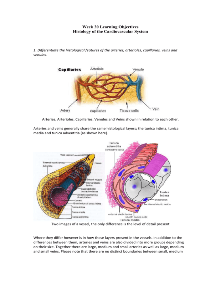
Week 20 Learning Objectives Histology of the Cardiovascular System 1. Differentiate the histological features of the arteries, arterioles, capillaries, veins and venules. Arteries, Arterioles, Capillaries, Venules and Veins shown in relation to each other. Arteries and veins generally share the same histological layers; the tunica intima, tunica media and tunica adventitia (as shown here). Two images of a vessel, the only difference is the level of detail present Where they differ however is in how these layers present in the vessels. In addition to the differences between them, arteries and veins are also divided into more groups depending on their size. Together there are large, medium and small arteries as well as large, medium and small veins. Please note that there are no distinct boundaries between small, medium and large vessels. These vessels tend to continue into each other and portions of the vessels may be intermediates between small, medium and large. Large Arteries Details - These arteries have tremendous elasticity which is required to maintain constant hematologic pressure. During systole, these arteries exhibit their elasticity and stretch in order to reduce blood pressure. During diastole, the arteries rebound and increase blood pressure. This allows the vessels to “buffer” the blood pressure, keeping it as constant as possible. Additionally, large arteries have a supplementary vessel known as a vaso vasorium. This vessel provides additional blood flow to the outer layers of the large artery since the artery’s own blood flow cannot supply this outer region due to its distance away from the bloodstream. Histology – They have a large amount of subendothelial connective tissue. The tunica intima contains also contains a large amount of connective tissue as well as smooth muscle. The tunica media is the thickest of the three layers and is wrapped with a thick band of smooth muscle. The tunica adventitia is the thinnest layer of this vessel and consists mainly of collagen fibers which act to prevent the artery from stretching beyond its normal physiological limit. Cross Section of a Large Artery (Aorta) TI – Tunica Intima, TM – Tunica Media, TA – Tunica Adventitia , VV – Vaso Vasorium ART - Artefact Medium Arteries Details – Medium arteries are less elastic compared to large arteries but are relatively more muscular (taking size into regard). These arteries need more smooth muscle since they are more involved with vasodilation and vasoconstriction compared to large arteries. Histology – The tunica intima is much thinner than in large arteries containing fewer smooth muscle cells and elastic tissue. The tunica media is predominantly made up of smooth muscle which spirals around the vessel. The smooth muscle here is used to exert control over the diameter of the vessel during vasodilation and vasoconstriction. The tunica adventitia of the medium arteries is relatively larger compared to the large arteries. This layer consists mainly of collagen fibers which fulfil a similar role to the ones in the large arteries. Cross Section of a Medium Artery TM – Tunica Media, TA – Tunica Adventitia, ART – Artefact Note: The Tunica Intima is present (the darker inner line of the vessel), just not labelled Small Arteries Details – Small arteries are very similar in constitution to medium arteries, just categorically smaller. Histology - The media is still muscular and has up to 8-10 layers of smooth muscle cells. The adventitia becomes thinner and the external elastic membrane disappears. The intima becomes smaller and the internal elastic membrane also eventually disappears as the artery becomes an arteriole. Cross Section of a Small Artery END – Endothelial Cells, INT – Internal Elastic Lamina, AD – Adipose Cell, EF – Elastic Fiber (part of Tunica Adventitia), N – Smooth Muscle Nuclei (part of Tunica Media), TM – Tunica Media, TA – Tunica Adventitia, EXT – External Elastic Lamina Arterioles Details – Arterioles are vessels which branch out from arteries and lead into capillaries. Histology – Arterioles are very small and only consist of a few layers of muscle cells (usually around two layers only). An Arteriole (middle) next to a Vaso Vasorium (left) END – Endothelial Cell, N – Nucleus of Smooth Muscle, RBC – Red Blood Cell Capillaries Details - Capillaries are the smallest diameter vessels and the site of exchange of metabolites between blood and tissues. These vessels are small enough to allow the passage of blood cells only in a single-file line. Histology - Capillaries consist of a single layer of endothelial cells and their basement membrane. The endothelial cells are joined together by tight junctions. At intervals, these tight junctions are interrupted, leaving small spaces allowing the passage of fluid between blood and ECF. These interruptions do not occur in the brain, and the lack thereof is responsible for the blood-brain barrier present in most of the brain. Two Capillaries and Two Blood Vessels (likely to be Arterioles – not sure) Bv – Blood Vessels, C – Capillaries, S – Nerve Cell Body, NF – Nerve Fiber, END – Endothelial Cell Nucleus Venules Details – Venules are small vessels that bridge the capillaries to small veins. Histology – Unlike arterioles, venules have three layers. An inner endothelium, a middle layer consisting of smooth muscle and an outer layer made of connective tissue. The layer of smooth muscle however is not as thick or as well developed as an arteriole, therefore venules do not have the same well toned shape as arterioles. An Arteriole and a Venule compared A – Arteriole, V – Venule, AD – Adipose Tissue Large Veins Details – Large veins are designed to carry large amounts of blood back to the heart. These vessels operate at a much lower pressure compared to arteries of similar classification and have other features such as valves and facilitate the use of muscle pumps in order to move blood. Veins in general have much less elasticity because they do not have to operate at higher pressure levels. Large veins do not contain valves as enough forward blood flow is available to prevent backflow. Also, in general veins look much more irregular in shape compared to the consistently circular/ovular arteries. Histology – The tunica intima of a large vein consists of the endothelial lining as well as some connective tissue and muscle cells. The tunica media is relatively thin compared to arteries and only has a small amount of smooth muscle. The tunica adventitia is very thick and consists of a large number of connective tissue (collagen), a small amount of elastic tissue and bundles of longitudinally arranged smooth muscle. A partial cross section of a large vein TM – Tunica Media, TA – Tunica Adventitia, SM – Smooth muscles of the tunica adventitia, CT – Connective tissue Note: the Tunica Intima is not labelled in this image but can be seen as the dark lining between the TM and the Lumen Medium Veins Details – Medium veins have a similar constitution to large veins, just smaller. These vessels however have valves, as they operate at low pressures they require semilunar valves to prevent backflow. Histology – Tunica intima consists of a very thin subendothelial layer and some smooth muscle cells. The tunica media consists of a small amount of circumferentially oriented smooth muscle. The tunica adventitia makes up the bulk of the vessel and is made up of connective tissue, a small amount of elastic tissue and bundles of longitudinally oriented smooth muscle. A cross section of a medium vein containing a valve A – Artery, AD – Adipose Tissue, TA- Tunica Adventitia, TM – Tunica Media, V – Valve Note that the Tunica Intima is not labelled again but is the very thin dark lining of the vessel inside the Tunica Media Small Veins Details – Small veins are very similar to medium veins, just smaller. Histology – Small veins are characteristically known for having very small tunica medias, possessing only a tiny amount of smooth muscle. 2. In a table, summarise the key histological features and functions of the different blood vessels listed above.
