Dendritic cells in lymph organs are the neuro-immune cross
advertisement
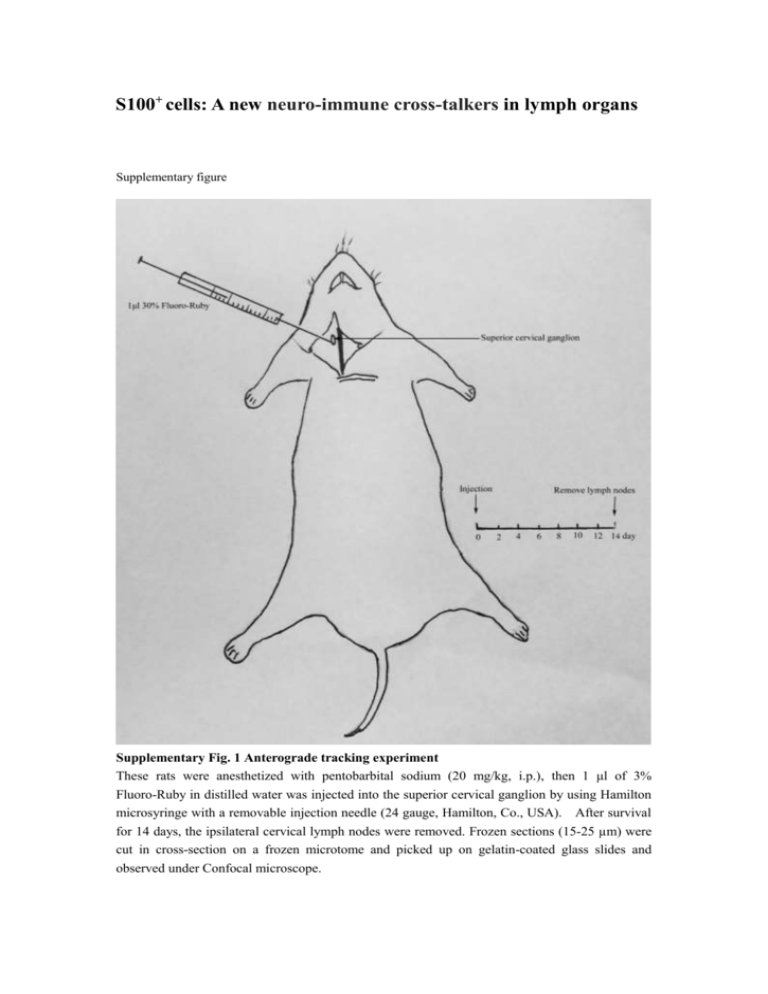
S100+ cells: A new neuro-immune cross-talkers in lymph organs Supplementary figure Supplementary Fig. 1 Anterograde tracking experiment These rats were anesthetized with pentobarbital sodium (20 mg/kg, i.p.), then 1 μl of 3% Fluoro-Ruby in distilled water was injected into the superior cervical ganglion by using Hamilton microsyringe with a removable injection needle (24 gauge, Hamilton, Co., USA). After survival for 14 days, the ipsilateral cervical lymph nodes were removed. Frozen sections (15-25 µm) were cut in cross-section on a frozen microtome and picked up on gelatin-coated glass slides and observed under Confocal microscope. Supplementary Fig. 2 Control experiment In order to prove the labeling of sympathetic nerve fibers is the result of axonal transport of FR dye in neurons, not as the diffusion FR dye, we have done this control experiment. After cutting superior cervical ganglion, 1 μl of 3% Fluoro-Ruby was injected into the point of removing ganglion. The animal was allowed to recover and was sacrificed typically 9-14 days later. The animal was then perfused with neutral buffered formaldehyde (10% formalin or 4% paraformaldehyde in 0.1 M neutral phosphate buffer.) The cervical lymph nodes were removed and postfixed for at least overnight in the same fixative solution plus 20% sucrose for cryo-protection. The lymph nodes were then cut on a freezing sliding microtome or cryostat into sections usually between 15 and 20 microns in thickness. The results showed that Fluoro-Ruby distributes in the diffusion between cells, and not surrounds the cells as experimental group. Supplementary Fig. 3 The results of retrograde tracing We also retrograde tracing the sympathetic neurons in SCG by injected Fluoro-Gold to cervical lymph nodes. The labeled cells in SCG can be seen (arrows in A and B).This experiment proved that the sympathetic nerve fibers projected from SCG directly contact with some cells in cervical lymph node. Supplementary Fig. 4 Ultrastructural of lymph node: the electron micrograph shown the presence of unmyelinated axons adjacent to the innervated cells (arrows in A, B). There is a gap junction between this axon endings and cytomembrane, partially synaptic vesicles, mitochondria and smooth endoplasmic reticulum rich in denuded axon endings, rich in (arrows in C and D). These results give an indirect evidence of synaptic structure in lymph organs. Movie 1:The three-dimensional reconstructions of the labeled nerve fibers by FR and cell membrane staining: Red fluorescence is labeled nerve fibers, and green fluorescence is labeled the plasma-membrane with fluorescent dye DiO. Movie 2: This is a partial enlarged film from movie 1, Red fluorescence is labeled nerve fibers, and green fluorescence is labeled the plasma-membrane with fluorescent dye DiO. This movie clearly showed 0.1-0.2μm labeled nerve ending in cell membrane.


