Karyotype Analysis of Human Chromosomes
advertisement

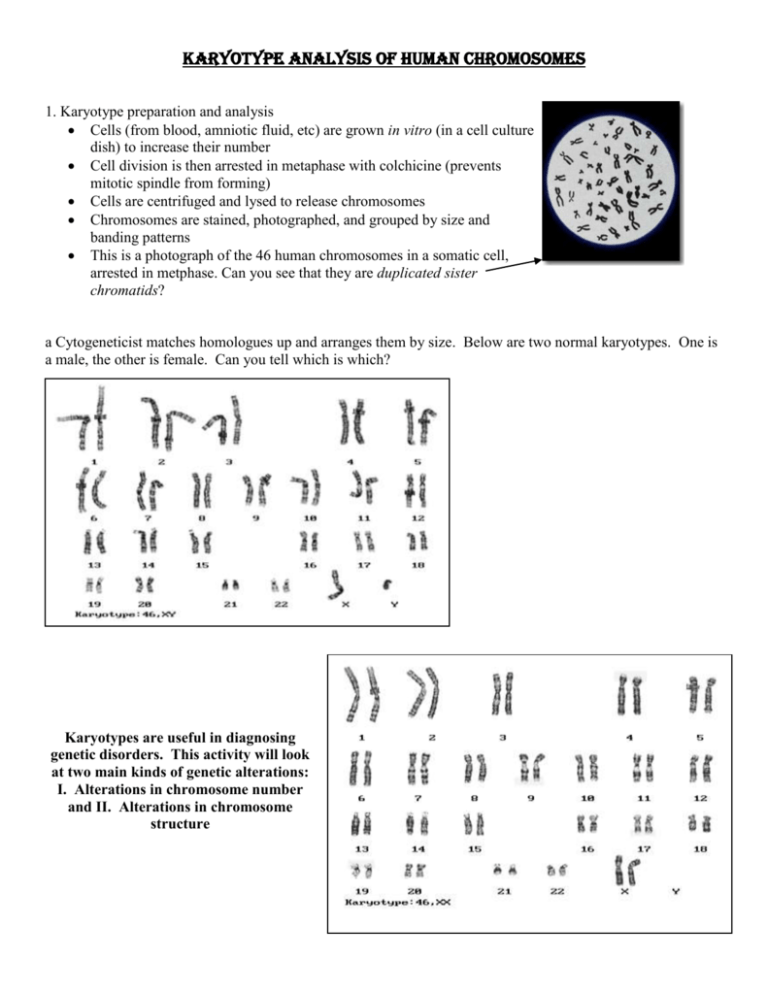
Karyotype Analysis of Human Chromosomes 1. Karyotype preparation and analysis Cells (from blood, amniotic fluid, etc) are grown in vitro (in a cell culture dish) to increase their number Cell division is then arrested in metaphase with colchicine (prevents mitotic spindle from forming) Cells are centrifuged and lysed to release chromosomes Chromosomes are stained, photographed, and grouped by size and banding patterns This is a photograph of the 46 human chromosomes in a somatic cell, arrested in metphase. Can you see that they are duplicated sister chromatids? a Cytogeneticist matches homologues up and arranges them by size. Below are two normal karyotypes. One is a male, the other is female. Can you tell which is which? Karyotypes are useful in diagnosing genetic disorders. This activity will look at two main kinds of genetic alterations: I. Alterations in chromosome number and II. Alterations in chromosome structure I. Alterations in chromosome number: Nondisjunction occurs when either homologues fail to separate during anaphase I of meiosis, or sister chromatids fail to separate during anaphase II. The result is that one gamete has 2 copies of one chromosome and the other has no copy of that chromosome. (The other chromosomes are distributed normally.) If either of these gametes unites with another during fertilization, the result is aneuploidy (abnormal chromosome number) A trisomic cell has one extra chromosome (2n +1) = example: trisomy 21. (Polyploidy refers to the condition of having three homologous chromosomes rather then two) A monosomic cell has one missing chromosome (2n - 1) = usually lethal except for one known in humans: Turner's syndrome (monosomy XO). The frequency of nondisjunction is quite high in humans, but the results are usually so devastating to the growing zygote that miscarriage occurs very early in the pregnancy. If the individual survives, he or she usually has a set of symptoms - a syndrome - caused by the abnormal dose of each gene product from that chromosome. We break these disorders into two main groups: Human disorders due to chromosome alterations in autosomes (Chromosomes 1-22). o Nondisjunction of the sex chromosomes (X or Y chromosome): Human disorders due to chromosome alterations in autosomes (Chromosomes 1-22). There only 3 trisomies that result in a baby that can survive for a time after birth; the others are too devastating and the baby usually dies in utero. A. . Down syndrome (trisomy 21): The result of an extra copy of chromosome 21. People with Down syndrome are 47, 21+. Down syndrome affects 1:700 children and alters the child's phenotype either moderately or severely: characteristic facial features, short stature; heart defects susceptibility to respiratory disease, shorter lifespan prone to developing early Alzheimer's and leukemia often sexually underdeveloped and sterile, usually some degree of mental retardation. Down Syndrome is correlated with age of mother but can also be the result of nondisjunction of the father's chromosome 21 B. Patau syndrome (trisomy 13): serious eye, brain, circulatory defects as well as cleft palate. 1:5000 live births. Children rarely live more than a few months. B. Edward's syndrome (trisomy 18): almost every organ system affected 1:10,000 live births. Children with full Trisomy 18 generally do not live more than a few months. Nondisjunction of the sex chromosomes (X or Y chromosome): Can be fatal, but many people have these karyotypes and are just fine! A. Klinefelter syndrome: 47, XXY males. Male sex organs; unusually small testes, sterile. Breast enlargement and other feminine body characteristics. Normal intelligence. B. 47, XYY males: Individuals are somewhat taller than average and often have below normal intelligence. At one time (~1970s), it was thought that these men were likely to be criminally aggressive, but this hypothesis has been disproven over time. C. Trisomy X: 47, XXX females. 1:1000 live births healthy and fertile - usually cannot be distinguished from normal female except by karyotype D. Monosomy X (Turner's syndrome): 1:5000 live births; the only viable monosomy in humans - women with Turner's have only 45 chromosomes!!! XO individuals are genetically female, however, they do not mature sexually during puberty and are sterile. Short stature and normal intelligence. (98% of these fetuses die before birth) II. Alterations in chromosome structure: 1. Deletion: a portion of one chromosome is lost during cell division. That chromosome is now missing certain genes. When this chromosome is passed on to offspring the result is usually lethal due to missing genes. Example - Cri du chat (cry of the cat): A specific deletion of a small portion of chromosome 5; these children have severe mental retardation, a small head with unusual facial features, and a cry that sounds like a distressed cat. Figure 1 Left, photograph of 5th Chromosome with a p arm deletion. Right, Diagram of the deletion. (Genetic,2003) 2. Duplication: Gene duplication (or chromosomal duplication or gene amplification) is any duplication of a region of DNA that contains a gene; it may occur as an error in homologous recombination, a retrotransposition event, or duplication of an entire chromosome.[1] The second copy of the gene is often free from selective pressure — that is, mutations of it have no deleterious effects to its host organism. Thus it mutates faster than a functional singlecopy gene, over generations of organisms. A duplication is the opposite of a deletion. Duplications arise from an event termed unequal crossing-over that occurs during meiosis between misaligned homologous chromosomes. The chance of this happening is a function of the degree of sharing of repetitive elements between two chromosomes. The product of this recombination are a duplication at the site of the exchange and a reciprocal deletion.[ Example - Fragile X: the most common form of mental retardation. The X chromosome of some people is unusually fragile at one tip - seen "hanging by a thread" under a microscope. Most people have 29 "repeats" at this end of their Xchromosome, those with Fragile X have over 700 repeats due to duplications. Affects 1:1500 males, 1:2500 females. 3. Translocation: a fragment of a chromosome is moved ("trans-located") from one chromosome to another - joins a non-homologous chromosome. The balance of genes is still normal (nothing has been gained or lost) but can alter phenotype as it places genes in a new environment. Can also cause difficulties in egg or sperm development and normal development of a zygote. Acute Myelogenous Leukemia is caused by this translocation: Objectives: 1. What is a karyotype, and how is this analysis performed? 2. What is nondisjunction? 3. Distinguish between the chromosomal alterations that are involved in the following human disorders: Down syndrome, Patau's syndrome, Edwards syndrome Klinefelters syndrome, XYY syndrome Trisomy X, Monosomy X 5. Distinguish between the 3 different alterations in chromosome structure, and how Cri du Chat, Fragile X, and Acute myelogenous leukemia occur.


