Mycobacterium
Order: Actinomycetales
Family: Mycobactericeae
They are widely distributed in nature.
Few no is pathogenic for man & animal.
Some are parasitic to cold blooded animal, others are saprophytic in nature.
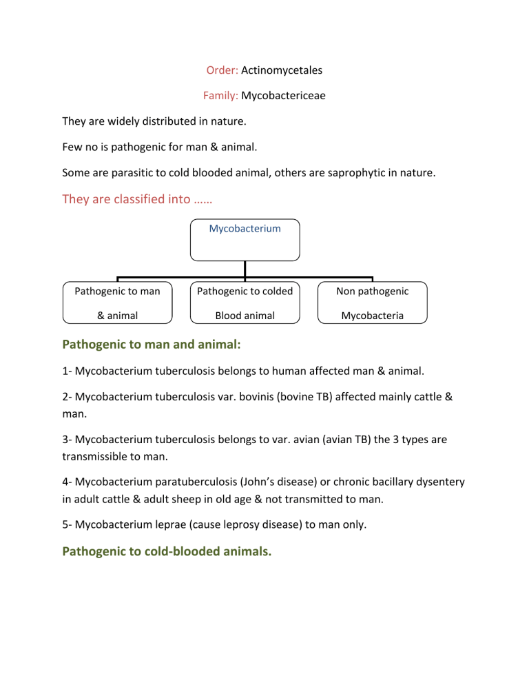
They are classified into ……
Mycobacterium
Pathogenic to man
& animal
Pathogenic to colded
Blood animal
Non pathogenic
Mycobacteria
Pathogenic to man and animal:
1- Mycobacterium tuberculosis belongs to human affected man & animal.
2- Mycobacterium tuberculosis var. bovinis (bovine TB) affected mainly cattle & man.
3- Mycobacterium tuberculosis belongs to var. avian (avian TB) the 3 types are transmissible to man.
4- Mycobacterium paratuberculosis (John’s disease) or chronic bacillary dysentery in adult cattle & adult sheep in old age & not transmitted to man.
5- Mycobacterium leprae (cause leprosy disease) to man only.
Pathogenic to cold-blooded animals.
Saprophytic mycobacterium:
It is also, called atypical or anomyous myco.
They are found in the soil-water-dust-milk.
They are classified according to Runyon & Boenick into…..
Atypical mycobacterium
Saprophytic myco.
Photochromogenes produced yellow pigment when exposed to light
Scotochromogenes produced yellow pigment in the absence of light
Non chromogenes don’t produce pigmentation in the presence or absence of light
They produce tuberculosis like nodules in man & animals specially cattle, camel, pig.
When any bacteria of members of this group stained with suitable dye as strong carbol fuchsin they aren’t decolorized by the action of acid diluted in alcohol or water. So it called acid fast bacteria.
Ziehl neelsen stain specific for TB.
They are characterized by presence of waxy-lipoid material in high concentration in cell wall.
Require the use of heat during staining to dissolve the waxy lipoid material in cell wall of bacteria.
This waxy lipoid material in the tissue of host (Irritates the tissue of man & animal resulting in the formation of TB nodules).
Mycobacterium tuberculosis
Include 3 spp. which is human TB - bovine TB (mammalian TB)-avian TB.
Morphology:
1- Slender rods.
2- Size = 2.5 - 3.5 x 0.4 – 0.6 u in thickness.
Human TB long – thin and beaded.
Bovine TB short, thick and plump (length = width).
Avian TB pleomorphic …… some are short, thick & plump while other is long, thin & beaded but majority appear filamentous.
3- They are non-motile, non-sporulated, non- capsulated.
4- Acid fast resist decolourization with 3% HCl in absolute alcohol or 20% H
2
SO
4
in water.
5- Stained by Ziehl Neelson stain within 10 min.
6- The organism is gram + ve bacteria but very difficult to be stained by gram stain, as stain must left on preparation for about 24 hr.
Culture characteristic:
1- Aerobic MO so, most of infection in lung.
2- Optimum temp. of mammalian type = 37 ○ c & Avian type = 40 ○ c.
3- Bovine type is more difficult to grow than the human type therefore bovine TB is termed dysgenic bacteria (difficult growth) and Eugenic term is given to human
TB (easily growth).
4- Avian TB is faster in growth than both human & bovine TB and given more eugenic term.
5- Doesn’t grow on ordinary nutrient agar media it required specific media for its growth such as:
Dorset egg media
Lowenstein Jensen media
Petreganani's media
6- Addition of 5% glycerin to media, enhance the growth of human & avian type, but has no such effect on the bovine TB.
7- Colonies appear usually within 2-4 weeks, according to type.
Avian type ………….. After 2 weeks
Human type ……….. After 3 weeks
Bovine type ……….. After 4 weeks
8- Growth of TB…
The growth of human TB
The colonies appear as large in number Eugonic, dry, tough, irregular, wrinkled and appears red brick brown pigmentation in old age.
The growth of bovine TB
Colonies appear less in number , moist, granular & easily to broken up colonies.
The growth of avian TB
Colonies appear large in no., large colonies, convex, smooth, glistening or shiny colony with different pigmentation varies from yellow to orange pigmentation.
Biochemical reaction:
1TB MO inactive to enzymatic action (poor enzymatic activity).
2Give slight acidic without gases in fermentation of glucose
2- Catalase + ve 3- Niacine + ve
Typing of bacterium TB:
It is based on morphology & culture character.
Isn’t accurate in distinguishing or in differentiation of 3 spp. Of TB.
The most accurate method is by animal inoculation test.
G.pig Rabbit Chicken
Bovine TB +++ +++ - - -
Human TB +++ +
Avian TB - - ++
- - -
+++
Diagnosis:
1- Direct microscopically examination:
Films are prepared from caseous purulent portion of sputum or affected lesion & stain by ziehl neelsons stain.
The morphological appearance of the organism is described.
In TB urinary tract: first 3voided (post urine) urine
Urine centrifuge 3000 rpm/30 min. & take deposit.
In TB meningitis CSF.
TTT of the sample…
1- Antiformin method:
If MO is scanty (few in no.) or combined with other MO or if a pure culture is required, the antiformin method is used.
Antiformin is composed of equal parts of Na chlorinate + 15% Na hydroxide.
Antiformin is diluted to 1:6 then added to the sample with ratio 1:4 or 1:3 then wait 1hr at 37c giving chance or antiformin to kill all MO in sample leaving TB after 1hr.
2- Petroffs method:
Tuberculin test:
(delayed type of hypersensitivity)
Tuberculin is preparation containing specific protein extract of tubercle bacilli which on injection into an infected animals, allergic symptoms are set up making a reaction.
Immunization against TB:
1- Using B.C.G. vaccine (Bacille Calmette Guerin).
2- Diaplyte vaccine:
Human type which fat has been extracted with formaline.
3- Spahlinger vaccine:
Dead vaccine prepared by growing TB on media enriched with body fluids & left naturally to die the immunization power is very weak & not commonly used.
4- Vole bacillus:
Mycobacterium murius when injected into G.pig produces resistance against both human or bovine tubercle bacilli.
Immunization power is similar to that produced by BCG.
