Spin polarized transport in semiconductors – Challenges for
advertisement
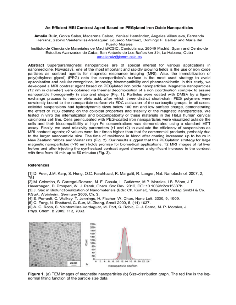
An Efficient MRI Contrast Agent Based on PEGylated Iron Oxide Nanoparticles Amalia Ruiz, Gorka Salas, Macarena Calero, Yenisel Hernández, Angeles Villanueva, Fernando Herranz, Sabino Veintemillas-Verdaguer, Eduardo Martínez, Domingo F. Barber and María del Puerto Morales Instituto de Ciencia de Materiales de Madrid/CSIC, Cantoblanco, 28049 Madrid, Spain and Centro de Estudios Avanzados de Cuba, San Antonio de Los Baños km 3½, La Habana, Cuba amaliaruiz@icmm.csic.es Abstract Superparamagnetic nanoparticles are of special interest for various applications in nanomedicine. Nowadays, one of the most important and rapidly growing fields is the use of iron oxide particles as contrast agents for magnetic resonance imaging (MRI). Also, the immobilization of poly(ethylene glycol) (PEG) onto the nanoparticles's surface is the most used strategy to avoid opsonisation and cellular recognition, improving biocompatibility and pharmacokinetic. In this study, we developed a MRI contrast agent based on PEGylated iron oxide nanoparticles. Magnetite nanoparticles (12 nm in diameter) were obtained via thermal decomposition of a iron coordination complex to assure nanoparticle homogeneity in size and shape (Fig. 1). Particles were coated with DMSA by a ligand exchange process to remove oleic acid, after which three distinct short-chain PEG polymers were covalently bound to the nanoparticle surface via EDC activation of the carboxylic groups. In all cases, colloidal suspensions had hydrodynamic sizes below 100 nm and low surface charge, demonstrating the effect of PEG coating on the colloidal properties and stability of the magnetic nanoparticles. We tested in vitro the internalization and biocompatibility of these materials in the HeLa human cervical carcinoma cell line. Cells preincubated with PEG-coated iron nanoparticles were visualized outside the cells and their biocompatibility at high Fe concentrations was demonstrated using a standard MTT assay. Finally, we used relaxivity parameters (r1 and r2) to evaluate the efficiency of suspensions as MRI contrast agents; r2 values were four times higher than that for commercial products, probably due to the larger nanoparticle size. The time of residence in blood after coating increased up to hours in New Zealand rabbits and Wistar rats (Fig. 2). Our results suggest that this PEGylation strategy for large magnetic nanoparticles (>10 nm) holds promise for biomedical applications. T2 MRI images of rat liver before and after injecting the synthesized contrast agent showed a significant increase in the contrast with time from 10 min up to 50 minutes (Fig. 3). References [1] D. Peer, J.M. Karp, S. Hong, O.C. Farokhzad, R. Margalit, R. Langer, Nat. Nanotechnol. 2007, 2, 751 [2] M. Colombo, S. Carregal-Romero, M. F. Casula, L. Gutiérrez, M.P. Morales, I.B. Böhm, J.T. Heverhagen, D. Prosperi, W. J. Parak, Chem. Soc Rev. 2012, DOI:10.1039/c2cs15337h. [3] J. Gao in Biofunctionalization of Nanomaterials (Eds: Ch. Kumar), Wiley-VCH Verlag GmbH & Co. KGaA, Weinheim, Germany 2005, Ch. 3. [4] S. Perrault, C. Walkey, T. Jennings, H. Fischer, W. Chan, Nano Lett. 2009, 9, 1909. [5] C. Fang, N. Bhattarai, C. Sun, M. Zhang, Small 2009, 5, (14) 1637. [6] A. G. Roca, S. Veintemillas-Verdaguer, M. Port, C. Robic, C. J. Serna, M. P. Morales, J. Phys. Chem. B 2009, 113, 7033. Figure 1. (a) TEM images of magnetite nanoparticles (b) Size-distribution graph. The red line is the lognormal fitting function of the particle size data. Figure 2. (e, f) ICP quantification of iron concentration in blood after injection into e) rabbits and f) rats. NP-DMSA-PEG-NH2 [○], NP-DMSA-PEG-(NH2)2 [■] and NP-DMSAPEG-Prop-(NH2)2) [∆]. Figure 3. T2 MR images of rat liver before and after injecting the synthesized contrast agent. In the left animal injected with NP-DMSA-PEG(NH2)2 and in the right control animal (a)T2 image after 10 minutes of injection.(b)T2 image after 50 minutes of injection.

