O84 Left ventricular systolic strain and rotation is reduced in incident
advertisement
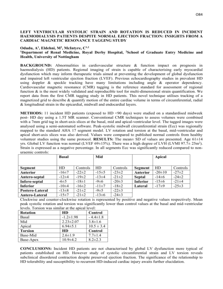
O84 LEFT VENTRICULAR SYSTOLIC STRAIN AND ROTATION IS REDUCED IN INCIDENT HAEMODIALYSIS PATIENTS DESPITE NORMAL EJECTION FRACTION: INSIGHTS FROM A CARDIAC MAGNETIC RESONANCE TAGGING STUDY Odudu, A1, Eldehni, M2, McIntyre, C1,2 1 Department of Renal Medicine, Royal Derby Hospital, 2School of Graduate Entry Medicine and Health, University of Nottingham BACKGROUND: Abnormalities in cardiovascular structure & function impact on prognosis in haemodialysis (HD) patients. Regional imaging of strain is capable of characterising early myocardial dysfunction which may inform therapeutic trials aimed at preventing the development of global dysfunction and impaired left ventricular ejection fraction (LVEF). Previous echocardiography studies in prevalent HD using doppler & speckle tracking have many limitations including angle & operator dependency. Cardiovascular magnetic resonance (CMR) tagging is the reference standard for assessment of regional function & is the most widely validated and reproducible tool for multi-dimensional strain quantification. We report data from the first CMR tagging study in HD patients. This novel technique utilises tracking of a magnetized grid to describe & quantify motion of the entire cardiac volume in terms of circumferential, radial & longitudinal strain in the epicardial, midwall and endocardial layers. METHODS: 11 incident HD patients (exposed to HD <90 days) were studied on a standardised midweek post- HD day using a 1.5T MR scanner. Conventional CMR techniques to assess volumes were combined with a 7mm grid tag in short-axis slices at the basal, mid and apical-ventricular level. The tagged images were analysed using a semi-automated software. Peak-systolic midwall circumferential strain (Ecc) was regionally mapped to the standard AHA 17 segment model. LV rotation and torsion at the basal, mid-ventricular and apical short-axis slices was also derived. Values were compared to published normal controls from healthy volunteer studies using the same protocol. RESULTS: The mean± SD of values are presented. Age 61±14 yrs. Global LV function was normal (LVEF 69±15%). There was a high degree of LVH (LVMI 97.7± 25m2). Strain is expressed as a negative percentage. In all segments Ecc was significantly reduced compared to nonuraemic controls: Basal Mid Apical HD Controls HD Controls Controls Segment Segment HD -16±7 -22±2 -15±5 -23±2 -27±2 Anterior Anterior -20±10 -12±4 -19±2 -13±4 -21±2 -14±6 -24±2 Antero-septal Septal -6±5 -18±1 -9±6 -20±3 -15±6 -21±4 Infero-septal Inferior -10±4 -16±2 -11±7 -18±2 -17±9 -25±3 Inferior Lateral -13±8 -21±2 -9±5 -22±3 Postero-Lateral -15±7 -21±2 -13±6 -24±3 Antero-Lateral Clockwise and counter-clockwise rotation is represented by positive and negative values respectively. Mean peak systolic rotation and torsion was significantly lower than control values at the basal and mid-ventricular levels. Torsion was similar at the apical level: Rotation HD Control Basal -1.2±1.98 - 4.4±1.8 Mid 2.23±2.07 3.8±1.6 Apical 6.94±5.1 10.5 ± 3.4 Torsion HD Control Base-Mid 2.6±1.9 7.7±1.4 Base-Apex 10.9±4.2 8.2±2.3 CONCLUSIONS: Incident HD patients are not characterised by global LV dysfunction more typical of patients established on HD. However study of systolic circumferential strain and LV torsion reveals subclinical disordered contraction despite preserved ejection fraction. The significance of the relationship to HD tolerability and susceptibility to recurrent HD-induced cardiac injury awaits further elucidation.
