continuous catheter impedance measurements
advertisement
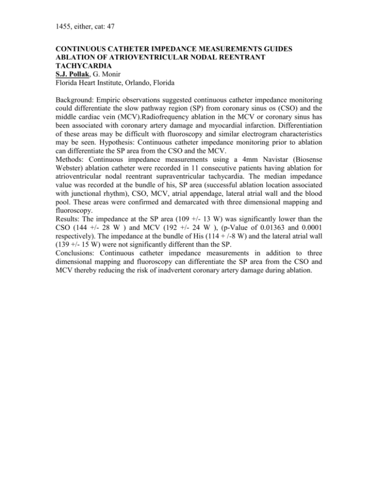
1455, either, cat: 47 CONTINUOUS CATHETER IMPEDANCE MEASUREMENTS GUIDES ABLATION OF ATRIOVENTRICULAR NODAL REENTRANT TACHYCARDIA S.J. Pollak, G. Monir Florida Heart Institute, Orlando, Florida Background: Empiric observations suggested continuous catheter impedance monitoring could differentiate the slow pathway region (SP) from coronary sinus os (CSO) and the middle cardiac vein (MCV).Radiofrequency ablation in the MCV or coronary sinus has been associated with coronary artery damage and myocardial infarction. Differentiation of these areas may be difficult with fluoroscopy and similar electrogram characteristics may be seen. Hypothesis: Continuous catheter impedance monitoring prior to ablation can differentiate the SP area from the CSO and the MCV. Methods: Continuous impedance measurements using a 4mm Navistar (Biosense Webster) ablation catheter were recorded in 11 consecutive patients having ablation for atrioventricular nodal reentrant supraventricular tachycardia. The median impedance value was recorded at the bundle of his, SP area (successful ablation location associated with junctional rhythm), CSO, MCV, atrial appendage, lateral atrial wall and the blood pool. These areas were confirmed and demarcated with three dimensional mapping and fluoroscopy. Results: The impedance at the SP area (109 +/- 13 W) was significantly lower than the CSO (144 +/- 28 W ) and MCV (192 +/- 24 W ), (p-Value of 0.01363 and 0.0001 respectively). The impedance at the bundle of His (114 + /-8 W) and the lateral atrial wall (139 +/- 15 W) were not significantly different than the SP. Conclusions: Continuous catheter impedance measurements in addition to three dimensional mapping and fluoroscopy can differentiate the SP area from the CSO and MCV thereby reducing the risk of inadvertent coronary artery damage during ablation.

