pmic7683-sup-0001-FigureS1
advertisement
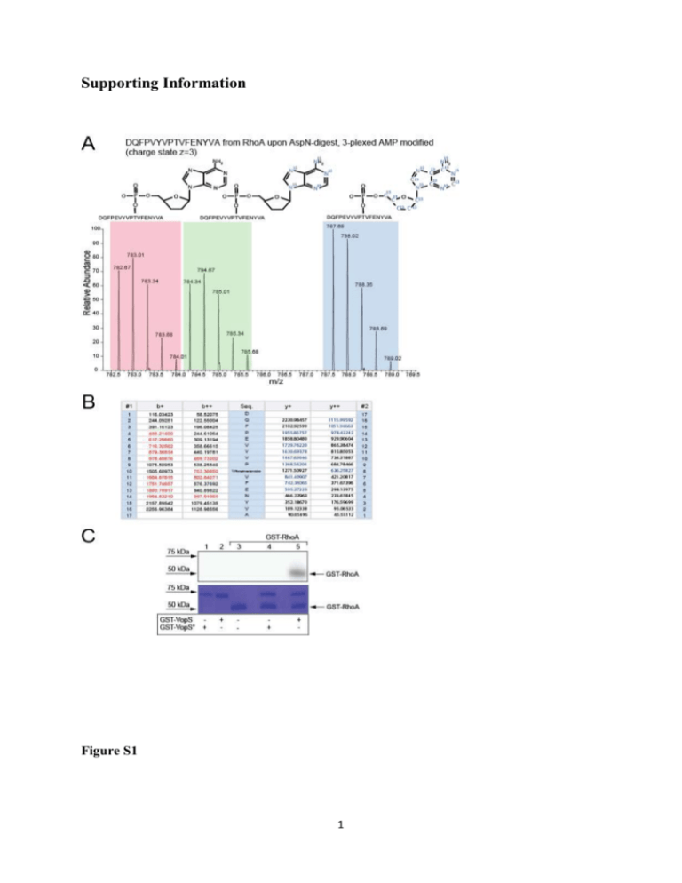
Supporting Information Figure S1 1 Figure S2 2 Figure S1: Workflow validation by confirming RhoA as AMPylation target of VopS. A) Target identification by AMPylation-specific reporter ion clusters. In vitro AMPylation assay was performed on purified RhoA and VopS in the presence of 3-plexed ATP. The sample was then analyzed by in-gel digest and LC-MS/MS. The mass spectrum shows the m/z of the 3-plexed AMPylated peptide DQFPVYVPTVFENYVA confirming RhoA as an AMPylation target of VopS as reported previously [1]. More details are shown in the Material and Method section provided as Supporting Information. B) Ion series of the AMPylated peptide of RhoA. The ion series are shown and b/y-ions covering the AMPylated residue are indicated. C) VopS-mediated AMPylation of RhoA. In vitro AMPylation assays were performed with purified GST-VopS or the inactive mutant GST-VopS° and purified GST-RhoA in the presence of α32P-ATP. AMPylated proteins were visualized by autoradiography (top). The SDS-gel used for autoradiography was then stained with Coomassie to visualize all proteins (bottom). Figure S2: Ion series and MS/MS spectrum of AMPylated vimentin peptide SLYSSSPGGGAYVTR. A) Ion series of detected fragment ions derived from SLY(AMP)SSSPGGGAYVTR. Samples derived from in vitro AMPylation assays with Bep2 using 3-plexed ATP were analyzed after in-gel digestion by LC-MS/MS. B) MS/MS spectrum of SLY(AMP)SSSPGGGAYVTR. From mapped ion series and fragment spectra tyrosine 3 in the peptide sequence is the most likely residue carrying the AMPylation PTM. Peaks corresponding masses to matching to neutral loss fragments of SLY(AMP)SSSPGGGAYVTR are highlighted. Singly charged precursors with neutral loss of the nucleoside group [M-249+H]+ and the base [M-135+H]+ were detected, a similar observation made by Li et al. [2]. Table S1: Peptides detected by in-gel digest and LC-MS analysis. List of all peptides detected by in-gel digest and LC-MS analysis over the linear gradient (0-60min) upon activity based assays using 3-plexed ATP in the presence of Bep2 and J774 mouse macrophage lysates. Peptide FDR was adjusted to 1% using the reverse decoy strategy. 3 4 Material and Methods DNA manipulations E. coli Expression Constructs - Bep2 of B. rochalimae was amplified with the primers prAH072 (CCGCTCGAGATGAAGAAAAGTGAAATGATGATA) and prAH073 (CCGCTCGA GTTAACAAACCATAGCTGTCGC) from genomic DNA of B. rochalimae and cloned via XhoI into pET15b to achieve pAH019. Using primers prAH110 (CAATTATATTGCACCTTTTAGGGAAGGTAATGGACG) and (CCTAAAAGGTGCAATATAATTGATAGAGCCAAATATTTTTG) prAH111 in site-directed mutagenesis [3], pAH051 encoding for the inactive site mutant of Bep2 was achieved. BiaA of B. rochalimae that is homologous to VbhA which is a small protein inhibiting the Fic protein VbhT of B. schoenbuchensis [4], was amplified using prAG0013 (GCCCATGGTGAAAAAAACAACTGATCATTCTAC) and prAG0014 (GCGGATCCTTA TAGTGTTGCATTGTCCATAAGAG) from genomic DNA of B. rochalimae and cloned via NcoI and BamHI restriction into pRSFDUET-1 to achieve pAG0056. The FIC-domain of Bep2 was amplified using prAG029 (GGGAATTCCATATGGATATTAACATC CCTTCTCC) and prAG035 (CGACCTCGAGTTAGTGATGGTGATGGTGATGTTCA CTCAAAGCAGCTAA TTTTTC) and introduced via NdeI and XhoI into pAG0056 to achieve pAG0061. In a next step, biaA was then mutated via site directed mutagenesis PCR to abolish its inhibitory activity GAATCACTCTTCATTCTAAAACG) using and prAG048 prAG047 (GCAATTGGAG (GAGTGATTCCTCCAATTG CATGTGTACTAATAG) to achieve pKP090. RhoA was amplified using prAH179 (GAAACTGGTGATTGTTGGTGATGGAGCCTGTG GAAAGACATGC) and prAH180 (CCACAGGCTCCATCACCAACAATCACCA GTTTCTTCCGGATGGC) from pRK5myc-RhoA [5] and cloned via BamHI and XhoI into pGEX-6p-1 to achieve pAH049. Constructs for E. coli expression of GST-VopS30-387 and GST-VopSH348A30-387 were a kind gift of K. Orth [1]. 5 Preparation of cell lysates J774 mouse macrophages were cultured in DMEM supplemented with 10% FCS up to a confluence of 90%. Cells were trypsinized, resuspended extensively in 10mL DMEM supplemented with 10% FCS, pellet at 3000 rpm and resuspended in 400uL Lysis buffer (10mM Tris, pH=8.0, 150mM NaCl, 10mM MgCl2, 5mM βME). Cells were lysed by 3x 10 pulses of sonication and centrifuged for 15min at 4°C and 4500rpm. Supernatants were stored in aliquots at -80°C. Expression and purification of recombinant proteins For recombinant expression of Bep2 or its inactive mutant (Bep2°) of B. rochalimae, Cacompetent E. coli were transformed with pAH019 or pAH051 and cultured at RT in Terrific Broth media containing 200 mg/L Ampicillin. At an OD600=0.5 expression was induced with 100 μM IPTG (Promega). After 18 h, cultures were harvested, aliquoted and stored at -20°C. Frozen bacteria were thawed and lysed in AMPylation buffer (10 mM Tris pH=8.0, 150 mM NaCl, 10 mM MgCl2, 2.5 mM βME) supplemented with 2 mg DNaseI from bovine pancreas (Roche) and Complete EDTA-free Protease Inhibitor Cocktail (Roche) [40 l/ml of stock solution (1 tablet / 2 ml H2O]. For AMPylation assays with full lysates, bacteria were lysed with 3x 15pulses of sonication, centrifuged for 5 min at 13000 rpm and supernatents utilized for AMPylation assays. In order to purify Bep21-360, Ca-competent E. coli were transformed with pKP090 and cultured at RT in 6 L Terrific Broth media containing 50 mg/L Kanamycin. At an OD600=0.5, expression was induced with 100 μM IPTG (Promega). After 18 h, cultures were harvested, and stored at -20°C. Frozen pellets of bacteria were thawed and lysed using French Press and cell debris was removed by high speed centrifugation (1 h, 100 g, 4°C). Bep21-360 was purified using metal affinity utilizing Ni-NTA columns and elution with 200 mM imidazole. Peak fractions were pooled and loaded onto a Superdex 200 10/300 column (GE Healthacre) for size exclusion chromatography. 10 μL of the peak fractions were separated via SDS-PAGE using 12% SDS-gel and stained with Coomassie staining solution. Fractions containing Bep21-360 were pooled and the concentration was measured via absorbance at 280 nm with Nanodrop-1000 (Nanodrop Technologies, Wilmington, USA). Protein was stored at 4°C. For recombinant expression of GST-fusion proteins, Ca-competent E. coli were transformed with pVopS or pVopSH348A [1] or pAH049 and cultured at RT in Terrific Broth media containing 200 mg/L Ampicillin. At an OD600=0.5 expression was induced with 50 μM IPTG 6 (Promega). After 8 h, cultures were harvested and stored at -20°C. Frozen bacteria were thawed and lysed in PBS buffer supplemented with 5mM βME, 2 mg DNaseI from bovine pancrease (Roche) and Complete EDTA-free Protease Inhibitor Cocktail (Roche) [40 l/ml of stock solution (1 tablet / 2 ml H2O]. For AMPylation assays with full lysates, bacteria were lysed with 3x 15 pulses of sonication, centrifuged for 5 min at 13000 rpm and supernatents were used in AMPylation assays. In order to purify GST-fusion proteins, cells were lysed using French Press and cell debris was removed by high speed centrifugation (1 h, 100 g, 4°C). GST-VopS, GST-VopSH348A or GST-RhoA were purified using affinity chromatography utilizing GST-Trap columns (GE Healthcare) and elution with 10 mM Glutathion (Sigma) in AMPylation buffer (10 mM Tris pH=8.0, 150 mM NaCl, 10 mM MgCl2, 2.5 mM βME). Peak fractions were pooled and 10μL of the peak fractions were separated via SDS-PAGE using 12% SDS-gel and stained with Coomassie staining solution. The concentration of fractions containg VopS was measured via absorbance at 280 nm with Nanodrop-1000 (Nanodrop Technologies, Wilmington, USA). Protein was stored at 4°C. AMPylation assays In order to determine the size of potential target proteins, in vitro AMPylation assays with radioactive labeled α32P-ATP were performed using full cell lysates as described in previous studies [4]. To this end, aliquots of eukaryotic cell lysates were thawed on ice and bacterial lysates were freshly prepared. Then, 25 µL of eukaryotic cell lysates mixed with 1.5 µL of 7.5 mg/mL RNAseI (Roche), 25 µL freshly prepared E. coli lysates and 1 µL of α32P-ATP (Hartmann Analytics, SRP-207). Alternatively, 250 pmol of purified Bep21-360 were mixed with 1.5 µL of 7.5 mg/mL RNAseI, 1 µL of α32P-ATP and either AMPylation buffer, 200 pmol vimentin or 250 pmol of BSA. Samples were then incubated for 1 h at 30°C, reactions were stopped by the addition of 25 µL SDS-loading buffer and incubated for 5 min at 95°C. Samples were loaded onto pre-cast gradient SDS-gels (Bio-Rad). Electrophoresis was performed 150 V for 52 min. Proteins were fixed for 1 h in Fixation buffer (50% water, 40% MeOH, 10% glacial acid), gels were sealed in plastic bags and exposed on autoradioscreens overnight. Screens were developed using a Typhoon FLA 7000 system (GE Healthcare) 7 Target identification by mass spectrometry After target size determination via radioactive AMPylation assays, AMPylation assays were repeated using heavy isotope labeled ATP (3-plexed, Cambridge Isotope Laboratories), gel area from approximately 35 kDa to 70 kDa proteins was excised and divided into 5 pieces. Ingel trypsin digestion was adopted from Shevchenko et al. [6]. RhoA in-solution digest was adopted from Engel et al. (Ref. 3) using 0.5 μg AspN as a protease for protein digestion. Prior to mass spectrometry analysis the peptides were purified using C18 Microspin columns (Harvard Apparatus) according to the manufactures instruction. LC-MS/MS analysis was performed on a dual pressure LTQ-Orbitrap mass spectrometer (Thermo Electron), which was connected to an electrospray ion source (Proxeon Biosystems). Peptide separation was carried out using an easy nano-LC systems (Proxeon Biosystems) equipped with an RP-HPLC column packed with C18 resin (Magic C18 AQ 3 μm; Michrom BioResources). A 0.2 μl/min linear gradient from 96% solvent A (0.15% formic acid, 2% acetonitrile) and 4% solvent B (98% acetonitrile, 0.15% formic acid) to 40% solvent B over 60 min was applied. The data acquisition mode was set to obtain one high-resolution MS scan in the FT part of the mass spectrometer at a resolution of 60,000 FWHM followed by MS/MS scans in the linear ion trap of the 20 most intense ions. Collision induced dissociation (CID) was triggered for precursors exceeding 100 ion counts at a normalized collusion energy of 35%. The ion accumulation time was set to 300 ms (MS) and 10 ms (MS/MS). For peak detection and extraction of peptide intensities Progenesis (Nonlinear Dynamics) was used in default settings. Peptide identification was carried out using the Mascot and SEQUEST search tool and the mouse swissprot protein database containing forward and reversed-decoy protein sequences and containing target protein sequences. The search criteria were set as follows: full tryptic specificity was required (cleavage after lysine or arginine residues); 2 missed cleavages were allowed; carbamidomethylation (C) was set as fixed modification; oxidation (M) and AMPylation (Phospoadenosine, T, Y) as variable modifications. For AMP-modifications, masses of the 15 N013C0-, 15 N5- and 15 N513C10-AMP labeled moieties were included in the search. The mass tolerance was set to 10 ppm for precursor ions and 0.6 Da for fragment ions. 8 References [1] Yarbrough, M. L., Li, Y., Kinch, L. N., Grishin, N. V., et al., AMPylation of Rho GTPases by Vibrio VopS disrupts effector binding and downstream signaling. Science 2009, 323, 269-272. [2] Li, Y., Al-Eryani, R., Yarbrough, M. L., Orth, K., Ball, H. L., Characterization of AMPylation on threonine, serine, and tyrosine using mass spectrometry. J Am Soc Mass Spectrom 2011, 22, 752-761. [3] Zheng, L., Baumann, U., Reymond, J. L., An efficient one-step site-directed and site-saturation mutagenesis protocol. Nucleic Acids Res 2004, 32. [4] Engel, P., Goepfert, A., Stanger, F. V., Harms, A., et al., Adenylylation control by intra- or intermolecular active-site obstruction in Fic proteins. Nature 2012, 482, 107-110. [5] Besson, A., Gurian-West, M., Schmidt, A., Hall, A., Roberts, J. M., p27(Kip1) modulates cell migration through the regulation of RhoA activation. Gene Dev 2004, 18, 862-876. [6] Shevchenko, A., Tomas, H., Havlis, J., Olsen, J. V., Mann, M., In-gel digestion for mass spectrometric characterization of proteins and proteomes. Nat Protoc 2006, 1, 2856-2860. 9
