Supplementary Figure 2 (doc 723 KB)
advertisement
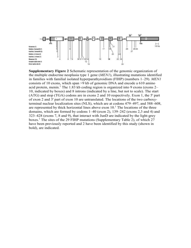
Supplementary Figure 2 Schematic representation of the genomic organization of the multiple endocrine neoplasia type 1 gene (MEN1), illustrating mutations identified in families with familial isolated hyperparathyroidism (FIHP) (numbers 1–29). MEN1 consists of 10 exons, which span >9 kb of genomic DNA and encode a 610 amino acid protein, menin.1 The 1.83 kb coding region is organized into 9 exons (exons 2– 10, indicated by boxes) and 8 introns (indicated by a line, but not to scale). The start (ATG) and stop (TGA) codons are in exons 2 and 10 respectively. Exon 1, the 5' part of exon 2 and 3' part of exon 10 are untranslated. The locations of the two carboxyterminal nuclear localization sites (NLS), which are at codons 479–497, and 588–608, are represented by thick horizontal lines above exon 10.1 The locations of the three domains, which are formed by codons 1–40 (exon 2), 139–242 (exons 2,3 and 4) and 323–428 (exons 7, 8 and 9), that interact with JunD are indicated by the light-grey boxes.1 The sites of the 29 FIHP mutations (Supplementary Table 2), of which 27 have been previously reported and 2 have been identified by this study (shown in bold), are indicated.
