RADIOLOGY CASE REPORT
advertisement
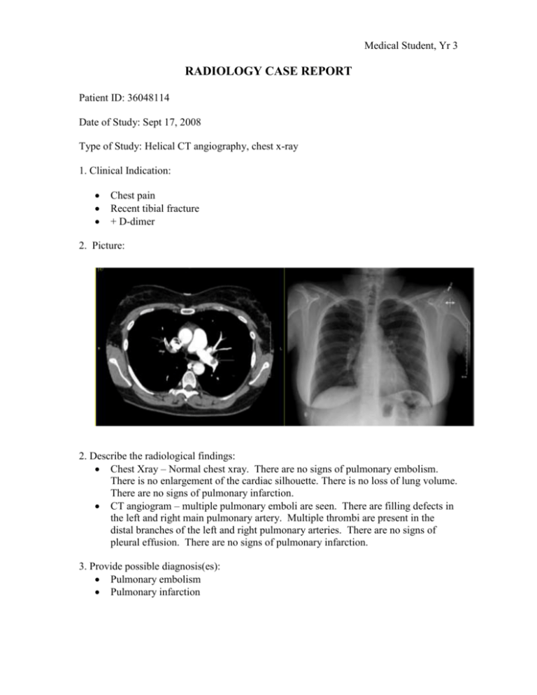
Medical Student, Yr 3 RADIOLOGY CASE REPORT Patient ID: 36048114 Date of Study: Sept 17, 2008 Type of Study: Helical CT angiography, chest x-ray 1. Clinical Indication: Chest pain Recent tibial fracture + D-dimer 2. Picture: 2. Describe the radiological findings: Chest Xray – Normal chest xray. There are no signs of pulmonary embolism. There is no enlargement of the cardiac silhouette. There is no loss of lung volume. There are no signs of pulmonary infarction. CT angiogram – multiple pulmonary emboli are seen. There are filling defects in the left and right main pulmonary artery. Multiple thrombi are present in the distal branches of the left and right pulmonary arteries. There are no signs of pleural effusion. There are no signs of pulmonary infarction. 3. Provide possible diagnosis(es): Pulmonary embolism Pulmonary infarction 4. What would you recommend next for this patient? Low-molecular Weight Heparin (LMWH) for 5 days Warfarin for 3 – 6 months following LMWH Monitor INR at regular intervals, to be followed by family doctor If onset of new pulmonary symptoms - Follow-up CXR to assess for atelectasis, pleural effusion, or pulmonary infarction resulting from 5. Is the use of this test/procedure appropriate? Yes. It is difficult to see PE on CXR. A CT chest with contrast is a more sensitive test for identifying suspected PE. 6. Is(are) there any alternate test(s)?1 Ventilation/Perfusion (V/Q) Lung scintigraphy – in this technique a radiotracer is injected intravenously to assess the patency of the pulmonary circulation. Perfusion defects are identified by a mismatch in the V/Q ratio. Pulmonary angiography – this technique is used if there is a high degree of suspicion of PE, but it cannot be definitively diagnosed with CTA. Pulmonary angiography requires intra-arterial contrast administration. Ultrasound – this can be done in the deep leg veins to check for the source of the pulmonary embolism 7. How would you explain to the patient about the possible risks and benefits of this test? Risks o Radiation exposure (~8 mSv) – the radiation dose that you will receive is higher than a chest xray. o Contrast toxicity – IV contrast must be given during this test. There have been reports of contrast nephropathy, or acute renal failure resulting from contrast administration. Your kidney function will be investigated prior to the CT scan by measuring your creatinine; if your kidney function is normal, the risk of contrast-induced nephropathy is low. Benefits o CTA is sensitive for detecting PE (~96%)2, therefore it is likely to pick up the presence of thrombus in the pulmonary arteries o CTA is a safe procedure that is quick to complete o The outcome of the CTA will help to guide management. If PE is detected on CTA, there is effective treatment. 8. What is the cost of this test? Approximately $500 - $600 The patient does not pay; the cost of this procedure is covered by the government. References: 1. Brant WE, Helms CA. (eds) Fundamentals of diagnostic radiology. 3rd edition. 2007 Lippincott Williams & Wilkins, Philadelphia. 2. Winer-Muram HT, Rydberg J, Johnson MS, et al. Suspected acute pulmonary embolism: evaluation with multi-detector row CT versus digital subtraction pulmonary arteriography. Radiology 2004; 233:806-815


