1BOM, 1BON - Chemistry Biochemistry and Bio
advertisement
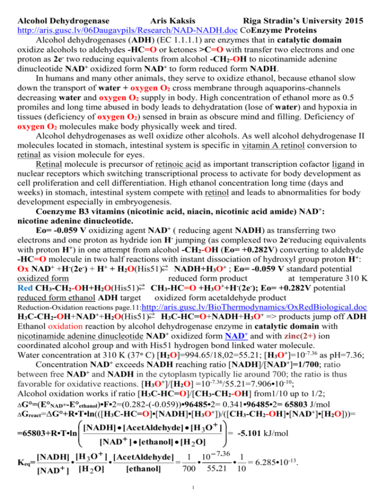
Alcohol Dehydrogenase
Aris Kaksis
Riga Stradin’s University 2015
http://aris.gusc.lv/06Daugavpils/Research/NAD-NADH.doc CoEnzyme Proteins
Alcohol dehydrogenases (ADH) (EC 1.1.1.1) are enzymes that in catalytic domain
oxidize alcohols to aldehydes -HC=O or ketones >C=O with transfer two electrons and one
proton as 2e- two reducing equivalents from alcohol -CH2-OH to nicotinamide adenine
dinucleotide NAD+ oxidized form NAD+ to form reduced form NADH.
In humans and many other animals, they serve to oxidize ethanol, because ethanol slow
down the transport of water + oxygen O2 cross membrane through aquaporins-channels
decreasing water and oxygen O2 supply in body. High concentration of ethanol more as 0.5
promiles and long time abused in body leads to dehydratation (lose of water) and hypoxia in
tissues (deficiency of oxygen O2) sensed in brain as obscure mind and filling. Deficiency of
oxygen O2 molecules make body physically week and tired.
Alcohol dehydrogenases as well oxidize other alcohols. As well alcohol dehydrogenase II
molecules located in stomach, intestinal system is specific in vitamin A retinol conversion to
retinal as vision molecule for eyes.
Retinal molecule is precursor of retinoic acid as important transcription cofactor ligand in
nuclear receptors which switching transcriptional process to activate for body development as
cell proliferation and cell differentiation. High ethanol concentration long time (days and
weeks) in stomach, intestinal system compete with retinol and leads to abnormalities for body
development especially in embryogenesis.
Coenzyme B3 vitamins (nicotinic acid, niacin, nicotinic acid amide) NAD+:
nicotine adenine dinucleotide.
Eo= -0.059 V oxidizing agent NAD+ ( reducing agent NADH) as transferring two
electrons and one proton as hydride ion H- jumping (as complexed two 2e-reducing equivalents
with proton H+) in one attempt from alcohol -CH2-OH (Eo= +0.282V) converting to aldehyde
-HC=O molecule in two half reactions with instant dissociation of hydroxyl group proton H+:
Ox NAD+ +H-(2e-) + H+ + H2O(His51) NADH+H3O+ ; Eo= -0.059 V standard potential
oxidized form
reduced form product
at temperature 310 K
+
Red CH3-CH2-OH+H2O(His51) CH3-HC=O +H3O +H (2e ); Eo= +0.282V potential
reduced form ethanol ADH target oxidized form acetaldehyde product
Reduction-Oxidation reactions page.11:http://aris.gusc.lv/BioThermodynamics/OxRedBiological.doc
H3C-CH2-OH+NAD++H2O(His51) H3C-HC=O+NADH+H3O+ => products jump off ADH
Ethanol oxidation reaction by alcohol dehydrogenase enzyme in catalytic domain with
nicotinamide adenine dinucleotide NAD+ oxidized form NAD+ and with zinc(2+) ion
coordinated alcohol group and with His51 hydrogen bond linked water molecule.
Water concentration at 310 K (37° C) [H2O]=994.65/18,02=55.21; [H3O+]=10-7.36 as pH=7.36;
Concentration NAD+ exceeds NADH reaching ratio [NADH]/[NAD+]=1/700; ratio
between free NAD+ and NADH in the cytoplasm typically lie around 700; the ratio is thus
favorable for oxidative reactions. [H3O+]/[H2O] =10-7.36/55.21=7.906•10-10;
Alcohol oxidation works if ratio [H3C-HC=O]/[CH3-CH2-OH] from1/10 up to 1/2;
ΔG°=(E°NAD+-E°ethanol)•F•2=(0.282-(-0.059))•96485•2= 0.341•96485•2= 65803 J/mol
ΔGreact=ΔG°+R•T•ln(([H3C-HC=O]•[NADH]•[H3O+])/([CH3-CH2-OH]•[NAD+]•[H2O]))=
[NADH] [ AcetAldehyde] [H O ]
3
= -5.101 kJ/mol
=65803+R•T•ln
[NAD ] [ethanol] [H 2 O]
[NADH] [ H 3O ] [ AcetAldehyde ] 1 10 7.36 1
Keq=
•
•
=
•
• = 6.285•10-13.
[ethanol]
700 55 .21 10
[NAD ] [H 2 O]
1
1 10 7.36 1
Keq=
•
• =11.2949•10-12•1/2=5.64746•10-12. ΔGre = -0.953 kJ/mol
700 55 .21 2
ΔGre=65803+8.3144•310•ln(11.295•10-12)= 65803+2577.46•(-27.509)= 65803-70904= -5101
ΔGre=65803+8.3144•310•ln(5.64745•10-12)= 65803+2577.46•(-25.8998)= 65803-66756 =-953
H
ADH oxidation reaction occurs in
..
H+
hydroxonium H..
ethanol
water medium driven by catalytic zinc
:O: H
H
O H :O: H
H Ser48
H H
His51 O
Zn2+ site moiety coordinated alcohol
H C C
H H
O
H
H C C O
H
H
H group -CH2-O-H as Brønsted acid
2+ H
H acetaldehyde
H H Zn
N
..
N
dissociates proton =>H+ which jump
Nicotin-amide
O
N
H
O
+
H
via two amino acids atoms Brønsted
:O
N
O P O
Adenine
+
O
H
H basis proper electron pair donors to H :
O P O
Adenine
O
N
in Ser-48 oxygen atom O:=>H+,
H
O
O
H
+
O
N O P O
H H N
N in His-51 nitrogen atom N:=>H , to
O
O
O P O
water molecule H2O:=>H+ forming
O
H H N
N
O N
N
hydroxonium ion H3O+ .
O
N
O
N
Bases oxygen atom O:=>H+ in Ser-48,
D-Ribose
nitrogen atom N:=>H+ in His-51 ,
D-Ribose
O H
H O
function as relay assistants promote
O H
H O
oxidation reaction and given free
ethanol
.. + ..
hydroxonium .. H
..
energy to jump proton H+ to water
+
H
N H
H H
O H
:O: H
H :O:
molecule H2O in water medium. That
H
H
H C C O
H
O
H
..
N
N
is Alcohol DeHydrogenase ADH
H C C
H H
2+
H
H
H
driven oxidation process by catalytic
Zn C
O H C His51 acetaldehyde
Hsite -CH2-O-H moiety coordinated by
Ser48 2
hydride
+
zinc Zn2+ ion.
NADH
ion
NAD
{
}
B3 vitamins (nicotinic acid, niacin, nicotinic acid amide); NAD structure: nicotine adenine dinucleotide.
Oxidized Form NAD+ + H- (2e-+H+)
His51
H
H
H H
:O:
+
H
..
H C C O
- H H O
2+ H
H
H H Zn
N
Nicotin-amide
:O
O
O P O
+
H
NADH Reduced Form Eo=-0.059V standard potential T=310 K
H
H
H
..
O
C C
H
H
H
Nicotin-amide
..
O
O
O P O
H :O: H
H
O
H
N
niacin
nicotinic acid amide
NAD
Nicotin adenine dinucleotide
Two reducing equivalents
hydrogen atoms 2H carrier
Adenine
Adenine
several OxRed enzyme
H
H
H
H
O
O
coenzyme B3 vitamin
N
N
O
O
O
O
Reduced form NADH + H+
O P O
O P O
H H N
H H N
N
N
destiny acidity increase.
O
O
Ordinary reduced form adding
N
O N
O
N
N
hydrogen atoms never acidized
water medium.
D-Ribose
D-Ribose
O H
O H
H O
H O
http://aris.gusc.lv/RedOxLehnigerHSGCO2-7-0512.xls strong reducing agent two electrons&proton (+2e-+H+) carrier
water soluble powered transferer of two electrons and one hydrogen ion as hydride H- ion
Half-reaction-OxRed systems
Data source
Eo (V) EM(V) E° (V) E°H2O(V) E°37(V)
+
+
-0.3429 -0.3740 -0.117
-0.0654 -0.0629
NADP +H3O +2e =NADPH+ H2O
-0.0614 -0.0590
NAD+ +H3O+ +2 e-=NADH + H2O
David Harris -0.3391 -0.3702 -0.113
Table 1. Standard Reduction Potentials Eo and EM of Some Biologically Important Half-Reactions, at 37 °C
for pH=7.36 and 8.37 (in mitochondria), E° at standard conditions 298.16 K, pH=0 for H+/ H reference
N
N
}
}
2
H
electrode E°=0.00 V, E°H2O corrected to water concentration [H2O] = 997.07/18.0153 = 55.3457 M from
equations where involved, and E°37. at body temperature conditions 310.16 K (37 °C) calculated from E° H2O
Data mostly from: 1. Loach, P.A. (I 976) In Handbook of Biochemistry and Molecular Biology,
2. 3rd edn (Fasman, G.D. ed.), Physical and Chemical Data, Vol. 1, pp. 122-130 e, CRC Press,
7. David A. Harris, "Bio-energetic at a Glance". b Blackwell Science Ltd ©, 1995, p.116.
NADH and NADPH Act with Dehydrogenases as Soluble Electron Carriers
Nicotin-amide adenine dinucleotide (NAD+ in its oxidized form) and its close analog nicotin-amide adenine
dinucleotide phosphate (NADP+) are composed of NAD+ joined Adenine ribose C2’-OH hydroxyl with
phosphate HO-PO32- esterified as ribose 2’C-O-PO32- (Fig. 3). Because the nicotinamide ring resembles pyridine,
these compounds are sometimes called pyridine nucleotides. The vitamin B3 ( niacin) is the source of the
nicotin-amide moiety in nicotin-amide nucleotides.
Both coenzymes undergo reversible
reduction of the nicotinamide ring (Fig. 3). As a substrate
molecule undergoes oxidation (dehydrogenation), giving up two 2 hydrogen H atoms, the oxidized form of the
nucleotide (NAD+ or NADP+) accepts a hydride ion (:H- the equivalent of a proton H+ and two 2 electrons e-)
and is transformed into the reduced form (NADH or NADPH). The second 2nd proton H+ dissociated from
alcohol -CH2-O-H is released to water H2O forms H3O+. The half-reaction for each type of nucleotide is :
(1) NADH
+ H2 O
E137 = -0.0590 V (David Harris)
NAD+ + H3O+ + 2e
+
+
(2) NADPH
+ H2 O
E237 = -0.0629 V (CRC)
NADP + H3O + 2e
Reduction of NAD+ or NADP+ converts the benzenoid ring of the nicotin-amide moiety (with a fixed positive
(+) charge on the ring nitrogen N) to the quinonoid form (with no charge ° on the nitrogen N). Note that the
reduced nucleotides absorb light at 340 nm: the oxidized forms do not (Fig. 13). The plus sign in the
abbreviations NAD+ and NADP+ does not indicate the no charge on these molecules (they are both negatively (-)
charged), but rather that the nicotin-amide ring is in its oxidized form, with a positive (+) charge on the
nitrogen N+ atom. In the abbreviations NADH and NADPH, the "H" denotes the added hydride ion. To refer to
these nucleotides without specifying their oxidation state, we use NAD and NADP. At T=310 K (37° C)
Figure 3.NAD and NADP NAD+ +H- (2e-+H+)
NADH; Eo=-0.059V standard potencial
C
(a) oxidized NAD+ +2e-+ H3O+↓(H-↓;+H2O)
Hydride transfer H-↓A
His51
H
H
H
H + :O:
H2O + H-↓side A
O ..
C O
- H
H
H
H
H
Zn
2+
H
H
Nicotin-amide
:O
O
O PO
N
+
O
P O
Adenine
O
H
O
H
H
N
N
N
N
O
N
}
D-Ribose
H
O
O H
↑A=log(Io/I)
NAD +
H
H
H
H
O
O
H
N
O
C H
N
N
H
Ribose
B side
↓ B side↓ H- + H2O
H
H
O
C H
N
N
H
Ribose
or
NADH+H2O reduced form product
(a) Nicotin-amide adenine di-nucleotide (NAD+) and its
phosphorylated analog NADP+ undergoes reduction to
NADH and NADPH, accepting a hydride :H- ion (two
electrons 2e- and one proton H+) from an oxidizable
substrate. The hydride :H- ion is added to either the front (the
A side) or the back (the B side) of the planar nicotin-amide
ring (seeTable2)
(a) oxidized NAD++ 2e-+H3O+
NADH+ H2O reduced
for NAD+ ribose C2’-OH hydroxyl in NADP+ is esterified
with phosphate HO-PO32- as ribose 2’C-O-PO32- ester
Absorbance measured A=a•C•l proportional to NADH concentration C into solution
Figure 3. (b) The UV absorption spectra of NAD+ and NADH.
Oxidized
Reduction of the nicotin-amide ring produces a new, broad
absorption band with a maximum at 340 nm. The production of
NADH during an enzyme-catalyzed reaction can be conveniently
Reduced
followed by observing the appearance of the absorbance at 340 nm;
extinction coefficient a= 6 200 M-1•cm-1 molar absorbance a=A/C/l
in Beer-Buger-Lambert’s law A=a•C•l shows good sensitivity.
NADH
220 240 260 280 300 320 340 360 380 (b) Wavelength (nm) —→
Table 2. Stereo specificity of Dehydrogenases That Employ NAD+ or NADP+ as Coenzymes
Enzyme
Coenzyme Stereo chemical specificity nicotin-amide ring (A or B)
Iso-citrate dehydrogenase
NAD+
A
alpha-Keto-glutarate dehydrogenase
NAD+
B
Glucose 6-phosphate dehydrogenase
NADP+
B
+
Malate dehydrogenase
NAD
A
Glutamate dehydrogenase
NAD+or NADP+
B
Glyceraldehyde 3-phosphate dehydrogenase NAD+
B
Lactate dehydrogenase
NAD+
A
Alcohol dehydrogenase
NADA
3
The total concentration of NAD+ + NADH in most tissues is about 10-5 M; that of NADP+ + NADPH is
about 10 times lower ↓. Human homeostasis at presence oxygen [O2]=6 10-5M in blood plasma tissues ratio of
NAD+ (oxidized) to NADH (reduced) is remarkable high 700/1, favoring hydride H- transfer from a substrate
to NAD+ to form NADH. By contrast, NADPH (reduced) is generally present in greater ↑ amounts than its
oxidized form, NADP+, favoring hydride H- transfer from NADPH to a substrate. This reflects the specialized
metabolic roles of the two 2 coenzymes: NAD+ generally functions in oxidations - usually as part of a catabolic
reaction; and NADPH is the usual coenzyme in reductions nearly always as part of anabolic reaction. A few
enzymes can use either coenzyme. but most show a strong preference for one over the other. This functional
specialization allows a cell to maintain two 2 distinct pools of electron carrier, with two 2 distinct functions, in
the same cellular compartment.
More than 200 enzymes are known to catalyze reactions in which NAD+ (or NADP+) accepts a hydride :H- ion
from a reduced substrate XH2, or NADPH (or NADH) donates a hydride :H- ion to an oxidized substrate X.
When NAD+ or NADP+ is reduced. the hydride :H- ion could in principle the transferred to either side of
the nicotin-amide ring: the front (A side) or the back (B side) as represented in Figure 3. Studies with
isotopically labeled * substrates have shown that a given enzyme catalyzes either an A-type or B-type transfer,
but not both. For example, yeast alcohol dehydrogenase and lactate dehydrogenase of vertebrate heart transfer
a hydride :H- ion to (or remove a hydride :H- ion from) the A side of the nicotin-amide ring: they are classed as
type A dehydrogenases to distinguish them from another group of enzymes that transfer a hydride :H- ion to (or
remove a hydride : H- ion from) the B side of the nicotin-amide ring (Table 2).
The association between a dehydrogenase and NAD or NADP is relatively loose; the coenzyme readily
diffuses from one enzyme to another, acting as a water-soluble carrier of electrons e- from one 1 metabolite to
another. For example, in the production of alcohol during fermentation of glucose by, yeast cells, a hydride :Hion is removed from glycer-aldehyde 3-phosphate by, one 1 enzyme (glycer-aldehyde 3-phosphate
dehydrogenase, a type B enzyme) and transferred to NAD+. The NADH produced then leaves the enzyme
surface and diffuses to another enzyme (alcohol dehydrogenase, a type A enzyme), which transfers a hydride
:H- ion to acet-aldehyde, producing ethanol: Reduced
(calculated) At T=310 K (37° C) human body
Red OHC-CHOH-CH2OPO3H-+2H2O+HPO42- 2-O3POOC-CHOHCH2OPO3H-+2e-+2H3O+; E1= -0.0273V
(Ox) NAD+ + H3O+ + 2e+ H2 O
E237 = -0.059 V (David Harris)
NADH
+
22(1) OHC-CHOH-CH2OPO3H +NAD +H2O+HPO4 → O3POOC-CHOHCH2OPO3H-+NADH+H3O+
ΔG° = 6.117 kJ/mol
Glyceraldehyde 3-phosphate →
acetaldehyde 3-phospho-glycerate (Carnegie Mellon Univ.)
(2) H3C-CH=O + NADH + H3O+
H3C-CH2-OH + NAD+ + H2O ΔG° = -55.498 kJ/mol (calculated)
Sum ΔG° == 6.117 kJ/mol + -55.498 kJ/mol = -49.381 kJ/mol
Notice that in the overall reaction there is no net production or consumption of NAD+ or NADH; the coenzymes
function catalytically and are recycled repeatedly without a net change in the concentration C of [NAD+]+[NADH].
↓ flavin mono-nucleotide FMN→
(semi-quinone)
O
N
H3 C
N
H3 C
N
N
H C H
H C O H
FMN H C O H
H
C
H C
O
O P
O
O P
O
FAD
H
H
H3 C
N
H3 C
Ribose
N
+
O
H
•
O
N
N O
isoalloxazine ring
O H
+e-+H+→FADH*(FMNH*)
H
+e-+H+→FADH2 (FMNH2)
H
O
N
N
N
H3 C
N
H3 C
Ribose
N
O
H
N
N
O
O
H
H
N
Adenine
Figure 4. Structures of oxidized and reduced
FAD and FMN. FMN consists of the structure
above the dashed line shown on the oxidized (FAD)
structure. The flavin nucleotides accept two
hydrogen 2H atoms (two electrons 2e- and two
protons 2H+), both of which appear in the flavin
ring system. When FAD or FMN accepts only one
1 hydrogen H atom, the semi-quinone, a stable free
radical, forms.
N O
H
(fully reduced)
←————————— Flavin adenine dinucleotide FA D
Flavo-proteins (Table 3) are enzymes that catalyze oxidation-reduction reactions using either flavin mononucleotide (FMN) or flavin adenine dinucleotide (FAD) as coenzyme (Fig. 4). These coenzymes are derived
from the vitamin riboflavin. The fused ring structure of flavin nucleotides (the isoalloxazine ring) undergoes
H
O
O H
4
reversible reduction, accepting either one 1 or two 2 electrons e- in the form of one 1 or two hydrogen 2H
atoms (each atom an electron e- plus a proton H+) from a reduced substrate. The fully reduced forms are
abbreviated FADH2 and FMNH2. When a fully oxidized flavin nucleotide accepts only one 1 electron e- (one
hydrogen H atom), the semi-quinone form of the isoalloxazine ring is produced, abbreviated FADH* and
FMNH*. Because flavo-proteins can participate in either one-1 or two electron 2e- transfers, this class of
proteins is involved in a greater diversity of reactions than the pyridine nucleotide-linked dehydrogenases.
Flavin Nucleotides Are Tightly Bound in Flavo-proteins
Enzyme
Flavin nucleotide
Enzyme
Fatty acyl-CoA dehydrogenase
FAD
Table 3. Four Enzymes
Di-hydro-lipoyl
dehydrogenase
FAD Glycerol 3-phosphate dehydrogenase
(Flavo-proteins) Flavin
Succinate dehydrogenase
FAD Thio-redoxin reductase
Nucleotide Coenzyme
NADH
dehydrogenase
Complex1
Glycolate dehydrogenase
FMN
dependant oxido-reductases
Like the nicotin-amide coenzymes (Fig. 14-15), the flavin nucleotides undergo a shift in a major absorption
band on reduction. Oxidized flavo-proteins generally have an absorption maximum near 570 nm; when
reduced, the absorption maximum shifts → to about 450 nm. This change can be used to assay reactions
involving a flavo-protein.
The flavin nucleotide in most flavo-proteins is bound rather tightly to the protein, and in some enzymes,
such as succinate dehydrogenase, it is bound covalently. Such tightly bound coenzymes are properly called
prosthetic groups. They do not transfer electrons e- by diffusing from one 1 enzyme to another second 2nd;
rather, they provide a means by which the flavo-protein can temporarily hold electrons e- while it catalyzes
electron e- transfer from a reduced substrate to an electron e- acceptor. One important feature of the flavoproteins is the variability in the standard reduction potential (E°) of the bound flavin nucleotide. Tight
association between the enzyme and prosthetic group confers on the flavin ring a reduction potential E
typical of that particular flavo-protein, sometimes quite different from that of the free flavin nucleotide. FAD
bound to succinate dehydrogenase, for example, has an EM = 0.0953 V compared with -0.3356 V for free
FAD. Flavo-proteins are often very complex: some have, in addition to a flavin nucleotide. tightly bound
inorganic ions (iron Fen+ or molybdenum Mon+, for example) capable of participating in electron e- transfer.
Summary for Biological Thermodynamics
Living cells constantly perform work W and thus require energy E for the maintenance of highly organized.
structures, for the synthesis or cellular components, for movement, for the generation of electric currents, for
the production of light, and for many other processes. Bio-energetic is the quantitative study of energy E
relationships and energy E conversions in biological systems. Biological energy E transformations obey the
laws of thermodynamics. All chemical reactions are influenced by two 2 forces: the tendency to achieve the
most stable bonding state (for which enthalpy, H, is a useful expression to show minimum reach at the most
stable bonding state ) and the tendency to achieve the highest ↑ degree of dispersed energy, T•S (called bound
energy), which measure is the entropy, S. The production of entropy in a reaction as positive difference
ΔS = Sproducts - Sreactants > 0 is entropy increase ↑ from Sreactants to Sproducts due to dispersion of energy among the
members of reaction. Heat amount dispersion at temperature T let us calculate entropy for evolved heat content
dispersion ΔSdispersed= -ΔHreaction/T. Total entropy change is sum ΔStotal = ΔSreaction+ ΔSdispersed of reaction and of
heat dispersion. The net driving force in a reaction is the free-energy G decrease ↓ from Greactants to Gproducts and
negative difference ΔGr = Gproducts - Greactants < 0, which represents the net effect of these two 2 factors:
ΔGr = ΔHr - T•ΔStotal = W. Cells require sources of free energy G to perform work W. Cells consume
compounds (reactants ,for example, glucose C6H12O6, oxygen O2 etc.) with high free energy content and
evolving compouns with low free energy content (CO2, H2O), as experimental discovered Antoine Lavoisier.
The standard transformed free-energy change, ΔG°, is a physical constant characteristic for a given reaction
and can be calculated from the equilibrium constant Keq for the reaction: ΔG° = -R•T•In(Keq). The actual freeenergy change, ΔG, is a variable, which depends on, ΔG° and on the concentrations C of reactants and
products: ΔG = ΔG° + R•T•ln([products]/[reactants]). When ΔG is large and negative ΔG < 0, the reaction
tends to go in the forward → direction; when it is large and positive ΔG > 0, the reaction tends to go in the
reverse ← direction; and when ΔG = 0, the system is at equilibrium. The free-energy change ΔG for a reaction
is independent of the pathway by which the reaction occurs only on initial(Greactants) and final (Gproducts) states.
Free-energy changes ΔG are additive; the net chemical reaction that results from the successive occurrence of
reactions sharing a metabolitecommon intermediate has an overall free-energy change ΔGmetabolite that is the
sum of the ΔGmetabolite = ΔGreaction1 + ΔGreaction2 values for the individual reactions reaction1 and reaction2.
5
ATP is the chemical link between catabolism and anabolism. It constitutes the energy currency of the living
cell. Its exoergonic conversion to ADP and Pi, or to AMP and PPi, is coupled to a large number of endoergonic
reactions and processes. In general, it is not ATP hydrolysis, but the transfer of a phosphoryl,
pyro-phosphoryl, or adenylyl group from ATP to a substrate or enzyme molecule that couples the energy of
ATP breakdown to endoergonic transformations of substrates. By these group transfer reactions, ATP
provides the energy for anabolic reactions, including the synthesis of informational molecules, and for the
transport of molecules and ions across membranes against <=> concentration C gradients and electrical
potential E gradients. Muscle contraction is one of several exceptions to this generalization; the conformational
changes that produce muscle contraction are driven by ATP hydrolysis directly.
Cells contain other metabolites with large, negativeΔG < 0, free energies of hydrolysis, including
phospho-enol-pyruvate, 1,3-bis-phospho-glycerate, and phospho-creatine. These high-energy compounds,
like ATP, have a high phosphoryl group transfer potential; they are good donors of the phosphoryl group.
Thio-esters also have high ↑ free energies G of hydrolysis.
Biological oxidation-reduction reactions can be described in terms of two 2 half-reactions (called RedOx
systems), each with a characteristic standard reduction potential, E°. When two 2 electro-chemical half-cells,
each containing the components (oxidized and reduced forms) of a half-reaction, are connected, electrons etend to flow → to the half-cell with the higher ↑ potential E. The strength of this tendency is proportional to the
difference between the two 2 reduction potentials (ΔE) and is a function of the concentrations C of oxidized
[Ox] and reduced [Red] species. The standard free-energy change ΔG° for an oxidation-reduction reaction is
directly proportional to the difference in standard reduction potentials ΔE° of the two 2 half-cells:
ΔG°=F•n•ΔE°=F•n• (ERred°-EOx°).
Many biological oxidation reactions are dehydrogenation in which one 1 or two 2 hydrogen H atoms
(electron e- and proton H+) are transferred from a substrate to a hydrogen H acceptor. Oxidation-reduction
reactions in cells involve specialized electron e- carriers. NAD and NADP are the freely diffusible coenzymes
of many dehydrogenases. Both NAD+ and NADP+ accept two 2 electrons e- and one 1 proton H+. FAD and
FMN, the flavin nucleotides, serve as tightly bound prosthetic groups of flavo-proteins. They can accept either
one 1 or two 2 electrons e-. In many organisms, a central energy-conserving process is the stepwise oxidation of
glucose to CO2, in which some of the energy of oxidation is conserved in ATP as electrons e- are passed to O2.
Further ReadingBio-energetic and Thermodynamics
1. Prigogine, R. Defey. "Chemical Thermodynamics". 1954, Longmans Green and co ©. Correct basic concepts for
Biochemical Thermodynamics.
2. S.Kortly and L.Shucha. Handbook of chemical equilibria in analytical chemistry. 1985.Ellis Horwood Ltd.©
3. 3rd edn (Fasman, G.D. ed.), Physical and Chemical Data, Vol. 1, pp. 130 e, The Chemical Rubber Publishing Co.
CRC Press ©
4. Atkins, P.W. (1984) The Second Law, Scientific American Books, Inc., New York. A well-illustrated and elementary
discussion of the second law and its implications.
5. Becker, W.M. (1977) Energy and the Living Cell: An Introduction to Bio-energetics, J.B. Lippincott Company,
Philadelphia.
A, clear introductory account of cellular metabolism, in terms of energetics.
6. Bergethon, P.R. (1998) The Physical Basis of Biochemistry, Springer Verlag, New York.
The excellent general references for physical biochemistry, with good discussions of the application of
thermodynamics to biochemistry.
7. Edsall, J.T. & Gutfreund, H. (198.'3) Bio-thermodynamics: The Study of Biochemical Processes at Equilibrium, John
Wiley & Sons, Inc., New York.
8. Harold, F.M. (1986) The Vital Force: A Study of Bio-energetics, W.H. Freeman and Company, New York.
A beautifully clear discussion of thermodynamics in biological processes.
9. Harris, D.A. (1995) Bio-energetics at a Glance, Blackwell Science, Oxford.
A short, clearly written account of cellular energetics, including basic concepts on thermodynamics.
10. Morowitz, H.J. (1978) Foundations of Bio-energetics, Academic Press, Inc., New York.
Clear, rigorous description of thermodynamics biology. Out of print.
11. Tinoco, L, Jr., Saner, K., & Wang, J.C. (1996) Physical Chemistry: Principles and Applications in Biological
Sciences, 3rd edn, Prentice-Ha1l, Inc., Upper Saddle River, NJ. Thermodynamics.
12. van Holde, K.E., Johnson, W.C., & Ho, P.S. (1998) Principles of Physical Biochemistry,
Prentice-Hall, Inc., Upper Saddle River N.J.
6
