Leaf Structure Lab: Monocot & Dicot Analysis
advertisement
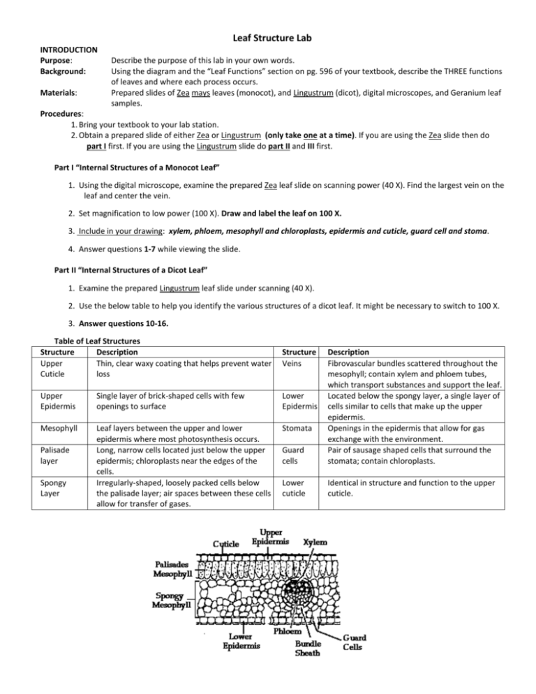
Leaf Structure Lab INTRODUCTION Purpose: Background: Materials: Describe the purpose of this lab in your own words. Using the diagram and the “Leaf Functions” section on pg. 596 of your textbook, describe the THREE functions of leaves and where each process occurs. Prepared slides of Zea mays leaves (monocot), and Lingustrum (dicot), digital microscopes, and Geranium leaf samples. Procedures: 1. Bring your textbook to your lab station. 2. Obtain a prepared slide of either Zea or Lingustrum (only take one at a time). If you are using the Zea slide then do part I first. If you are using the Lingustrum slide do part II and III first. Part I “Internal Structures of a Monocot Leaf” 1. Using the digital microscope, examine the prepared Zea leaf slide on scanning power (40 X). Find the largest vein on the leaf and center the vein. 2. Set magnification to low power (100 X). Draw and label the leaf on 100 X. 3. Include in your drawing: xylem, phloem, mesophyll and chloroplasts, epidermis and cuticle, guard cell and stoma. 4. Answer questions 1-7 while viewing the slide. Part II “Internal Structures of a Dicot Leaf” 1. Examine the prepared Lingustrum leaf slide under scanning (40 X). 2. Use the below table to help you identify the various structures of a dicot leaf. It might be necessary to switch to 100 X. 3. Answer questions 10-16. Table of Leaf Structures Structure Description Upper Thin, clear waxy coating that helps prevent water Cuticle loss Structure Veins Upper Epidermis Single layer of brick-shaped cells with few openings to surface Lower Epidermis Mesophyll Leaf layers between the upper and lower epidermis where most photosynthesis occurs. Long, narrow cells located just below the upper epidermis; chloroplasts near the edges of the cells. Irregularly-shaped, loosely packed cells below the palisade layer; air spaces between these cells allow for transfer of gases. Stomata Palisade layer Spongy Layer Guard cells Description Fibrovascular bundles scattered throughout the mesophyll; contain xylem and phloem tubes, which transport substances and support the leaf. Located below the spongy layer, a single layer of cells similar to cells that make up the upper epidermis. Openings in the epidermis that allow for gas exchange with the environment. Pair of sausage shaped cells that surround the stomata; contain chloroplasts. Lower cuticle Identical in structure and function to the upper cuticle. Part III “The Stomata” 1. Obtain a geranium leaf and using forceps, peal a small amount of epidermis from the lower surface of the leaf. 2. It should be clear and thin. If it is green it is too thick. 3. Place this piece of epidermis on a slide and make a wet mount. 4. Examine under the microscope and draw it on 400x or higher. Label stoma and guard cells. DATA COLLECTION Be sure to include all PROPERLY labelled microscope images and drawings in the section. DATA ANALYSIS 1. How many cell layers make up the upper epidermis? 2. What is the function of the cuticle? 3. What type of tissue comprises the vein? 4. How can you differentiate between these tissues of the vein? (Hint: Think about color and shape) 5. What are the functions of the tissue that comprise a vein? 6. Where are most of the guard cells and stoma? 7. What is the function of the stomata? 8. How thick (how many cell layers) is the palisade layer? 9. What structures are present in the palisades cells? 10. Why are these found in this layer? 11. If you were to travel down the xylem of the leaf where do you think you would end up? 12. What color is the leaf phloem? This might help you answer question # 5. 13. What is the function of the phloem in the leaf? 14. What color is the xylem of the leaf? 15. What is the function of the xylem of the leaf? 16. How does the Lingustrum (Dicot) differ from the Zea (monocot) leaf? CONCLUSION Address the purpose: Discuss what you learned in this lab in relation to the purpose Error Analysis: Suggestions/Modifications:

