doc file - What If
advertisement
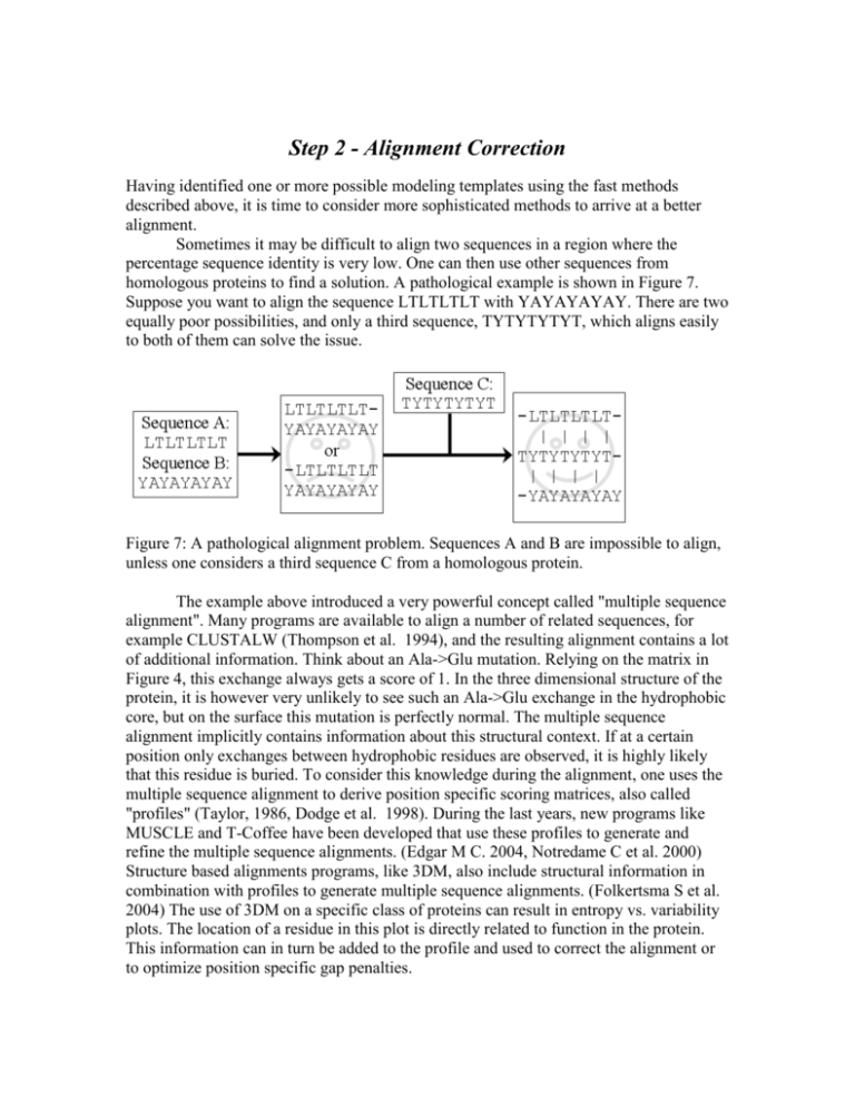
Step 2 - Alignment Correction Having identified one or more possible modeling templates using the fast methods described above, it is time to consider more sophisticated methods to arrive at a better alignment. Sometimes it may be difficult to align two sequences in a region where the percentage sequence identity is very low. One can then use other sequences from homologous proteins to find a solution. A pathological example is shown in Figure 7. Suppose you want to align the sequence LTLTLTLT with YAYAYAYAY. There are two equally poor possibilities, and only a third sequence, TYTYTYTYT, which aligns easily to both of them can solve the issue. Figure 7: A pathological alignment problem. Sequences A and B are impossible to align, unless one considers a third sequence C from a homologous protein. The example above introduced a very powerful concept called "multiple sequence alignment". Many programs are available to align a number of related sequences, for example CLUSTALW (Thompson et al. 1994), and the resulting alignment contains a lot of additional information. Think about an Ala->Glu mutation. Relying on the matrix in Figure 4, this exchange always gets a score of 1. In the three dimensional structure of the protein, it is however very unlikely to see such an Ala->Glu exchange in the hydrophobic core, but on the surface this mutation is perfectly normal. The multiple sequence alignment implicitly contains information about this structural context. If at a certain position only exchanges between hydrophobic residues are observed, it is highly likely that this residue is buried. To consider this knowledge during the alignment, one uses the multiple sequence alignment to derive position specific scoring matrices, also called "profiles" (Taylor, 1986, Dodge et al. 1998). During the last years, new programs like MUSCLE and T-Coffee have been developed that use these profiles to generate and refine the multiple sequence alignments. (Edgar M C. 2004, Notredame C et al. 2000) Structure based alignments programs, like 3DM, also include structural information in combination with profiles to generate multiple sequence alignments. (Folkertsma S et al. 2004) The use of 3DM on a specific class of proteins can result in entropy vs. variability plots. The location of a residue in this plot is directly related to function in the protein. This information can in turn be added to the profile and used to correct the alignment or to optimize position specific gap penalties. When building a homology model, we are in the fortunate situation of having an almost perfect profile - the known structure of the template. We simply know that a certain alanine sits in the protein core and must therefore not be aligned with a glutamate. Multiple sequence alignments are nevertheless useful in homology modeling, for example to place deletions or insertions only in areas where the sequences are strongly divergent. A typical example for correcting an alignment with the help of the template is shown in Figures 8 and 9. Although a sequence alignment gives the highest score for alignment 1 in Figure 8, a simple look at the structure of the template reveals that alignment 2 actually is correct, because it leads to a small gap, compared to a huge hole associated with alignment 1. Figure 8. Example of a sequence alignment where a three-residue deletion must be modeled. While alignment 1, dark grey, appears better when considering just the sequences (a matching proline at position 5), a look at the structure of the template leads to a different conclusion (Figure 9). Figure 9. Correcting an alignment based on the structure of the modeling template (C-trace shown in black). While the alignment with the highest score (dark gray, also in Figure 8) leads to a big gap in the structure, the second option (light gray) creates only a tiny hole. This can easily be accommodated by small backbone shifts.
