NEW TREND IN NUCLEAR MEDICINE, "CODED APERTURE
advertisement
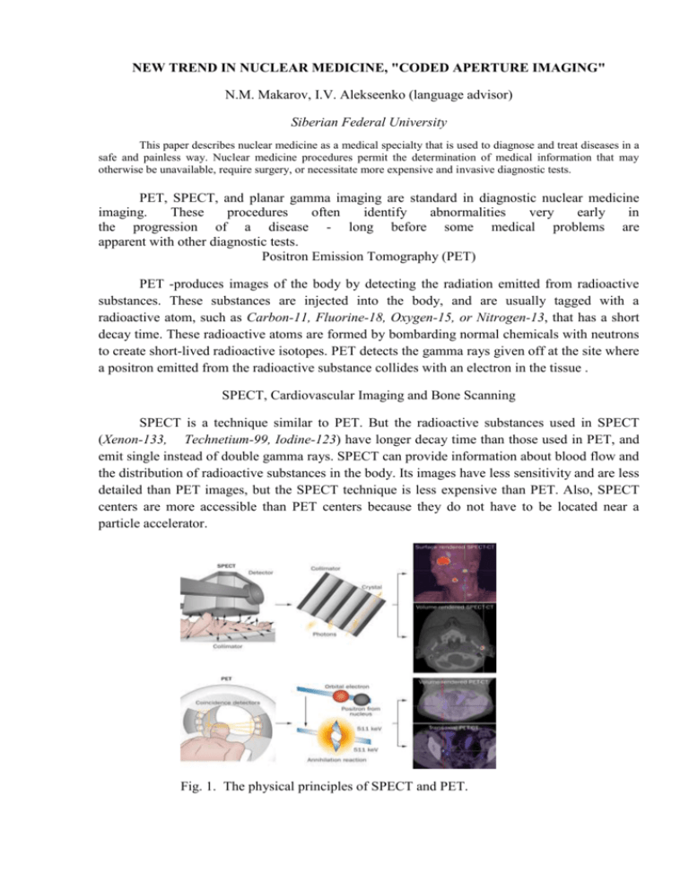
NEW TREND IN NUCLEAR MEDICINE, "CODED APERTURE IMAGING" N.M. Makarov, I.V. Alekseenko (language advisor) Siberian Federal University This paper describes nuclear medicine as a medical specialty that is used to diagnose and treat diseases in a safe and painless way. Nuclear medicine procedures permit the determination of medical information that may otherwise be unavailable, require surgery, or necessitate more expensive and invasive diagnostic tests. PET, SPECT, and planar gamma imaging are standard in diagnostic nuclear medicine imaging. These procedures often identify abnormalities very early in the progression of a disease - long before some medical problems are apparent with other diagnostic tests. Positron Emission Tomography (PET) PET -produces images of the body by detecting the radiation emitted from radioactive substances. These substances are injected into the body, and are usually tagged with a radioactive atom, such as Carbon-11, Fluorine-18, Oxygen-15, or Nitrogen-13, that has a short decay time. These radioactive atoms are formed by bombarding normal chemicals with neutrons to create short-lived radioactive isotopes. PET detects the gamma rays given off at the site where a positron emitted from the radioactive substance collides with an electron in the tissue . SPECT, Cardiovascular Imaging and Bone Scanning SPECT is a technique similar to PET. But the radioactive substances used in SPECT (Xenon-133, Technetium-99, Iodine-123) have longer decay time than those used in PET, and emit single instead of double gamma rays. SPECT can provide information about blood flow and the distribution of radioactive substances in the body. Its images have less sensitivity and are less detailed than PET images, but the SPECT technique is less expensive than PET. Also, SPECT centers are more accessible than PET centers because they do not have to be located near a particle accelerator. Fig. 1. The physical principles of SPECT and PET. We know that, a form of photon collimation for the latter two is used in all these techniques. As a result, this equipment suffers from very low sensitivity. A common collimator system has sensitivity of only 0.1% or even less. The aim of this paper is to demonstrate how significantly is improved sensitivity for a coded aperture imaging system over collimator systems, with retaining reasonable resolution and to show the possibility of using coded apertures and the conventional gamma camera to image gamma rays of high energy in nuclear medicine. Coded aperture imaging is a technique for producing images of radiation emitting objects by using a mask (coded aperture). A radiation will cast a shadow onto a position sensitive detector, thus encoding the spatial information contained in the source. The resulting shadowgram can be deconvolved with a suitable decoding algorithm to reconstruct the original source distribution. The sensitivity improvement is used for coded aperture methods over a "pinhole" camera, which size is like the size of each individual coded aperture. The choice of aperture patterns determines the spatial resolution as well as the system response function. Coded aperture techniques are used to: • provide improved system sensitivity over collimator systems. The sensitivity improvement is proportional to the total open area of the basic coded aperture pattern if the patter opens fraction is the same; • image 511 keV gammas using commercial gamma cameras; • complement colimator systems in nuclear medicine imaging; Coded aperture imaging techniques provide high sensitivity in diagnostic nuclear medicine imaging, compared to collimator systems and it can produce 3-D images with spatial resolutions in the several millimeter region (5 mm or less) for high energy gamma rays. References 1. Zimmermann R. Nuclear Medicine: Radioactivity for Diagnosis and Therapy = La Médecine nucléaire. La radioactivité au service du diagnostic et de la thérapie. // LezJulis: EDP Sciences, 2007. 2. "How Nuclear Medicine Works" by Craig Freudenrich, Ph.D. 3. D. Vaughan A heathy citizenry: Gifts of the New Era, A Vital Legacy: Biological and Environmental Research in the Atomic Age. 4. L.Zhang,R.C.Lanza, B.K.P Horn - "High Energy 3-D Nuclear Medicine Imaging using Coded Apertures with a Conventional Gamma Camera"//NE43-715A,Cambridge,MA 02115.

