1476-4598-13-223-S1
advertisement

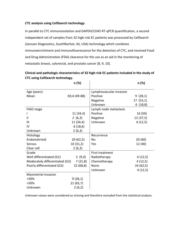
CTC analysis using CellSearch technology In parallel to CTC immunoisolation and GAPDH/CD45 RT-qPCR quantification, a second independent set of samples from 32 high-risk EC patients was processed by CellSearch (Janssen Diagnostics, SouthRaritan, NJ, USA) technology which combines immunoenrichment and immunofluorescence for the detection of CTC, and received Food and Drug Administration (FDA) clearance for the use as an aid in the monitoring of metastatic breast, colorectal, and prostate cancer [8, 9, 10]. Clinical and pathologic characteristics of 32 high-risk EC patients included in the study of CTC using CellSearch technology. n (%) n (%) Age (years) Mean 69,4 (49-88) FIGO stage I 11 (34,4) II 2 (6,3) III 11 (34,4) IV 6 (18,6) Unknown 2 (6,3) Histology Endometrioid 20 (62,5) Serous 10 (31,2) Clear cell 2 (6,3) Grade Well differentiated (G1) 3 (9,4) Moderately differentiated (G2) 7 (21,8) Poorly differentiated (G3) 22 (68,8) Myometrial invasion <50% >50% Unknown Lymphovascular invasion Positive Negative Unknown Lymph node metastasis Positive Negative Unknown 9 (28,1) 17 (53,1) 6 (18,8) 16 (50) 12 (37,5) 4 (12,5) Recurrence No Yes 20 (60) 12 (40) First treatment Radiotherapy Chemotherapy None Unknown 4 (12,5) 4 (12,5) 24 (62,5) 4 (12,5) 9 (28,1) 21 (65,7) 2 (6,2) Unknown values were considered as missing and therefore excluded from the statistical analysis. Briefly, samples were drawn into 10ml evacuated blood draw tubes (CellSave Preservative tubes, Janssen Diagnostics), maintained at room temperature, and processed within 96 hours of collection. The semiautomated CellSearch technology performed an immunomagnetic enrichment of nucleated cells expressing EpCAM from 7.5ml of blood, and a fluorescent labeling with DAPI nuclear marker, CD45-APC for cells of hematogenous origin, and CK-PE cytokeratin staining for epithelial cells. Images of stained cells were acquired by a semiautomated fluorescence microscopy system (Celltracks Analyzer II), and two experimented reviewers selected the CTC, according to a round-oval morphology, nucleated (DAPI+ and ≥ 4µm), lacking CD45 and expressing CK-PE (CK8, 18 and 19) cells from the gallery of objects proposed by the system. CTC counting indicated that 21.9% (n=7) of patients had positivity for CTC identification with a range of 1 to 10 CTC. Interestingly, patients with high grade tumors were those with more CTC compared to low grade tumors (31.5% in grade 3 tumors vs. 10% in grade 1 or 2 tumors, data not shown), and CTC levels were higher in patients with metastatic FIGO Stage IV compared to earlier stages. Quantification of CTC based on CellSearch technology. Representative examples of CTC are shown in upper and middle left panels while a non-CTC of hematopoietic origin is shown in the lower left panel; images are merge of cytokeratin (CK) staining indicative of epithelial origin, DAPI for nucleated cells and CD45 as marker of hematopoietic cells. The quantification of CTC with CellSearch technology in non-metastatic high-risk versus metastatic and recurrent EC patients is shown in the right panel, demonstrating the presence of CTC almost restricted to FIGO Stage IV patients. We finally analyzed in a subset of patients included in the CellSearch study, the RTqPCR expression levels of the panel of biomarkers (n=22). Clinical and pathologic data of this subset of patients is as follows: n (%) Age (years) Mean FIGO Stage I II III IV Histology Endometrioid Serous Grade G1 G2 G3 Myometrial <50% invasión >50% Unknown 68.63 9 (40.9) 1 (4.5) 8 (36.4) 4 (18.2) 15 (68.2) 7 (31.8) 2 (9.1) 4 (18.2) 16 (72.7) n (%) Lymphovascular Positive invasion Negative Unknown Lymph node Positive metastasis Negative Negative Unknown Recurrence Yes No First treatment RT CTX None Unknown 6 (27.3) 15 (68.2) 1 (4.5) 10 (45.5) 12 (54.5) 7 (31.8) 15 (68.2) 4 (18.2) 1 (4.5) 17 (77.3) 1 (4.5) 6 (27.3) 15 (68.2) 1 (4.5) Unknown values were considered as missing and therefore excluded from the statistical analysis. Among genes included in the panel, we found significant higher levels of GDF15 (p=0,02) and the EMT related markers ETV5 and TGFB1 in patients with 1 or more CTC quantified by CellSearch, further indicating the relevance of the plasticity phenotype in the process of endometrial cancer dissemination and metastasis. High GDF15, ETV5 and TGFB1 expression correlated with positive detection of CTC by CellSearch technology in high-risk EC patients. White and grey boxes represent the levels of expression in the group of patients negative and positive for CTC detection by CellSearch technology, respectively. (Mann-Whitney test, *p<0.05). Globally, these results confirmed the presence of CTC in high-risk endometrial cancer patients. The relatively low percentage of positive CTC patients, mainly concentrated in Stage IV disseminated disease, could be due to the stringent criteria used by CellSearch. CTC identifying with CellSearch technology is mainly based on the size of the cytokeratin (CK8, 18+, and/or 19+) signal that should be ≥ 4µm and on the location of the DAPI signal, which should be at least 50% inside the cytokeratin signal. According to these parameters, we hypothesize that CellSearch technology might be discarding tumor cell fragments and multivesicular bodies carrying RNA as a cargo. Furthermore, significant proportion of CTC that express other cytokeratins different than CK8, 18 and/or 19 are also rejected but, on the contrary, may be isolated with EpCAM magnetic-beads and contribute to the RT-qPCR gene expression levels of the candidate genes. We are currently evaluating the possible contribution of multivesicular bodies purified from blood samples of high-risk EC patients to the CTC-phenotype.


