Human_Immunodeficiency_Virus_(HIV)
advertisement
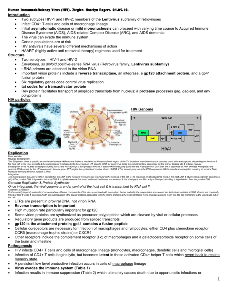
Human Immunodeficiency Virus (HIV). Ziegler. Katelyn Rogers. 04.05.10. Introduction Two subtypes HIV-1 and HIV-2; members of the Lentivirus subfamily of retroviruses Infect CD4+ T-cells and cells of macrophage lineage Initial asymptomatic disease or mild mononucleosis can proceed with varying time course to Acquired Immune Disease Syndrome (AIDS), AIDS-related Complex Disease (ARC), and AIDS dementia The virus can evade the immune system Certain populations are at risk HIV antivirals have several different mechanisms of action HAART (highly active anti-retroviral therapy) regimens used for treatment Structure Two serotypes – HIV-1 and HIV-2 Enveloped, ss diploid positive-sense RNA virus (Retrovirus family, Lentivirus subfamily) t-RNA primers are attached to the virion RNA Important virion proteins include a reverse transcriptase, an integrase, a gp120 attachment protein, and a gp41 fusion protein Six regulatory genes code control virus replication tat codes for a transactivator protein Rev protein facilitates transport of unspliced transcripts from nucleus; a protease processes gag, gag-pol, and env polyproteins HIV particles HIV Genome Replication From websiteReverse transcription: The SU protein binds a specific rec on the cell surface Membrane fusion is mediated by the hydrophobic region of the TM protein or membrane fussion can also occur after endocytosis, depending on the virus & the host cellThe virus uncoats & the nucleocapsid is released into the cytoplasmA specific tRNA for each virus binds the complimentary sequences on the primer binding site & initiates reverse transcriptionThe reverse transcriptase (RT) acts as the RNAdDNAp & also possess RNase H activity The first jump paris with the R sequence of DNA with the R sequence of RNARNase H degrades the genomic RNA except for the +P sequence in the env gene RT begins the synthesis of positive strand of DNAThe second jump pairs the PBS sequencesBoth strands are elongated, creating ds proviral DNA molecule with long terminal repeats (LTRs). Integration: The matrix protein may play a role in transport of the DNA to the nucleus The provirus is circular in the nucleus of the cellThe integrase creats staggered nicks in the host DNA & at proviral recognition sequences (att)The proviral DNA is ligated to the host DNA & a hybrid molecule is formed Mismatched bases are removed & the short gaps are filled in by a DNA pol, resulting in 4bp deletion from the proviral DNA. Genome Replication & Protein Synthesis: Once integrated, the viral genome is under control of the host cell & is transcribed by RNA pol II Assembly & Release: Viral assembly is poorly understood process where different components of the virus associated with each other, before and after the polyproteins are cleaved into individuals proteins 2RNA strands are covalently linked at their 5’ ends & associated with the nucleoprotein the capsid proteins associated with the matrix proteins & the nucleoproteins The envelope proteins insert into the cell membrane & the virus buds out of the cell. LTRs are present in proviral DNA, not virion RNA Reverse transcription is important High mutation rate particularly important for gp120 Some virion proteins are synthesized as precursor polypeptides which are cleaved by viral or cellular proteases Regulatory gene products are produced from spliced transcripts gp120 is the attachment protein; gp41 contains a fusion peptide Cellular coreceptors are necessary for infection of macrophages and lympocytes; either CD4 plus chemokine receptor CCR5 (macrophage-trophic strains) or CXCR4 Other receptors include the complement receptor (Fc) of macrophages and a galactocerebroside receptor on some cells of the brain and intestine Pathogenesis HIV infects CD4+ T cells and cells of macrophage lineage (monocytes, macrophages, dendritic cells and microglial cells) Infection of CD4+ T cells begins lytic, but becomes latent in those activated CD4+ helper T cells which revert back to resting memory state A persistent low level productive infection occurs in cells of macrophage lineage Virus evades the immune system (Table 1) Infection results in immune suppression (Table 2) which ultimately causes death due to opportunistic infections or 1 malignancies Figure 1: Pathogenesis of HIV. HIV causes lytic and latent infection of CD4 T cells, persistent infection of cells of the monocyte/macrophage family, and disrupts neurons. The outcomes of these actions are immunodeficiency and AIDS dementia. Clinical Diseases Initial disease may be asymptomatic or a mild mononucleosis and/or meningitis Disease proceeds with a varying time course through several stages including AIDS-related complex (ARC), AIDS, and AIDS dementia A disease latency period occurs for a varying length of time following acute infection Figure 2: Time course and stages of HIV disease. A long latent period follows the initial mononucleosis-like symptoms. The progressive decrease in CD4 T lymphocytes, even during the latency period, allows opportunistic infections. The stages (World 2 Health Organization [WHO] and Centers for Disease Control [CDC]) in HIV disease are defined by the CD4 T-cell levels and opportunistic diseases. Epidemiology HIV is transmitted through body fluids (blood, semen, vaginal secretions, and breast milk) IV drug abusers, homosexual and heterosexual multipartner sexually active people, prostitutes and children born to HIV+ mothers are at risk Diagnosis AIDS is a clinical diagnosis done in the presence of HIV specific antibodies ELISA tests (SUDS or OraQuick Rapid HIV Antibody Test) for antibody to envelope glycoproteins (gp120 or gp41) for routine screening Western blots are the most popular confirmation test used to detect antibody to viral proteins (Fig. 3). To be considered positive, Abs to at least two bands, including p24, gp41 or gp120/160 must be present Both viral RNA and p24 antigen (capsid protein) is detected in lymphocytes during acute phase of disease and later when virus replication begins in earnest again (Fig. 2) Viral load (amount of viral RNA in blood) allows monitoring of disease progression and efficacy of therapy (Fig. 4) Figure 4 is below: Figure 3: Detection of anti-HIV antibodies by Western Blot Treatment and Prevention Nucloside analogue RT inhibitors like Abacavir, azidothymidine (AZT), dideoxyinosine (ddI), dideoxycytodine (ddC), stavudine (d4T), lamivudine (3TC) and Tenofovir (nucleotide RTi) and promote premature chain termination of DNA synthesis Non-nucleoside RT inhibitors like Nevirapine (NVP), delvaridine (DLV), and efavirenz bind to reverse transcriptase causing a conformational change resulting in lost enzyme activity Amprenavir, indinavir, nelfinavir, ritonavir and saquinavir inhibit HIV protease Enfuviritide inhibits viral envelope-cell membrane fusion AZT is recommended for treatment of mildly symptomatic people and infected pregnant women Triple therapy with two nucleosides plus a protease inhibitor, highly active antiretroviral therapy (HAART), is the regimen of choice for people with more serious disease; no one combination is best for all patients 3 Education, blood screening procedures and infection control procedures used in prevention Vaccines are under development (Table 4). Questions: 1. Fill in the blanks: gp 120 is the _____________protein & gp41 contains a ____________. 2. HIV infects all of the following cells except? a. Dendritic cells b. Microglial cells c. Macrophages d. Monocytes e. Plasma cells 3. What type of drug is dideoxyinosine? a. Nucleoside RTi b. NNRTi c. Nucleotide RTi d. HIV protease inhibitor e. Fusion inhibitor 4. What type of drug is Tenovir in the options of Q3? 5. How about Amprenavir? Answers: 1. Attachment, fusion 2. E 3. A 4 4. 5. C D 5
