02 Acute Leukemia
advertisement
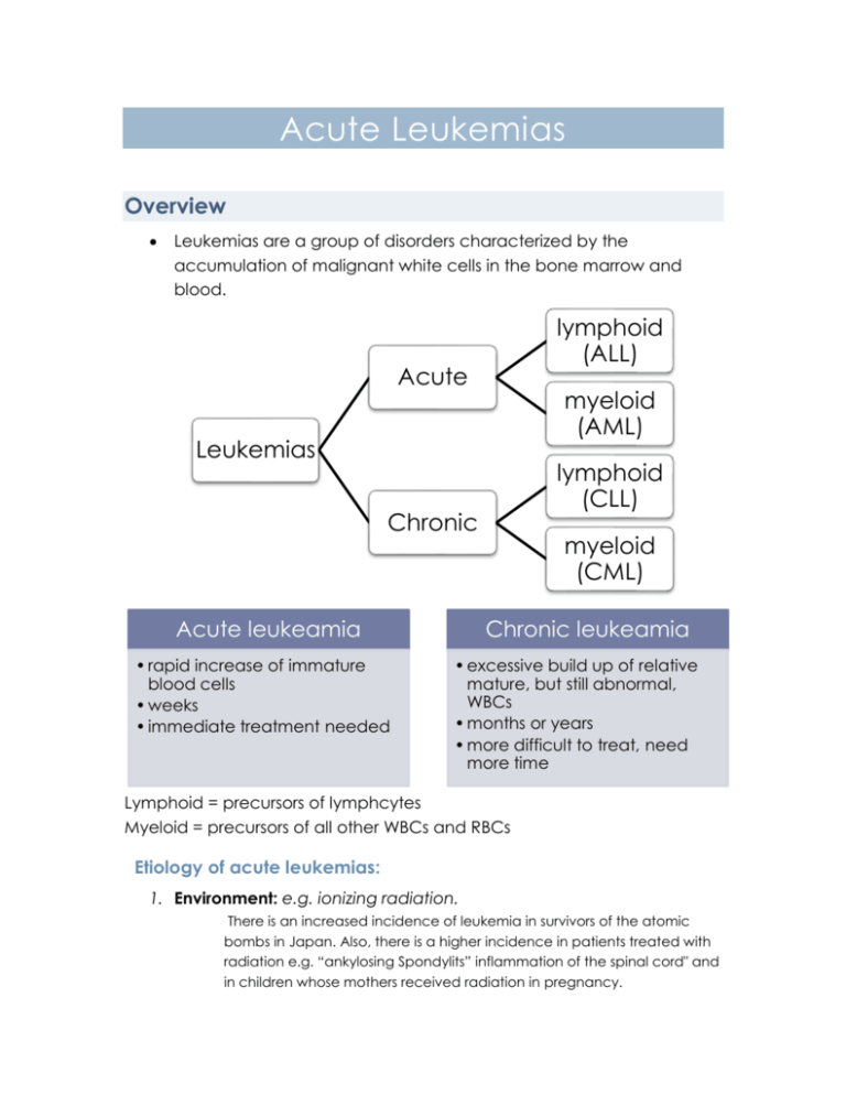
Acute Leukemias Overview Leukemias are a group of disorders characterized by the accumulation of malignant white cells in the bone marrow and blood. lymphoid (ALL) Acute myeloid (AML) Leukemias lymphoid (CLL) Chronic Acute leukeamia • rapid increase of immature blood cells • weeks • immediate treatment needed myeloid (CML) Chronic leukeamia • excessive build up of relative mature, but still abnormal, WBCs • months or years • more difficult to treat, need more time Lymphoid = precursors of lymphcytes Myeloid = precursors of all other WBCs and RBCs Etiology of acute leukemias: 1. Environment: e.g. ionizing radiation. There is an increased incidence of leukemia in survivors of the atomic bombs in Japan. Also, there is a higher incidence in patients treated with radiation e.g. “ankylosing Spondylits” inflammation of the spinal cord" and in children whose mothers received radiation in pregnancy. 2. Chemicals: e.g. Alkylating agents, used to treat malignant neoplasms. 3. Congenital: e.g. Down's syndrome, Bloom’s syndrome 4. Marrow failure syndrome e.g. Fanconis anemia (3,4 are primary factors) 5. Infections: Viral a. EB virus: in Burkitt's lymphoma. b. T cell leukemia/lymphoma virus: An HTLV virus "Human T Lymphocyte Leukemia": a rare virus present in Japan and the Caribbean. Clinical Features of acute leukemias The accumulation of malignant WBCs mainly lead to 2 things, of which arise most of the clinical symptoms of the disease: o Bone marrow failure, due to increase abnormal WBCs too much that displaces the normal progenitor marrow cells, causing: Anemia (due to decreased RBC production) presenting as pallor, lethargy, and dyspnea. Bleeding (due to decreased platelet production) usually purpura, less commonly internal bleeding. Increased susceptibility to Infection (due to decreased normal WBC production) present as fever, mouth ulcers, pharyngeal ulcers, pneumonia, septicemia, skin infection. o Infiltration of organs by malignant leukocytes, causing: Hepatomegaly and splenomegaly Lymphadenopathy Bone, meninges, and testes are usually involved in ALL and are common sites of relapse Gums and skin in AML Laboratory Findings Peripheral Blood: o WBC increased, decreased or normal. o Anemia. o Decreased platelets. Bone marrow: o Increased number of blasts (immature blood cells) Blasts in blood and bone marrow: Normally <5% 5-20%: pre-leukemic >20%: leukemic How to differentiate between acute Myeloid leukemia (AML) and acute Lymphoblastic leukemia (ALL)? A) Morphologically: ALL usually shows no differentiation (except ALL of B-lymphocytes (B-ALL), whereas AMLs are usually differentiated somehow into granulocytes or monocytes. Auer Rods: needle like structures of accumulated granules fond in some AML but not ALL. o Called azurophilic granules because they can be stained by azure stain. AML cells are bigger than ALL cells. B) Cytochemical stains AML are stained by Sudan black and perioxidase, ALL are not Both are stained bypass. C) Immunlogical Monoclonal antibodies: o Myeloid markers: e.g.CD13, CD33. o T-cell lymphoblasts: e.g. CD3 o B cell lymphoblasts: e.g.CD19. o Common ALL lymphoblasts: CALLA (CD 10). A) Acute Lymphoproliferative Leukemia (ALL) Usually seen in childhood, most common 2-10 years. A second peak is seen in middle age and elderly. Classification Classification based on morphology (FAB) or immunological. 1) FAB classification Frequency - In children - In adults Morphology L1 L2 L3 80% 50% 20% 50% 3% of all - Homogenous -Small size -Heterogeneous -Variable size - more nuclei, more cytoplasm -Vacuolated cytoplasm -Basophilic (burkitt cell type) L2 can be misdiagnosed with AML of typesM2 & M1. And can be differentiated by sudan black (+ve in M1 & M2 AML and –ve in L2 ALL) Immunological Classification: ALL is classified according to its immunohistiochemicistry into: 1. Common ALL (C-ALL): both CD10 and CD19 are +ve" a. Most common type (65% of all cases) b. Associated with the C-ALL antigen (CALLA). c. Associated with good prognosis.(20-27 years survival) d. Staining with PAS show block positivity. (in 70-80% of cases) 2. B-lymphocyte ALL (B-ALL): a. Rare: only 3% of cases. b. Poor prognosis. c. Associated with L3 morphology. d. Show B cell antigens e.g. Sig (Surface immunoglobulin) e. It has several subtypes, of which early pre B-ALL "CD19 is + and CD 10 Is –ve 3. T-ALL: a. b. c. d. e. f. g. 10 -20% of cases. Associated with: Older age group. More common in boys. Mediastinal mass. (lymphadenopathy) Paranuclear block positivity for acid phosphatase stain. (+) for T cell antigens e.g. Erosette. "CD3" , "CD7", CD1a and CD5 h. High incidence of CNS relapse and high WBC presentation. Prognostic factors: Parameter Good Poor WBC <20.000 >20.000 Sex Female Male Age 2-10 years <2 or > 10 Morphologic type L1 L3 Immunophenotype Common ALL B Chromosomal Hyperdiploidy (Increase no, of chromosomes) 9:11 (translocation) 4:11 With Hyperdiploidy Other Remission in 4 weeks. CNS involvement (central nervous system). Acute lymphblastic leukemia ccarrying t(9,22) is not related to chronic myeloid leukemia with t(9,22) & have poor prognosis. 2. Acute Myeloproliferative Leukemia Morphologic (FAB) classification: Mo: Myloblastic with No differentiation (without granules) M1: Myeloblastic with poor differentiation (some granules) Similar to ALL in morphology. M2: Myeloblastic with differentiation. "Auer rods can be found" M3: Promyelocytic (many granules) - Diagnostic feature: t(15,17) - Multiple auer rods known as phagote cells Very rare M4: Myelomonocytic. Mixture between myoblast & monoblast M5: Monocytic. M5a: Non mature - Undifferentiated monoblastic M5b: Mature - High nucleus:cytoplasm ratio M6: Ertholeukemia: abnormal erythroid precursors o Very difficult to diagnose, due to similarity with sickle cell anemia & thalassemia. M7: Megakaryoblastic. o More than 20% of non-erythroid cells are megakaryoblasts. Nowadays they use WHO classification which uses molecular level as well ACUTE LYMPHOBLASTIC LEUKEMIA ( ALL) Which cells predominate in ALL? Lymphoblast, with 80% of B-cell origin In what population is ALL found? Children What are some presenting symptoms? Fatigue, weakness, lymphadenopathy. Excessive bleeding, easy bruisabilitv, bone pain, and joint pain What is the prognosis? With current treatment, 5-year survival rates are close to 80%. How are the subgroups classified? Based on morphology and cell-surface markers ACUTE MYELOBLASTIC LEUKEMIA (AML) Which cells predominate in AML? Myeloblasts and early promyelocytes What abnormal granule may be present in blasts? Auer rods In what population is it found? Adults What are some abnormalities on physical exam? Petechiae, lymphadenopathy, hepatosplenomegaly, and thickened gums What is the response to therapy? Not as good as ALL; about 50% How are the subgroups classified? Based on morphology and cytochemical characteristics Treatment of Acute Leukemia 1- Kill the leukemic clones by chemotherapy a. This will kill leukemic clones as well as normal cells in the bone marrow, so the bone marrow will become hypoplastic. 2- Leave the patient for 2-3 weeks, then observe whether there is relapse (the leukemic cells came back) or remission, where normal hematopoiesis occurrs and Hb, RBC count and platelet improves. 3- In AML, we don’t wait for consolidation, instead we give chemotherapeutic agents and go for stem cell transplantation. 4- In ALL we give consolidation and then continue maintenance for 2 years. 5- If there is CNS relapse, we give intrathecal methotrexate or cranial irradiation & continue for maintance for 2 years. 6- If relapse happens within the period of induction of remission, this indicates poor prognosis.
