Apicectomy/Surgical Endodontics Referral Guide
advertisement
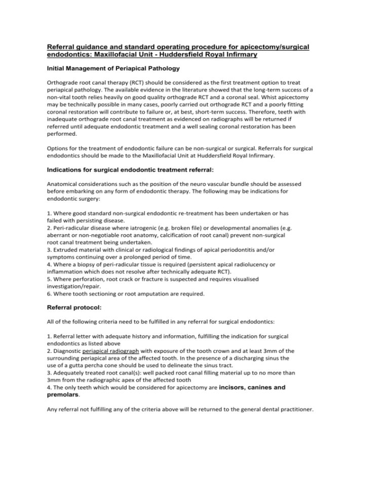
Referral guidance and standard operating procedure for apicectomy/surgical endodontics: Maxillofacial Unit - Huddersfield Royal Infirmary Initial Management of Periapical Pathology Orthograde root canal therapy (RCT) should be considered as the first treatment option to treat periapical pathology. The available evidence in the literature showed that the long-term success of a non-vital tooth relies heavily on good quality orthograde RCT and a coronal seal. Whist apicectomy may be technically possible in many cases, poorly carried out orthograde RCT and a poorly fitting coronal restoration will contribute to failure or, at best, short-term success. Therefore, teeth with inadequate orthograde root canal treatment as evidenced on radiographs will be returned if referred until adequate endodontic treatment and a well sealing coronal restoration has been performed. Options for the treatment of endodontic failure can be non-surgical or surgical. Referrals for surgical endodontics should be made to the Maxillofacial Unit at Huddersfield Royal Infirmary. Indications for surgical endodontic treatment referral: Anatomical considerations such as the position of the neuro vascular bundle should be assessed before embarking on any form of endodontic therapy. The following may be indications for endodontic surgery: 1. Where good standard non-surgical endodontic re-treatment has been undertaken or has failed with persisting disease. 2. Peri-radicular disease where iatrogenic (e.g. broken file) or developmental anomalies (e.g. aberrant or non-negotiable root anatomy, calcification of root canal) prevent non-surgical root canal treatment being undertaken. 3. Extruded material with clinical or radiological findings of apical periodontitis and/or symptoms continuing over a prolonged period of time. 4. Where a biopsy of peri-radicular tissue is required (persistent apical radiolucency or inflammation which does not resolve after technically adequate RCT). 5. Where perforation, root crack or fracture is suspected and requires visualised investigation/repair. 6. Where tooth sectioning or root amputation are required. Referral protocol: All of the following criteria need to be fulfilled in any referral for surgical endodontics: 1. Referral letter with adequate history and information, fulfilling the indication for surgical endodontics as listed above 2. Diagnostic periapical radiograph with exposure of the tooth crown and at least 3mm of the surrounding periapical area of the affected tooth. In the presence of a discharging sinus the use of a gutta percha cone should be used to delineate the sinus tract. 3. Adequately treated root canal(s): well packed root canal filling material up to no more than 3mm from the radiographic apex of the affected tooth 4. The only teeth which would be considered for apicectomy are incisors, canines and premolars. Any referral not fulfilling any of the criteria above will be returned to the general dental practitioner. What the patient can expect from us: A full diagnosis and treatment plan conveyed to the referring dentist by letter. If the referral needs meet the criteria above, patients will be accepted for treatment and placed on a treatment waiting list. The quality of treatment provided will meet the standard operating procedures of the Maxillofacial Unit, Huddersfield Royal Infirmary (see below). The patient will be discharged back to the referring dentist once surgical endodontic treatment is complete. They will be reviewed at one year to assess ongoing success or failure of treatment. Standard operating procedure for Apicectomy/Surgical Endodontics: Operative management 1. Medication There is no current evidence to support the use of peri/post-operative antibiotics but the use of Chlorhexidine mouthwash to reduce plaque formation may be beneficial. Oral analgesia preoperatively could be helpful in the management of post-operative pain. 2. Anaesthesia Local anaesthesia should be the method of choice. The only role for general anaesthesia would be in cases where the size of the periradicular lesion/cyst makes surgery in an outpatient conscious environment unsafe. 3. Magnification The use of magnification with surgical loupes (3-4x) will be the standard of practice in this unit. Although the operating microscope/endoscope has been shown to improve outcomes significantly, this has been primarily restricted to molar apicectomy, a service which would not be provided in this unit. 4. Soft tissue management This will vary depending on a number of factors: Access to and size of the periradicular lesion Periodontal status Status of tooth crown/existing restorations Adjacent anatomical structures and Aesthetic considerations 5. Osteotomy and periradicular curettage The removal of bone, following an assessment of the root-length and its axis, should allow adequate access to the root end. The soft tissue in the periradicular region should be removed by curettage and histopathological assessment of the excised tissue should be reserved for cystic lesions or any atypical findings intra-operatively. As most periradicular pathology is inflammatory in origin, vital anatomical structures (e.g. mental nerve) should be preserved. 6. Root-end resection, preparation and filling Resection of the root should be carried out as close to 90 degrees to the long axis of the tooth as possible, to reduce the number of exposed dentinal tubules. At least 3mm of root end should be resected with a rotating bur. The resected root surface should be examined under magnification for cracks, canal irregularities and a smooth, even finish. The resected root-end canal should be prepared ensuring all gutta percha root canal filling material is removed using angled ultrasonic diamond tips to a depth of 3-4mm, incorporating the entire three-dimensional pathological pulp space. The prepared root-end should be adequately isolated and dry prior to filling. The material utilised in this unit is reinforced zinc-oxide eugenol cement (IRM) 7. Post-operative instructions and care This would be the standard care pathway as for any other minor oral surgical procedures, unless otherwise specified by the operator. Assessment of outcome Evaluation of success/failure of apicectomy is an essential component of clinical governance to this service. The data and findings can be used to inform local commissioners to the future and scope of our surgical practice. The recommended clinical and radiographic follow-up required would be at: 3 months, and 1 year. But longer may be required where healing is incomplete or there is a history of trauma. Radiographic examination endeavours to achieve the same angulation as the preoperative radiograph. The possible outcomes should be categorised as: a. Successful – clinical resolution of signs and symptoms, and radiological evidence of resolution and normal periodontal ligament width or a slight increase (<2x normal periodontal ligament space). b. Incomplete – clinical resolution of signs and symptoms, and partial regeneration of periapical bone (due to scar formation). c. Uncertain – vague symptoms: mild discomfort, feeling of pressure and fullness around the treated tooth; partial radiographic evidence of periapical bone regeneration d. Failure – persistent or worsening clinical signs and symptoms with no radiographic evidence of periapical bone regeneration. Revision apicectomy should only be considered in exceptional circumstances as it has been associated with a very low success rate (36%). References 1. Guidelines for Surgical Endodontics. GE Evans, K Bishop and T Renton. The Faculty of Dentistry, Royal College of Surgeons England 2012. 2. https://www.wales.nhs.uk/sites3/page.cfm?orgid=967&pid=63796. 3. Guy’s, King’s and St. Thomas’ Dental Institute Referral Guidelines. 4. Outcome of endodontic surgery: a meta-analysis of the literature--part 1: Comparison of traditional root-end surgery and endodontic microsurgery.Setzer FC, Shah SB, Kohli MR, Karabucak B, Kim S. J Endod. 2010 Nov;36(11):1757-65 5. Outcome of endodontic surgery: a meta-analysis of the literature--Part 2: Comparison of endodontic microsurgical techniques with and without the use of higher magnification.Setzer FC, Kohli MR, Shah SB, Karabucak B, Kim S. J Endod. 2012 Jan;38(1):1-10
