Sarcomere Coloring
advertisement

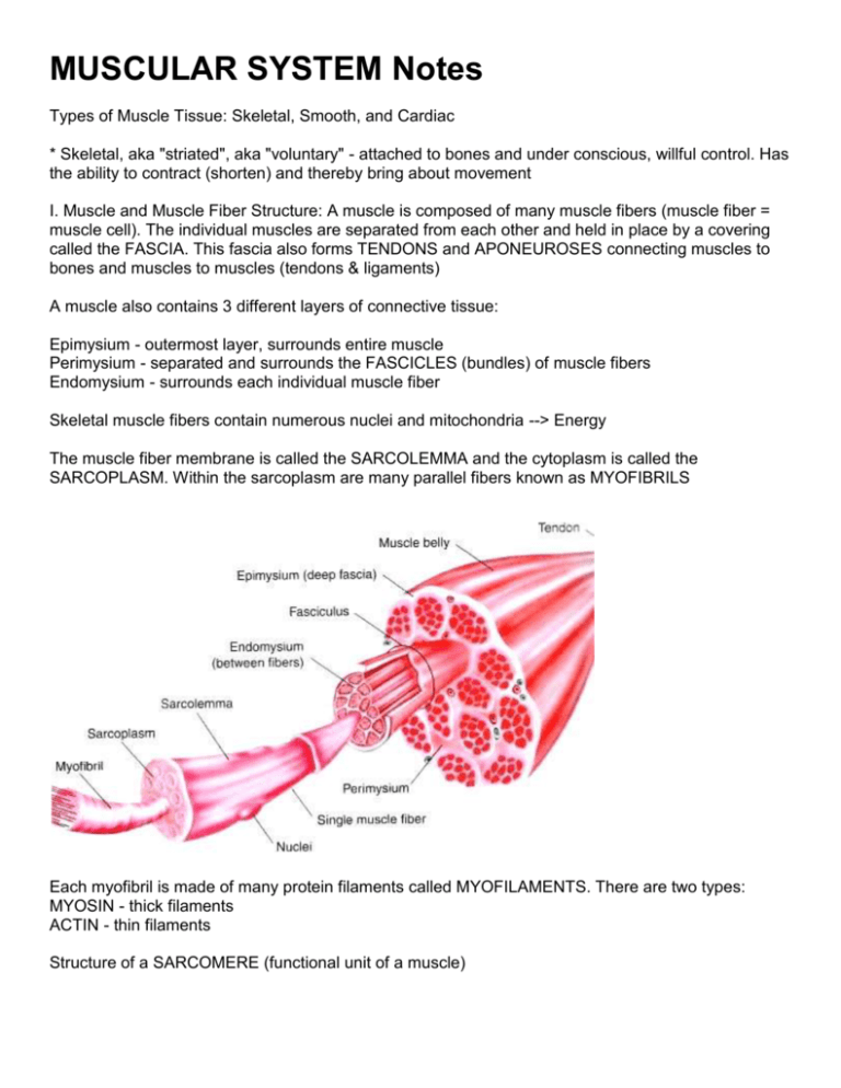
MUSCULAR SYSTEM Notes Types of Muscle Tissue: Skeletal, Smooth, and Cardiac * Skeletal, aka "striated", aka "voluntary" - attached to bones and under conscious, willful control. Has the ability to contract (shorten) and thereby bring about movement I. Muscle and Muscle Fiber Structure: A muscle is composed of many muscle fibers (muscle fiber = muscle cell). The individual muscles are separated from each other and held in place by a covering called the FASCIA. This fascia also forms TENDONS and APONEUROSES connecting muscles to bones and muscles to muscles (tendons & ligaments) A muscle also contains 3 different layers of connective tissue: Epimysium - outermost layer, surrounds entire muscle Perimysium - separated and surrounds the FASCICLES (bundles) of muscle fibers Endomysium - surrounds each individual muscle fiber Skeletal muscle fibers contain numerous nuclei and mitochondria --> Energy The muscle fiber membrane is called the SARCOLEMMA and the cytoplasm is called the SARCOPLASM. Within the sarcoplasm are many parallel fibers known as MYOFIBRILS Each myofibril is made of many protein filaments called MYOFILAMENTS. There are two types: MYOSIN - thick filaments ACTIN - thin filaments Structure of a SARCOMERE (functional unit of a muscle) Actin and Myosin filaments are arranged in an overlapping pattern of light ("I" bands) and dark ("A" bands). In the middle of each "I" band is a line called a "Z" line. The section of a myofibril from one Zline to the next Z-line is the SARCOMERE. *** The arrangement of these sarcomeres next to each other produces the STRIATIONS of the skeletal muscle fibers. Each Myofibril is surrounded by a network of channels called SARCOPLASMIC RETICULUM. TRANVERSE TUBULES pass through the fibers - see p174 II. How Muscles Work with the Nervous System NEUROMUSCULAR JUNCTION - where a NERVE FIBER and muscle fiber come together. A.K.A. Myoneural junction. MOTOR NEURON ENDINGS - nerve fiber caries the impulse that stimulates the muscle fibers MOTOR END PLATE - specialized part of muscle fiber membrane (sarcolemma) located at the neuromuscular junction, has many folds SYNAPTIC CLEFT - An actual "gap" or cleft which exits between the motor neuron endings and the motor end plate. SYNAPTIC VESICLES - numerous vesicles in motor neuron endings, where neurotransmitters are stored before being released into the synaptic cleft. NEUROTRANSMITTER - substance that is released from nerve endings into synaptic cleft. Stimulates an impulse. In this case, a "muscle impulse". One of the major neurotransmitters is ACETYLCHOLINE. This brings about muscle contractions. CHOLINESTERASE is an enzyme that breaks down acetylcholine III. Events in Muscle Contraction: Nerve impulse stimulates the release of a neurotransmitter (acetylcholine) from synaptic vesicles into synaptic cleft ' stimulates muscle impulse ' impulse spreads across sarcolemma and into fiber along the T-tubules ' this impulse causes an increase in the cisternae's permeability to calcium ions. The S.R. has a high conc. of Ca++. Calcium ions diffuse into the sarcoplasm ' the Ca++ causes the formation of "cross bridges" between the actin and myosin filaments ' the filaments slide between each other ' this shortens the myofibrils which in turn shorten the muscle fibers, which shortens the muscles "Calcium Pump" returns CA++ into the S.R. (requires energy- ATP) Enzyme Cholinesterase stops action of Acetylcholine IV. ENERGY SOURCE: Provided by ATP around myofibrils. ATP is produced by cellular respiration which occurs in the mitochondria (requires O2 and glucose) * Creatine Phosphate provides energy for the regeneration of ATP * Only 25% of energy produced during cellular respiration is used in metabolic processes - the rest is in the form of HEAT. This is what produces our body heat and maintains body temperature. More muscle activity = more heat ATP = adenosine triphosphate | ADP = adenosine diphosphate V: Other Terms 1. Threshold Stimulus 2. All-or-None Response 3. Motor Unit 5. Recruitment 6. Muscle Tone 7. Muscular Hypertrophy 8. Muscular Atrophy 9. Muscle Fatigue 10. Muscle Cramp LABEL PRACTICE Fascicle Endomysium (2) Bone Muscle Fiber Epimysium Perimysium Tendon Name:___________________________________Date:__________ Study Guide - Musclular System (Chapter 8) 1. Name and describe the three different layers of connective tissue in a muscle. 2. Myofibrils are composed primarily of two protein filaments called _______________________ and __________________________ 3. What is a motor unit? 4. Name and describe the parts of a neuromuscular junction. 5. What do acetylcholine and cholinesterase do? 6. Bundles of muscular fibers are called ________________________ 7. The more active your muscles are, the more body heat they release. True or False? 8. Name the following structures A. ______________________ Network of membranous channels that surround and run parallel to the myofibrils B. ______________________Thick protein filaments within the A-bands C. ______________________Protein filament that slides inward, toward the middle of a sarcomere, during a muscle contraction D. ______________________ Conducts a muscle impulse deep into a sarcoplasm, to the cisternae E. ______________________The segment of a myofibril between two Z-lines 9. What causes muscles to appear striated? 10. Neurotransmitters are stored in vesicles found in the ____________________________ 11. Creatine phosphate serves to do what? (It supplies energy for what?) 12. What causes muscle fatigue and muscle cramps? 14. What is a threshold stimulus? 15. It is the function of the _________________________ in muscle contractions to supply energy for muscle fiber contractions. 16. What does the "all-or-none response" mean? 17. A "partial, but sustained, contraction" describes what? 18. The ________________________ appearance of skeletal muscles results from the arrangement of sarcomeres. 19. ____________________________ has the function of transmitting a muscle impulse into the interior of the cell. 20. ___________% of energy released by cellular respiration is available for use by metabolic processes - the rest is lost as body heat. 21. A substance called a ______________________ crosses the synaptic cleft and stimulates the muscle fiber to contract. 22. A _______________________ can be described as cordlike and connecting muscles to bones, while an ___________________ would be described as a fibrous sheet of connective tissue connecting muscles to muscles. 23. Describe the events of a muscle contraction in order. What is the sliding filament model? 24. __________________________is the shrinkage of a muscle due to lack of use. 25. What molecules pass in and out of the sarcoplasmic reticulum as a muscle fiber contracts and releases? _________________________ 26. The A bands are [ light / dark ] whereas the I bands are [ light / dark ] 27. What are the three types of muscles? Which type(s) are voluntary? Which type(s) are striated? 28. Where do you find the motor end plate? 29. What is recruitment? 30. Muscle fibers are made of individual ___________________________ which are composed of ___________________ and ____________________ Name:__________________________________Date:_______ Sarcomere Coloring Color the individual myofilaments (A) purple, these are composed of both thick and thin filaments. Mitochondria (B) are dispersed through the muscle fibers, color all mitochondria pink. Recall that mitochondria supply energy needed for muscle contraction. Two types of transport systems are found within the muscle. The sarcoplasmic reticulum (E) is a network of tubes that run parallel to the myofilaments. Color this network green. The transverse tubules (C) run perpendicular to the filaments – color both yellow. The enter muscle fiber is surrounded by the sarcolemma (D), color this membrane brown. If expanded, the light and dark bands are shown as individual thick and thin filaments. Color the thick filaments (not labeled) red and the thin filaments blue. The Z line is the boundary between sarcomeres, named after its shape. Color the Z–line orange. Name:_________________________________________Date:_________ Muscle Anatomy Crossword Down 1. helps regenerate ATP, ___ phosphate 3. thick filaments of a muscle fiber 5. type of muscle that connects to bones, voluntary 6. store neurotransmitters 7. neurotransmitter used to cause muscle contraction 9. connects muscles to bones Down 10. individual muscle fiber 12. organelle that provides the energy needed for muscle contraction 13. connects bones to other bones 15. surrounds fascicles 18. thin filaments of a muscle fiber 19. minimal level of stimulus to cause a contraction 21. this superhero has huge muscles when he's angry; David Banner ACROSS 2. section of a myofibril from one Z line to the next Z line 4. bundle of muscle fibers 8. theory that explains how muscle contraction works; sliding __ theory 11. Outermost layer, surrounds entire muscle 14. describes muscles that are striped in appearance 16. muscle fiber membrane 17. space between a neuron and the muscle, synaptic ___ 20. overlapping patterns of actin and myosin; I and A ___ 22. membranous channels that surround the myofibrils; sarcoplasmic _____ 23. when muscles become tired 24. type of muscle found in the digestive tract, involuntary 25. type of muscle that makes up the heart


